Amplification of the 3q26.3 locus is associated with progression to invasive cancer and is a negative prognostic factor in head and neck squamous cell carcinomas
- PMID: 12163360
- PMCID: PMC1850746
- DOI: 10.1016/S0002-9440(10)64191-0
Amplification of the 3q26.3 locus is associated with progression to invasive cancer and is a negative prognostic factor in head and neck squamous cell carcinomas
Abstract
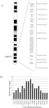
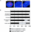
Amplification of the 3q26-q27 has a high prevalence in squamous cell carcinomas of mucosal origin, including those originating in the head and neck region. To elucidate its role as a prognostic tool in head and neck squamous cell carcinoma, a yeast artificial chromosome (YAC) contig spanning the entire 3q26-27 region was constructed. The minimal region of amplification was refined within a 1- to 2-Mb genomic segment contained within three overlapping, nonchimeric YAC clones using sequential fluorescent in situ hybridization analysis. These YAC clones containing the apex of amplification were used to develop a two-color fluorescence in situ hybridization assay and applied to the detection of 3q copy numbers in interphase nuclei on archival tumor tissue from 29 cases of normal mucosa, 20 of premalignant mucosa, and 50 of invasive head and neck squamous cell carcinomas. The presence of 3q amplification increased from 3% in normal mucosa to 25% in premalignant mucosa and 56% in invasive cancers (P < 0.01). In invasive tumors, low-level 3q amplification (3 to 4 X copy number) was identified in 18 of 50 primary head and neck cancers and high-level amplification (>4 X copy number) in 10 of 50 cases. With a median follow-up of 82.5 months, an increasing proportion of recurrences (32%, 72%, and 90%; P = 0.003) and cancer-related deaths (14%, 44%, and 70%; P = 0.006) were seen in patients with normal 3q copy number, low-level amplification, and high-level amplification, respectively. The 3-year disease-free (69%, 56%, and 10%; P = 0.001) and cause-specific (94%, 83%, and 40%; P = 0.01) survivals also decreased from normal copy number to low-level and high-level amplification. Only high-level amplification at 3q remained a significant prognostic variable on multivariate analysis including common prognostic predictors for both disease-free (relative risk, 5.1; 95% confidence interval = 1.9 to 13.9) and cause-specific survival (relative risk, 7.6; 95% confidence interval = 1.9 to 29.6). The findings suggest that the 3q copy number status is an important marker for tumor progression and prognostication in patients with head and neck squamous cell carcinoma.
Figures


References
-
- Knuutila S, Bjorkqvist AM, Autio K, Tarkkanen M, Wolf M, Monni O, Szymanska J, Larramendy ML, Tapper J, Pere H, El-Rifai W, Hemmer S, Wasenius VM, Vidgren V, Zhu Y: DNA copy number amplifications in human neoplasms: review of comparative genomic hybridization studies. Am J Pathol 1998, 152:1107-1123 - PMC - PubMed
-
- Bockmuhl U, Wolf G, Schmidt S, Schwendel A, Jahnke V, Dietel M, Petersen I: Genomic alterations associated with malignancy in head and neck cancer. Head Neck 1998, 20:145-151 - PubMed
Publication types
MeSH terms
Substances
Grants and funding
LinkOut - more resources
Full Text Sources
Medical
Miscellaneous

