. 2002 Aug;161(2):381-9.
doi: 10.1016/S0002-9440(10)64193-4.

Cardiomyogenic differentiation in cardiac myxoma expressing lineage-specific transcription factors

Affiliations

Cardiomyogenic differentiation in cardiac myxoma expressing lineage-specific transcription factors

Hiroaki Kodama et al. Am J Pathol. 2002 Aug.

Abstract

We investigated five cases of cardiac myxoma and one case of cardiac undifferentiated sarcoma by light and electron microscopy, in situ hybridization, immunohistochemical staining, and reverse transcriptase-polymerase chain reaction for cardiomyocyte-specific transcription factors, Nkx2.5/Csx, GATA-4, MEF2, and eHAND. Conventional light microscopy revealed that cardiac myxoma and sarcoma cells presented variable cellular arrangements and different histological characteristics. Ultrastructurally, some of the myxoma cells exhibited endothelium-like or immature mesenchymal cell differentiation. Immunohistochemistry for Nkx2.5/Csx, GATA-4, and eHAND was slightly to intensely positive in all myxoma cases. MEF2 immunoreactivity was observed in all cases including the case of sarcoma, thus suggesting myogenic differentiation of myxoma or sarcoma cells. In situ hybridization for Nkx2.5/Csx also revealed that all myxoma cells, but not sarcoma cells, expressed mRNA of the cardiac homeobox gene, Nkx2.5/Csx. Furthermore, nested reverse transcriptase-polymerase chain reaction from formalin-fixed, paraffin-embedded tissue was performed and demonstrated that the Nkx2.5/Csx and eHAND gene product to be detected in all cases, and in three of six cases, respectively. In conclusion, cardiac myxoma cells were found to express various amounts of cardiomyocyte-specific transcription factor gene products at the mRNA and protein levels, thus suggesting cardiomyogenic differentiation. These results support the concept that cardiac myxoma might arise from mesenchymal cardiomyocyte progenitor cells.


Figures

Figure 1.
Figure 1.
Light micrographs. Conventional histology, immunohistochemistry, and in situ hybridization of cardiac myxomas, sarcoma, and nontumoral cardiomyocytes. A: Light micrographs of cardiac myxoma (case 1), H&E stain. Cardiac myxoma cells forming abortive vascular structures or aggregated cell strands in the abundant myxoid matrix. Mononuclear inflammatory cells seen in the matrix and on the tumor cells. B: Immunohistochemical MEF2 distribution in cardiac myxoma (case 1). C: Immunohistochemical MEF2 distribution in nontumoral cardiomyocytes (case 1). D: Immunohistochemical MEF2 distribution in cardiac sarcoma (case 2). E: Immunohistochemical eHAND distribution in cardiac myxoma (case 1). F: Immunohistochemical eHAND distribution in cardiac myxoma with ring structure (case 3). G: Immunohistochemical GATA-4 distribution in cardiac myxoma (case 4). H: Immunohistochemical GATA-4 distribution in nontumoral cardiomyocytes (case 4). I: Immunohistochemical Nkx2.5/Csx distribution in cardiac myxoma (case 4). J: The Nkx2.5/Csx mRNA distribution demonstrated by in situ hybridization techniques in the cardiac myxoma (case 1). K: The eHAND mRNA distribution demonstrated by in situ hybridization techniques in cardiac myxoma (case 1). L: Negative control of Nkx2.5/Csx mRNA distribution demonstrated by in situ hybridization techniques in cardiac myxoma (case 1). Original magnifications, ×250.
Figure 2.
Figure 2.
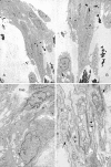
Transmission electron micrographs of the cardiac myxoma from case 1. A and B: Spindle-shaped myxoma cells forming cell strands in the myxoid stroma. Arrow, Cellular junctions between the adjacent tumor cells; arrowhead, rich intracytoplasmic filaments with a subplasmalemmal dense patch; M, macrophages with many lysosomes and hemosiderin deposition in the cytoplasm. C and D: Tumor cells forming a capillary in the myxoma tissue. Tumor cells aggregated forming cell clusters with many short villous cytoplasmic processes, many cellular junctions, and well-developed pericellular lamina. Cap, Tumor cells continuing the structure of a capillary blood vessel showing the lumen; arrow, Weibel-Palade bodies; arrowhead, cellular junctions between the adjacent cells; L, well-developed lamina around the cells. Original magnifications: ×2,500 (A); ×4,700 (B); ×16,000 (C); ×32,000 (D).
Figure 3.
Figure 3.
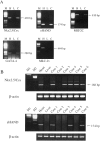
RT-PCR analysis of cardiomyocyte-specific transcription factors. A: RT-PCR analysis of Nkx2.5/Csx, eHAND, MEF2C, GATA-4, and MLC-2v transcript in cardiac myxoma from case 1. Total RNA was isolated from heart (H), liver (L), and cardiac myxoma (C) from case 1. After DNase1 treatment, RT-PCR was performed. The heart and liver were used as positive and negative controls. M, ΦXHaeIII molecular size marker. Cardiomyocytes and cardiac myxoma cells, but not hepatocytes, expressed Nkx2.5/Csx, eHAND, MEF2C, and GATA-4. Cardiac myxoma cells did not express MLC-2v. B: Nested RT-PCR analysis of Nkx2.5/Csx and eHAND. Total RNA was isolated from formalin-fixed, paraffin-embedded sections of the cardiac myxoma and sarcoma from cases 1 to 6, and nested RT-PCR was performed. The expression of Nkx2.5/Csx transcripts was detected in all samples, whereas eHAND transcripts were positive in three of six samples. M1 and M2 represent the ΦXHaeIII and 200-bp molecular size markers, respectively. Samples were identified according to the case’s number.

References

    1. Wold LE, Lie JT: Cardiac myxomas: a clinicopathologic profile. Am J Pathol 1980, 101:219-240 - PMC - PubMed
    1. Reynen K: Cardiac myxomas. N Engl J Med 1995, 333:1610-1617 - PubMed
    1. Burke AP, Virmani R: Cardiac myxoma: a clinicopathologic study. Am J Clin Pathol 1993, 100:671-680 - PubMed
    1. Tanimura A, Kitazono M, Nagayama K, Tanaka S, Kosuga K: Cardiac myxoma: morphologic, histochemical, and tissue culture studies. Hum Pathol 1988, 19:316-322 - PubMed
    1. Deshpande A, Venugopal P, Kumar AS, Chopra P: Phenotypic characterization of cellular component of cardiac myxoma. Hum Pathol 1996, 27:1056-1059 - PubMed

Publication types

MeSH terms