. 2002 Aug;161(2):459-70.
doi: 10.1016/S0002-9440(10)64202-2.

Susceptibility of cyclooxygenase-2-deficient mice to pulmonary fibrogenesis

Affiliations

Susceptibility of cyclooxygenase-2-deficient mice to pulmonary fibrogenesis

James C Bonner et al. Am J Pathol. 2002 Aug.

Abstract

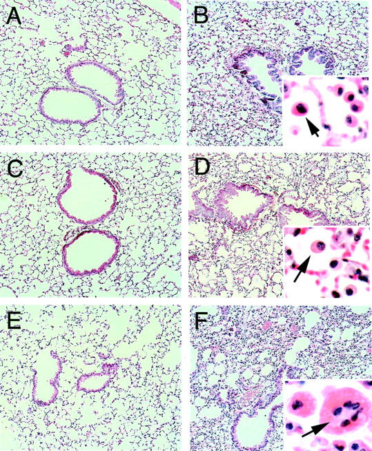
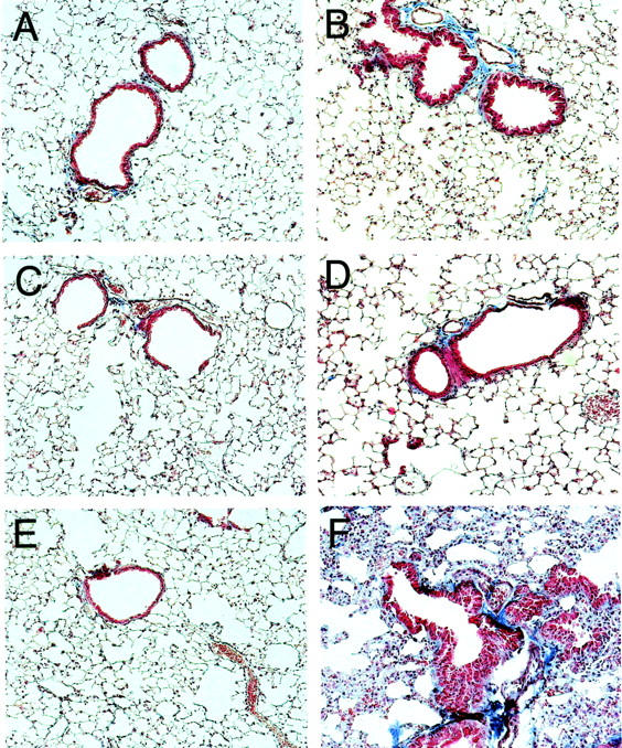
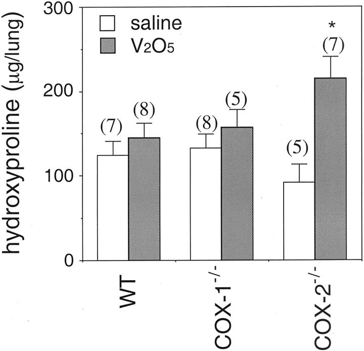
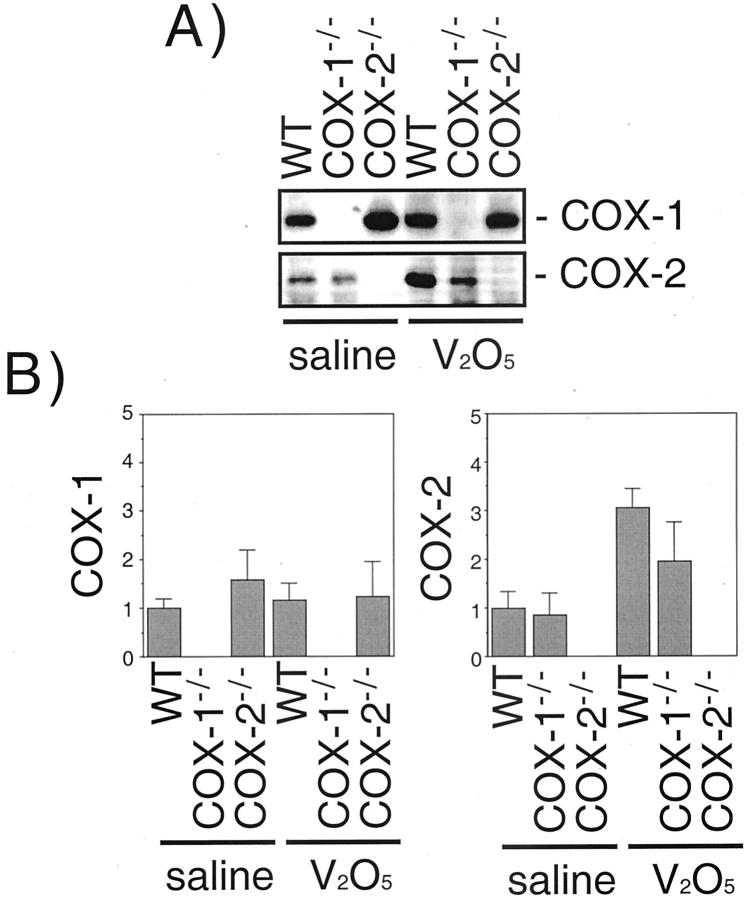
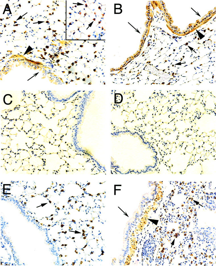
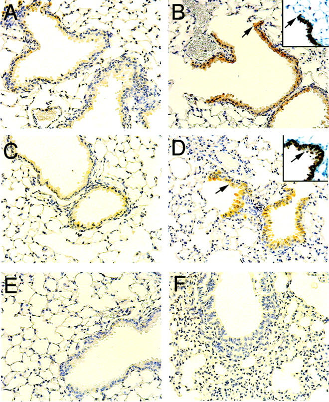
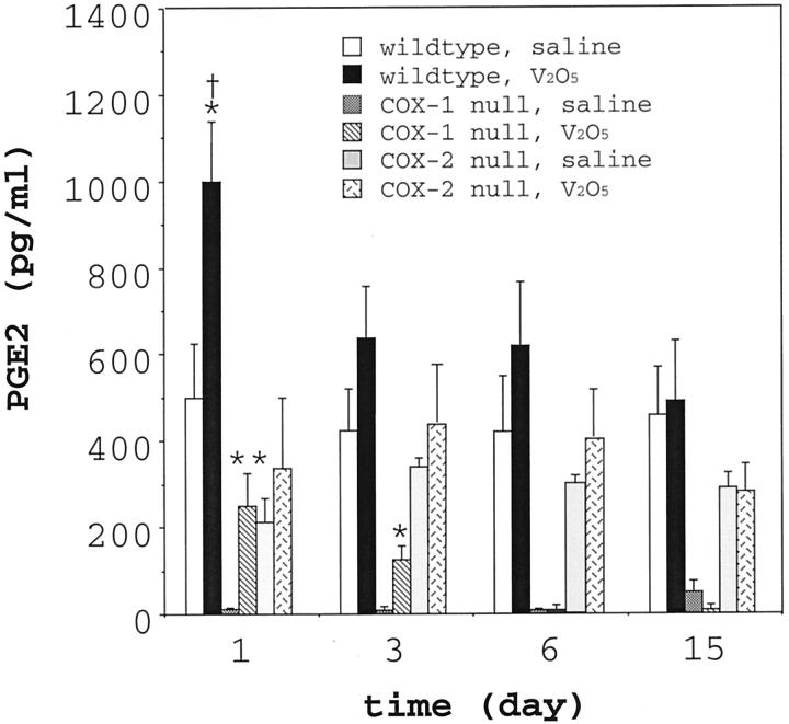
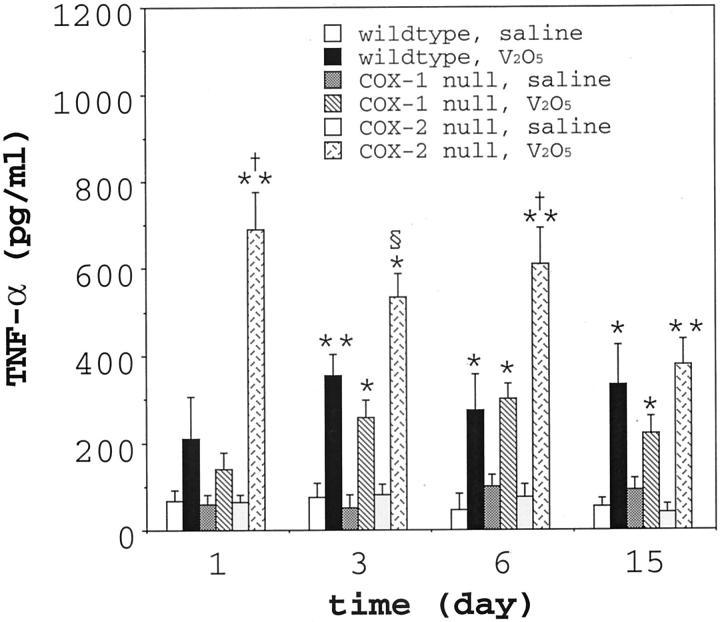
The cyclooxygenase (COX)-2 enzyme has been implicated as an important mediator of pulmonary fibrosis. In this study, the lung fibrotic responses were investigated in COX-1 or COX-2-deficient (-/-) mice following vanadium pentoxide (V(2)O(5)) exposure. Lung histology was normal in saline-instilled wild-type and COX-deficient mice. COX-2(-/-), but not COX-1(-/-) or wild-type mice, exhibited severe inflammatory responses by 3 days following V(2)O(5) exposure and developed pulmonary fibrosis 2 weeks post-V(2)O(5) exposure. Western blot analysis and immunohistochemistry showed that COX-1 protein was present in type 2 epithelial cells, bronchial epithelial cells, and airway smooth muscle cells of saline or V(2)O(5)-exposed wild-type and COX-2(-/-) mice. COX-2 protein was present in Clara cells of wild-type and COX-1(-/-) terminal bronchioles and was strongly induced 24 hours after V(2)O(5) exposure. Prostaglandin (PG) E(2) levels in the bronchoalveolar lavage (BAL) fluid from wild-type and COX-1(-/-) mice were significantly up-regulated by V(2)O(5) exposure within 24 hours, whereas PGE(2) was not up-regulated in COX-2(-/-) BAL fluid. Tumor necrosis factor-alpha was elevated in the BAL fluid from all genotypes after V(2)O(5) exposure, but was significantly and chronically elevated in the BAL fluid from COX-2(-/-) mice above wild-type or COX-1(-/-) mice. These findings indicate that the COX-2 enzyme is protective against pulmonary fibrogenesis, and we suggest that COX-2 generation of PGE(2) is an important factor in resolving inflammation.


Figures

Figure 1.
Figure 1.
Lung histopathology showing increased injury and inflammation in COX-2−/− mice relative to wild-type and COX-1−/− mice 3 days following intratracheal instillation of V2O5. A: Saline-instilled wild-type. B: V2O5-instilled wild-type. Inset shows mononuclear cell infiltration (arrow). C: Saline-instilled COX-1−/−. D: V2O5-instilled COX-1−/−. Arrow in inset indicates mononuclear cell. E: Saline-instilled COX-2−/−. F: V2O5-instilled COX-2−/−. Arrow in inset indicates multinucleated giant cell amid numerous mononuclear cells. All sections were stained with hematoxylin and eosin. Magnification, ×80, except for insets which are ×400.
Figure 2.
Figure 2.
Trichrome staining of lung sections showing enhanced fibrosis in COX-2−/− mice relative to wild-type and COX-1−/− mice 15 days following intratracheal instillation of V2O5. The magnification of each panel is ×80. A: Saline-instilled wild-type. B: V2O5-instilled wild-type. C: Saline-instilled COX-1−/−. D: V2O5-instilled COX-1−/−. E: Saline-instilled COX-2−/−. F: V2O5-instilled COX-2−/−.
Figure 3.
Figure 3.
Hydroxyproline content in lungs of wild-type, COX-1−/−, and COX-2−/− mice instilled with saline or V2O5. Lungs were harvested 15 days after instillation and digested for hydroxyproline content using the colorimetric assay described in Materials and Methods. Numbers in parentheses above each bar indicate number of animals in that group. *, P < 0.05 compared to saline.
Figure 4.
Figure 4.
COX-1 and COX-2 protein expression 24 hours following saline and V2O5 instillation in wild-type, COX-1−/−, and COX-2−/− mice. The abundance of COX-1 and COX-2 protein in the lungs was determined by Western blotting using immunospecific antibodies as described in Materials and Methods. A: Representative Western blot. B: Scanning densitometry of COX-1 and COX-2 expression. Data are the mean ± range of two separate experiments.
Figure 5.
Figure 5.
Immunohistochemistry showing COX-1 localization in type II epithelial cells, airway smooth muscle cells, and airway epithelium. A: Saline-instilled wild-type showing numerous COX-1 positive cells residing within the alveolar walls of the lung parenchyma (arrows); the inset panel shows the results of SP-A immunostaining that confirmed these cells as type II pneumocytes. Airway epithelial cells (barbed arrow) and airway smooth muscle cells (arrowhead) were also positive for COX-1. B: V2O5-instilled wild-type showing COX-1 localized in type II epithelial cells (arrows), airway epithelium (barbed arrows), and airway smooth muscle (arrowhead). C: Saline-instilled COX-1 null. D: V2O5-instilled COX-1 null. E: Saline-instilled COX-2 null showing COX-1 localized in type II cells (arrows) but not in Clara cells of terminal bronchiole. F: V2O5-instilled COX-2 null showing COX-1 localized in airway epithelium (barbed arrow), airway smooth muscle (arrowhead), and type II cells (arrows). Note the absence of COX-1 immunostaining in the COX-1 null mice (C and D). Magnification of all panels ×160.
Figure 6.
Figure 6.
Immunohistochemistry showing COX-2 localization in Clara cells of the distal airways and terminal bronchioles. A: Saline-instilled wild-type showing weak COX-2 immunostaining in Clara cells. B: V2O5-instilled wild-type showing increased COX-2 immunostaining in epithelial cells of terminal bronchioles that were confirmed as Clara cells by immunostaining with a CC10 antibody (inset). Arrows indicate the distal end of the terminal bronchiole in serial sections stained for COX-2 or CC10. C: Saline-instilled COX-1 null. D: V2O5-instilled COX-1 null showing increased COX-2 immunostaining in Clarac cells of terminal bronchioles (inset shows CC10 immunostaining on a serial section and arrows indicate the same bronchiole that is positive for both COX-2 and CC10). E: Saline-instilled COX-2 null. F: V2O5-instilled COX-2 null. Note the absence of COX-2 immunostaining in the COX-2 null mice. All panels ×160.
Figure 7.
Figure 7.
PGE2 levels in BAL fluid in wild-type, COX-1−/−, and COX-2−/− mice following instillation with saline or V2O5. BAL fluid was collected at the indicated time points post-instillation and PGE2 measured by an enzyme immunoassay. Each V2O5 group represents 5 to 6 animals and each saline group represents 3 to 4 animals. *, P < 0.05 vs. all saline-instilled groups. **, P < 0.01 vs. all saline-instilled groups. , P < 0.05 vs. COX-1−/− or COX-2−/− V2O5 group.
Figure 8.
Figure 8.
TNF-α levels in BAL fluid in wild-type, COX-1−/− and COX-2−/− mice following instillation with saline or V2O5. BAL fluid was collected at the indicated time points post-instillation and TNF-α was measured by ELISA. Each V2O5 group represents 5 to 6 animals and each saline group represents 3 to 4 animals. *P < 0.05 vs. all saline-instilled groups. **, P < 0.01 vs. all saline-instilled groups. §, P < 0.05 vs. wild-type V2O5 group. , P < 0.01 vs. wild-type V2O5 group.

References

    1. Smith WL, Dewitt DL: Prostaglandin endoperoxidase H synthases-1 and -2. Adv Immunol 1996, 62:167-215 - PubMed
    1. Gavett SH, Madison SL, Scarborough PE, Qu W, Boyle JE, Chulada PC, Tiano HF, Lee CA, Langenbach R, Roggli VL, Zeldin DC: Allergic lung responses are increased in prostaglandin H synthase-deficient mice. J Clin Invest 1999, 104:721-732 - PMC - PubMed
    1. Wilborn J, Crofford LJ, Burdick MD, Kunkel SL, Strieter RM, Peters-Golden M: Cultured lung fibroblasts isolated from patients with idiopathic pulmonary fibrosis have a diminished capacity to synthesize prostaglandin E2 and to express cyclooxygenase-2. J Clin Invest 1995, 95:1861-1868 - PMC - PubMed
    1. Vancheri C, Sortino MA, Tomaselli V, Mastruzzo C, Condorelli F, Bellistri G, Pistorio MP, Canonico PL, Crimi N: Different expression of TNF-α receptors and prostaglandin-E2 production in normal and fibrotic lung fibroblasts. Am J Respir Cell Mol Biol 2000, 22:628-634 - PubMed
    1. Boyle JE, Lindroos PM, Rice AB, Zhang L, Zeldin DC, Bonner JC: Prostaglanin-E2 counteracts interleukin-1β-stimulated up-regulation of platelet-derived growth factor α-receptor on rat pulmonary myofibroblasts. Am J Respir Cell Mol Biol 1999, 20:433-440 - PubMed