Review
. 2002 Aug 5;158(3):409-14.
doi: 10.1083/jcb.200205009. Epub 2002 Aug 5.

The cell biology of Listeria monocytogenes infection: the intersection of bacterial pathogenesis and cell-mediated immunity

Affiliations
Review

The cell biology of Listeria monocytogenes infection: the intersection of bacterial pathogenesis and cell-mediated immunity

Daniel A Portnoy et al. J Cell Biol. .

Abstract

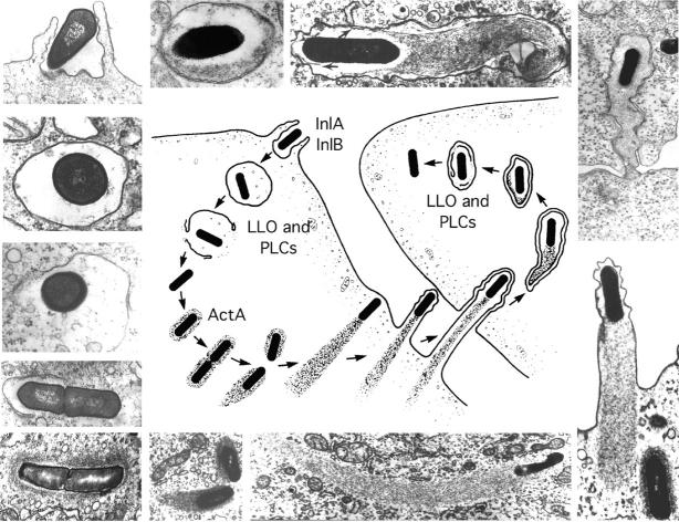
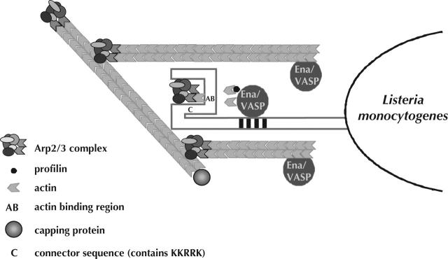
Listeria monocytogenes has emerged as a remarkably tractable pathogen to dissect basic aspects of cell biology, intracellular pathogenesis, and innate and acquired immunity. In order to maintain its intracellular lifestyle, L. monocytogenes has evolved a number of mechanisms to exploit host processes to grow and spread cell to cell without damaging the host cell. The pore-forming protein listeriolysin O mediates escape from host vacuoles and utilizes multiple fail-safe mechanisms to avoid causing toxicity to infected cells. Once in the cytosol, the L. monocytogenes ActA protein recruits host cell Arp2/3 complexes and enabled/vasodilator-stimulated phosphoprotein family members to mediate efficient actin-based motility, thereby propelling the bacteria into neighboring cells. Alteration in any of these processes dramatically reduces the ability of the bacteria to establish a productive infection in vivo.


Figures

Figure 1.
Figure 1.
Stages in the intracellular life-cycle of Listeria monocytogenes. (Center) Cartoon depicting entry, escape from a vacuole, actin nucleation, actin-based motility, and cell-to-cell spread. (Outside) Representative electron micrographs from which the cartoon was derived. LLO, PLCs, and ActA are all described in the text. The cartoon and micrographs were adapted from Tilney and Portnoy (1989).
Figure 2.
Figure 2.
Schematic representation of sequences of interest within LLO. The full length of the box represents the entire translated polypeptide of LLO expressed from the hly gene of Listeria monocytogenes. Shaded regions within the box represent stretches of amino acids of particular interest, but are not depicted to scale. The boundaries of the shaded regions are either indicated by the amino acid number directly above their depicted edge, or on either end of the detailed amino acid sequences contained within the region. MAPK consensus sites within and adjacent to the PEST-like sequence are underlined; NLS, a putative nuclear localization sequence, as predicted by the PSORTII program (http://psort.nibb.ac.jp/); PEST, the PEST-like region defined by PESTFind (http://at.embnet.org/embnet/tools/bio/PESTfind/), is represented as bold characters; SS, the signal sequence cleaved from the propeptide upon secretion; TMH1 and TMH2, transmembrane helixes discovered in PFO that pass through the target membrane upon pore formation (Tweten et al., 2001); UND, the conserved undecapeptide found in all cholesterol-dependent cytolysins. The mutation L461T increases hemolytic activity 10-fold at neutral pH (Glomski et al., 2002).
Figure 3.
Figure 3.
Model of L. monocytogenes actin-based motility. The eukaryotic Arp2/3 complex is activated by the NH2 terminus of the L. monocytogenes surface protein ActA (Welch et al., 1998). ActA, Mena/VASP proteins, and profilin may all contribute to Arp2/3-mediated nucleation by delivering monomer to the complex. At this point, actin clouds can be observed around the bacterium. Once an actin filament is nucleated, profilin may increase elongation rates. The Arp2/3 complex caps the pointed ends of filaments and eventually dissociates from ActA. Mena/VASP proteins interact with the barbed ends of growing filaments and compete with capping protein (Bear et al., 2002). Mena/VASP proteins (which bind ActA and barbed ends) remain concentrated at the bacterial-tail interface, whereas capping protein can be found throughout the tail. Rapidly growing barbed ends (protected from capping protein by Mena/VASP) are concentrated at the site necessary for force generation. At this point, the bacterium can be observed moving throughout the cytosol.

References

    1. Alvarez-Dominguez, C., R. Roberts, and P.D. Stahl. 1997. Internalized Listeria monocytogenes modulates intracellular trafficking and delays maturation of the phagosome. J. Cell Sci. 110:731–743. - PubMed
    1. Auerbuch, V., L. Lenz, and D. Portnoy. 2001. Developmment of a competitive index assay to evaluate the virulence of Listeria monocytogenes actA mutants during primary and secondary infection of mice. Infect. Immun. 69:5953–5957. - PMC - PubMed
    1. Bear, J.E., J.J. Loureiro, I. Libova, R. Fassler, J. Wehland, and F.B. Gertler. 2000. Negative regulation of fibroblast motility by Ena/VASP proteins. Cell. 101:717–728. - PubMed
    1. Bear, J.E., T.M. Svitkina, M. Krause, D.A. Schafer, J.J. Loureiro, G.A. Strasser, I.V. Maly, O.Y. Chaga, J.A. Cooper, G.G. Borisy, and F.B. Gertler. 2002. Antagonism between Ena/VASP proteins and actin filament capping regulates fibroblast motility. Cell. 109:509–521. - PubMed
    1. Beauregard, K.E., K. Lee, R.J. Collier, and J.A. Swanson. 1997. pH-dependent perforation of macrophage phagosomes by listeriolysin O from Listeria monocytogenes. J. Exp. Med. 186:1159–1163. - PMC - PubMed

Publication types

MeSH terms