. 2002 Aug 5;196(3):293-301.
doi: 10.1084/jem.20020308.

Blocking of HIV-1 infection by targeting CD4 to nonraft membrane domains

Affiliations

Blocking of HIV-1 infection by targeting CD4 to nonraft membrane domains

Gustavo Del Real et al. J Exp Med. .

Abstract

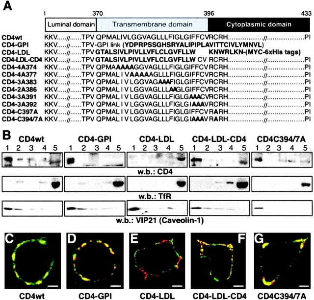
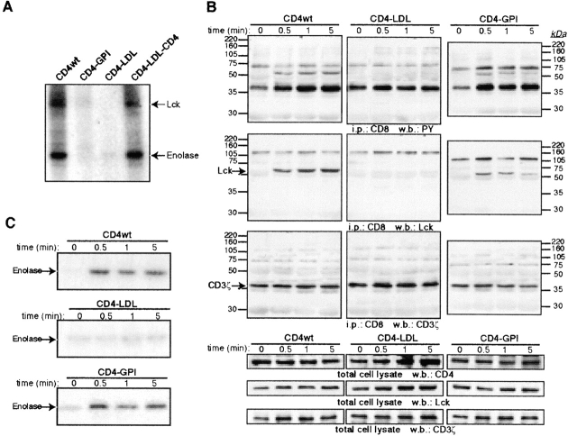
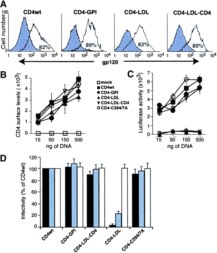
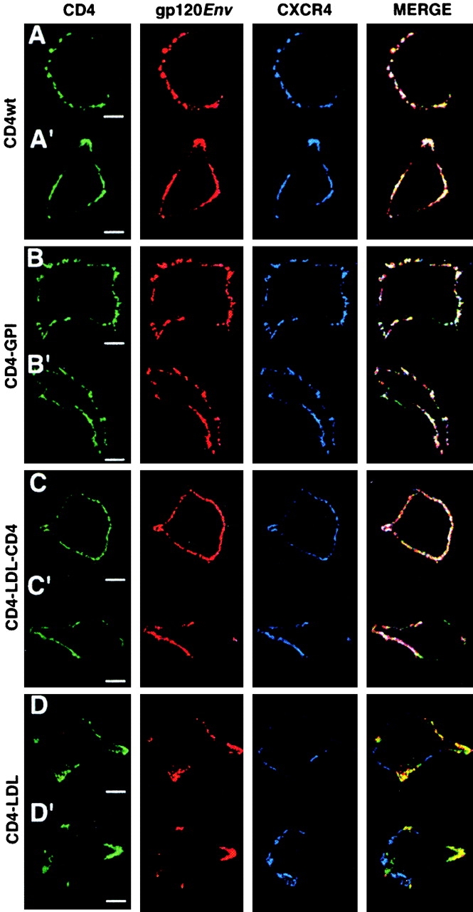
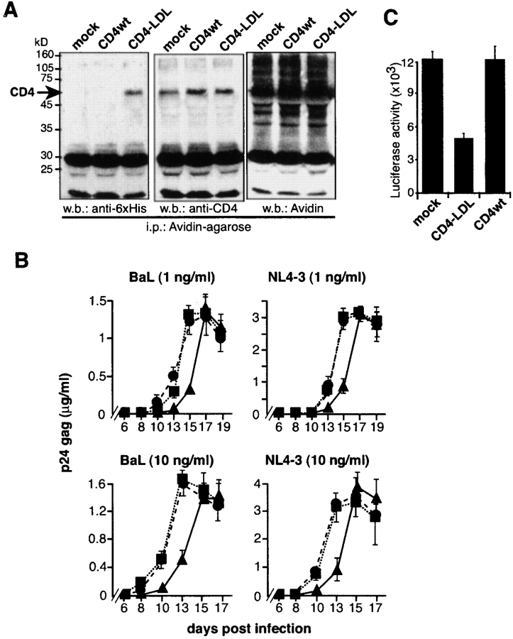
Human immunodeficiency virus (HIV)-1 infection depends on multiple lateral interactions between the viral envelope and host cell receptors. Previous studies have suggested that these interactions are possible because HIV-1 receptors CD4, CXCR4, and CCR5 partition in cholesterol-enriched membrane raft domains. We generated CD4 partitioning mutants by substituting or deleting CD4 transmembrane and cytoplasmic domains and the CD4 ectodomain was unaltered. We report that all CD4 mutants that retain raft partitioning mediate HIV-1 entry and CD4-induced Lck activation independently of their transmembrane and cytoplasmic domains. Conversely, CD4 ectodomain targeting to a nonraft membrane fraction results in a CD4 receptor with severely diminished capacity to mediate Lck activation or HIV-1 entry, although this mutant binds gp120 as well as CD4wt. In addition, the nonraft CD4 mutant inhibits HIV-1 X4 and R5 entry in a CD4(+) cell line. These results not only indicate that HIV-1 exploits host membrane raft domains as cell entry sites, but also suggest new strategies for preventing HIV-1 infection.


Figures

Figure 1.
Figure 1.
Partitioning of CD4 mutants into distinct membrane domains. (A) The scheme shows the amino acid sequence of the CD4 mutants generated. Mutations or foreign sequences added to the CD4 extracellular domain are indicated in bold. (B) HEK-293 cells expressing CD4 mutants were fractionated in flotation gradients and CD4 partitioning was analyzed by Western blot. Fraction 1 represents the top and fraction 5 represents the bottom of the gradient. Filters were hybridized with anti-TfR and anti-VIP21 (caveolin-1) as controls for nonraft- and raft-associated proteins, respectively. (C–G) Confocal microscopy of CD4 mutant–expressing cells stained with cholera toxin β subunit (green) and anti-CD4 antibody (red). Yellow staining indicates colocalization of the molecules. The two-color overlay shows the representative cells for (C) CD4wt, (D) CD4–GPI, (E) CD4–LDL, (F) CD4–LDL–CD4, and (G) CD4–C394/397A (n = 50/mutant). Bar, 5 μm.
Figure 2.
Figure 2.
Raft partitioning, but not association, is a requisite for CD4-induced Lck activation. (A) HEK-293 cells coexpressing the indicated mutants and Lck were anti-CD4 precipitated. CD4-associated tyrosine kinase activity was determined in an IVK assay using enolase as a substrate. The migration of both Lck and enolase is indicated by arrows. (B) HEK-293 cells coexpressing Lck, CD8ζ and CD4wt, and CD4–GPI or CD4–LDL were incubated with anti-CD4 for the times indicated. Additional cross-linking was induced with a secondary anti–mouse antibody. After lysis, anti-CD8 immunoprecipitates were blotted sequentially with antiphosphotyrosine (PY), anti-Lck, and anti-CD3ζ antibodies as indicated. CD4, Lck, and CD8ζ expression levels for each condition were determined by Western blot. (C) IVK assay of anti-Lck immunoprecipitates from lysates in B. Results represent two independent experiments.
Figure 3.
Figure 3.
The nonraft CD4–LDL mutant does not allow HIV-1 entry. (A) HEK-293 cells expressing CD4 chimeras were incubated alone (filled) or with recombinant gp120 (open). Anti-gp120 antibody fluorescence intensity was then recorded in the CD4+ gated cells by flow cytometry. The percentage of gp120-binding CD4+ cells is indicated in each panel. A representative experiment is shown (n = 3). (B) HEK-293 cells were transfected with different amounts of cDNA as indicated, and CD4 immunoreactivity on the cell surface was analyzed by flow cytometry. Cell surface levels of CD4 were calculated by multiplying the percentage of CD4+ cells and the mean fluorescence intensity for each condition. Data represent the mean ± SD of duplicate points (n = 4). (C) The cells in B were mixed with HIV-1IIIB env–expressing HEK-293 cells and cell–cell fusion events were measured. Luciferase activity values were normalized using a promoterless renilla plasmid. (D) Single-round infection experiments were performed in HEK-293-CCR5 cells expressing the indicated CD4 mutants using a replication-defective NL4-3 virus pseudotyped with HIVNL4–3 (solid bar), HIVAda (gray bar), or VSV-G (open bar) envelopes. Cell infection, detected as an increase in luciferase activity, was normalized considering CD4wt as 100%. Data represent mean ± SD of duplicate points (n = 4).
Figure 4.
Figure 4.
Raft partitioning of the CD4 extracellular domain is required for gp120-induced lateral association of CD4 and CXCR4. HEK-293 cells expressing (A and A′) CD4wt, (B and B′) CD4–GPI, (C and C′) CD4–LDL–CD4, and (D and D′) CD4–LDL were incubated with recombinant gp120IIIB and copatched with anti-gp120 (red), anti-CD4 (green), and anti-CXCR4 (blue) as indicated, and then analyzed by confocal microscopy. The three-color overlay is shown in the merge panel. Representative cells are shown (n = 50–60/mutant). Bar, 5 μm.
Figure 5.
Figure 5.
Expression of the nonraft CD4–LDL mutant prevents HIV-1 entry in CD4+ cells. (A) Mock-, CD4wt-, or CD4–LDL-transfected MT-2-CCR5 cells were biotinylated to analyze cell surface CD4 expression. Biotin-labeled proteins were precipitated with agarose-coupled avidin and sequentially blotted with anti-6xHis and anti-CD4 antibodies. Total biotinylated proteins in the same immunoprecipitates were developed with avidin. (B) The cells in A were exposed to R5 (BaL) or X4 (NL4-3) HIV-1 viral strains at the indicated doses and productive infection followed by measurement of p24 antigen levels. Data shown are mean ± SD of triplicate points (n = 3). ▪, mock; •, CD4wt; ▴, CD4–LDL. (C) Mock-, CD4wt-, and CD4–LDL-expressing MT-2-CCR5 cells were infected with a HIVNL4–3 env-pseudotyped replication-defective NL4-3 virus. Infected cells were determined 24 h later by measuring luciferase activity. Data are mean ± SD (n = 4).

References

    1. Simons, K., and D. Toomre. 2000. Lipid rafts and signal transduction. Nat. Rev. Mol. Cell Biol. 1:31–39. - PubMed
    1. Brown, D., and E. London. 1998. Functions of lipid rafts in biological membranes. Annu. Rev. Cell Dev. Biol. 14:111–136. - PubMed
    1. Harder, T., P. Scheiffele, P. Verkade, and K. Simons. 1998. Lipid domain structure of the plasma membrane revealed by patching of membrane components. J. Cell Biol. 141:929–942. - PMC - PubMed
    1. Rodgers, W., and J. Rose. 1996. Exclusion of CD45 inhibits activity of p56lck associated with glycolipid–enriched membrane domains. J. Cell Biol. 135:1515–1523. - PMC - PubMed
    1. Gomez-Mouton, C., J. Abad, E. Mira, R. Lacalle, E. Gallardo, S. Jimenez-Baranda, I. Illa, A. Bernad, S. Mañes, and C. Martinez-A. 2001. Segregation of leading-edge and uropod components into specific lipid rafts during T cell polarization. Proc. Natl. Acad. Sci. USA. 98:9642–9647. - PMC - PubMed

Publication types

MeSH terms

Substances