

Pancreatic lymph nodes are required for priming of beta cell reactive T cells in NOD mice
- PMID: 12163565
- PMCID: PMC2193939
- DOI: 10.1084/jem.20011353
Pancreatic lymph nodes are required for priming of beta cell reactive T cells in NOD mice
Abstract
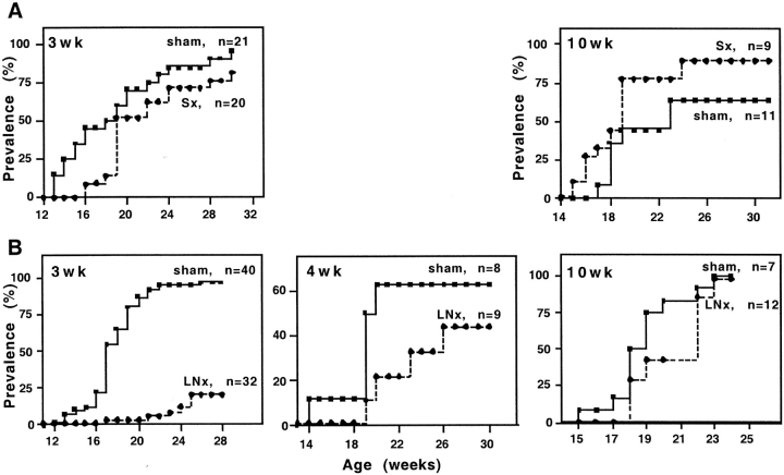
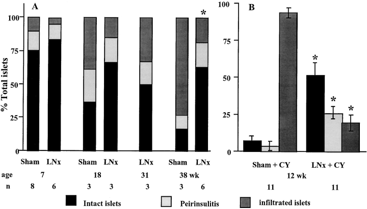
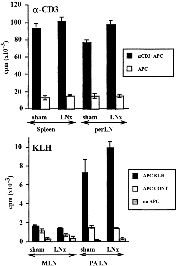
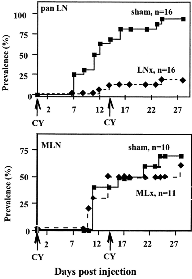
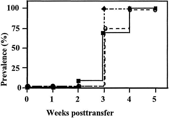
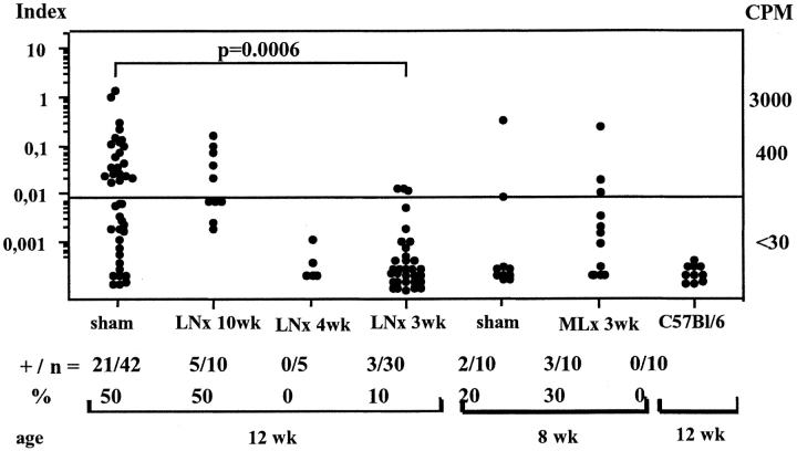
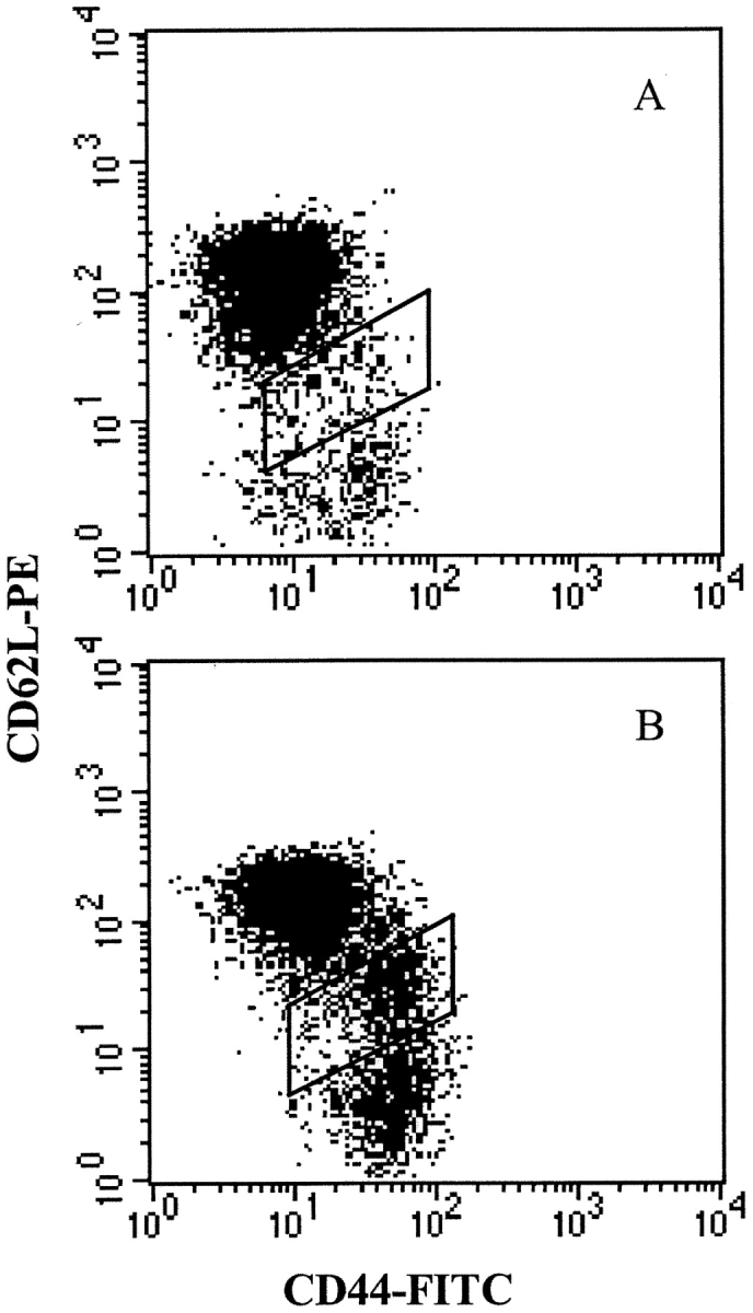
Nonobese diabetic (NOD) mice develop spontaneous autoimmune diabetes that results from the destruction of insulin secreting beta cells by diabetogenic T cells. The time and location of the encounter of autoantigen(s) by naive autoreactive T cells in normal NOD mice are still elusive. To address these issues, we analyzed diabetes development in mice whose spleen or pancreatic lymph nodes (panLNs) had been removed. Excision of panLNs (panLNx) at 3 wk protected mice against insulin autoantibodies (IAAs), insulitis, and diabetes development almost completely, but had no effect when performed at 10 wk. The protection afforded by panLNx at weaning was not due to modifications of the immune system, the absence of autoreactive T cells, or the increase in the potency of regulatory T cells. That panLNs are dispensable during adult life was confirmed by the capacity of 10-wk-old panLNx irradiated recipients to develop diabetes upon transfer of diabetogenic T cells. In contrast, splenectomy had no effect at any age. Partial excision of mesenteric LN at 3 wk did not prevent accelerated diabetes by cyclophosphamide as panLNx did. Thus, in normal NOD mice, autoreactive T cell initial priming occurs in LNs draining the target organ of the disease from 3 wk of age.
Figures
References
-
- Bach, J.F. 1995. Insulin-dependent diabetes mellitus as a β-cell targeted disease of immunoregulation. J. Autoimmun. 8:439–463. - PubMed
-
- Delovitch, T.L., and B. Singh. 1997. The nonobese diabetic mouse as a model of autoimmune diabetes: Immune dysregulation gets the NOD. Immunity. 7:727–738. - PubMed
-
- Shimizu, J., E. Carrasco-Marin, O. Kanagawa, and E.R. Unanue. 1995. Relationship between β cell injury and antigen presentation in NOD mice. J. Immunol. 155:4095–4099. - PubMed
-
- Masopust, D., V. Vezys, A.L. Marzo, and L. Lefrancois. 2001. Preferential localization of effector memory cells in nonlymphoid tissue. Science. 291:2413–2417. - PubMed
-
- Lee Reinhardt, R., A. Khoruts, R. Merica, T. Zell, and M.K. Jenkins. 2001. Visualizing the generation of memory CD4 T cells in the whole body. Nature. 410:101–105. - PubMed
Publication types
MeSH terms
Substances
LinkOut - more resources
Full Text Sources
Other Literature Sources
Medical
