

Phenotype, localization, and mechanism of suppression of CD4(+)CD25(+) human thymocytes
- PMID: 12163566
- PMCID: PMC2193942
- DOI: 10.1084/jem.20020110
Phenotype, localization, and mechanism of suppression of CD4(+)CD25(+) human thymocytes
Abstract
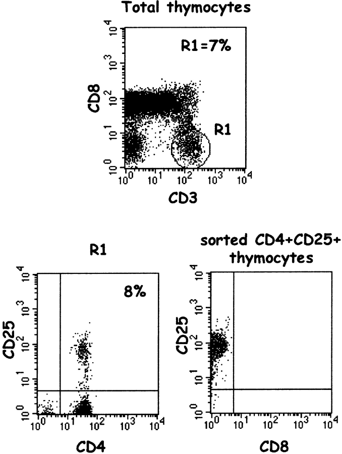
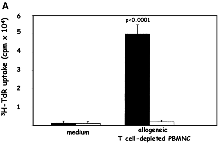
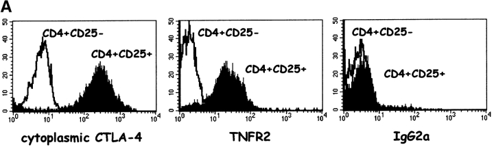
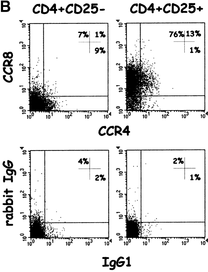
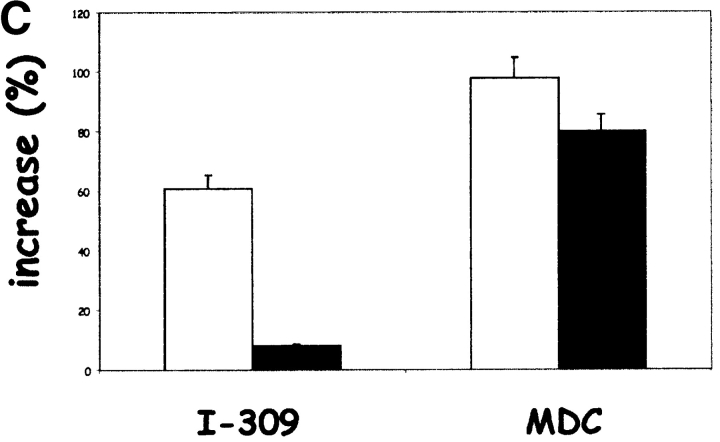
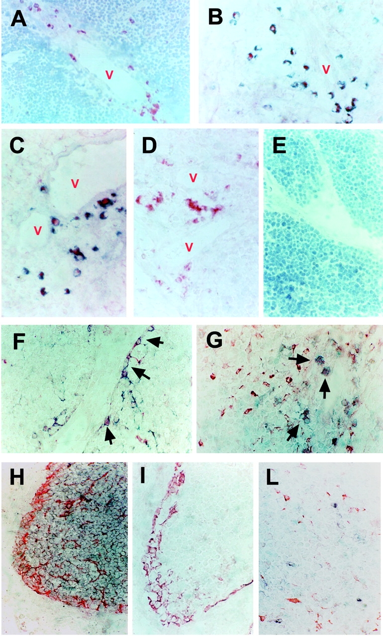
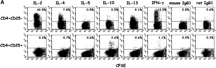
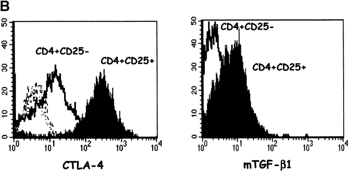
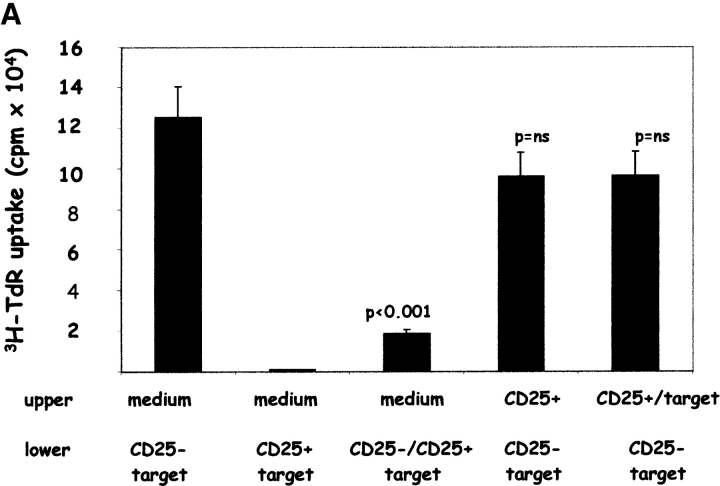
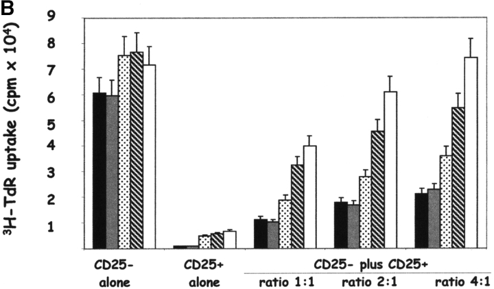
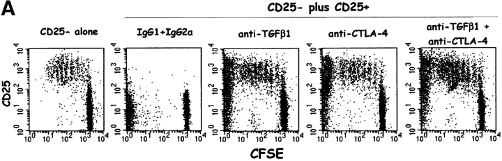
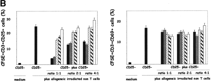
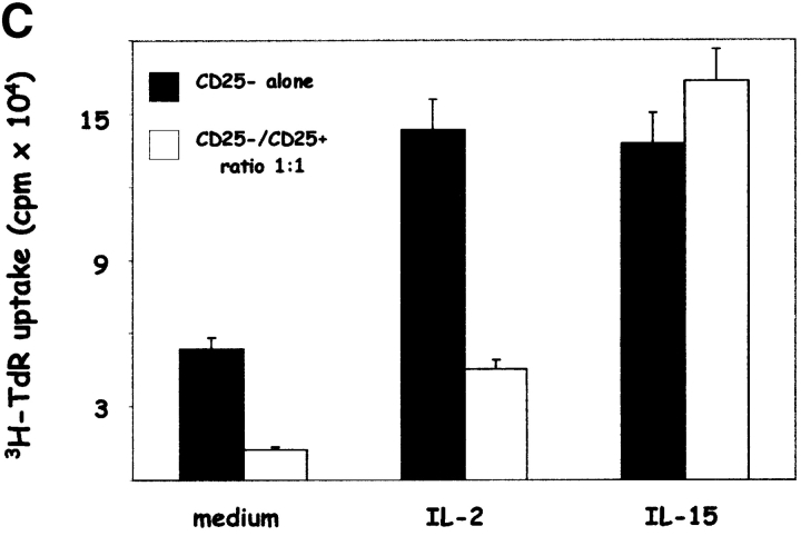
Phenotypic markers, localization, functional activities, and mechanisms of action in vitro of CD4(+)CD25(+) T cells, purified from postnatal human thymuses, were investigated. These cells showed poor or no proliferation in mixed lymphocyte culture (MLC), and suppressed in a dose-dependent fashion the proliferative response to allogeneic stimulation of CD4(+)CD25(-) thymocytes. Virtually all CD4(+)CD25(+) thymocytes constitutively expressed cytoplasmic T lymphocyte antigen (CTLA)-4, surface tumor necrosis factor type 2 receptor (TNFR2), and CCR8. They prevalently localized to perivascular areas of fibrous septa and responded to the chemoattractant activity of CCL1/I-309, which was found to be produced by either thymic medullary macrophages or fibrous septa epithelial cells. After polyclonal activation, CD4(+)CD25(+) thymocytes did not produce the cytokines interleukin (IL)-2, IL-4, IL-5, IL-13, interferon gamma, and only a very few produced IL-10, but all they expressed on their surface CTLA-4 and the majority of them also transforming growth factor (TGF)-beta1. The suppressive activity of these cells was contact dependent and associated with the lack of IL-2 receptor (IL-2R) alpha-chain (CD25) expression in target cells. Such a suppressive activity was partially inhibited by either anti-CTLA-4 or anti-TGF-beta1, and was completely blocked by a mixture of these monoclonal antibodies, which were also able to restore in target T cells the expression of IL-2R alpha-chain and, therefore, their responsiveness to IL-2. These data demonstrate that CD4(+)CD25(+) human thymocytes represent a population of regulatory cells that migrate in response to the chemokine CCL1/I-309 and exert their suppressive function via the inhibition of IL-2R alpha-chain in target T cells, induced by the combined activity of CTLA-4 and membrane TGF-beta1.
Figures

References
-
- Sakaguchi, S. 2000. Regulatory T cell: key controllers of immunologic self-tolerance. Cell. 101:455–458. - PubMed
-
- Shevach, E.M. 2000. Regulatory T cells in autoimmunity. Annu. Rev. Immunol. 18:423–449. - PubMed
-
- Suri-Payer, E., A.Z. Amar, R. McHugh, K. Natarajan, D.H. Margulies, and E.M. Shevach. 1999. Postéthymectomy autoimmune gastritis: fine specificity and pathogenicity of anti-H/K ATPase reactive T cells. Eur. J. Immunol. 29:669–677. - PubMed
Publication types
MeSH terms
Substances
LinkOut - more resources
Full Text Sources
Other Literature Sources
Research Materials
Miscellaneous
