. 2002 Aug 5;196(3):389-99.
doi: 10.1084/jem.20020399.

Donor-type CD4(+)CD25(+) regulatory T cells suppress lethal acute graft-versus-host disease after allogeneic bone marrow transplantation

Affiliations

Donor-type CD4(+)CD25(+) regulatory T cells suppress lethal acute graft-versus-host disease after allogeneic bone marrow transplantation

Petra Hoffmann et al. J Exp Med. .

Abstract

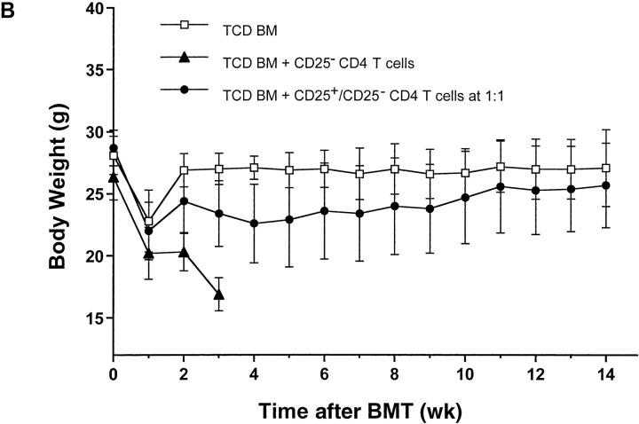
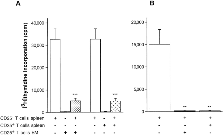
Acute graft-versus-host disease (aGVHD) is still a major obstacle in clinical allogeneic bone marrow (BM) transplantation. CD4(+)CD25(+) regulatory T (T(reg)) cells have recently been shown to suppress proliferative responses of CD4(+)CD25(-) T cells to alloantigenic stimulation in vitro and are required for ex vivo tolerization of donor T cells, which results in their reduced potential to induce aGVHD. Here we show that CD4(+)CD25(+) T cells isolated from the spleen or BM of donor C57BL/6 (H-2(b)) mice that have not been tolerized are still potent inhibitors of the alloresponse in vitro and of lethal aGVHD induced by C57BL/6 CD4(+)CD25(-) T cells in irradiated BALB/c (H-2(d)) hosts in vivo. The addition of the CD4(+)CD25(+) T(reg) cells at a 1:1 ratio with responder/inducer CD4(+)CD25(-) T cells resulted in a >90% inhibition of the mixed leukocyte reaction and marked protection from lethal GVHD. This protective effect depended in part on the ability of the transferred CD4(+)CD25(+) T cells to secrete interleukin 10 and occurred if the T(reg) cells were of donor, but not host, origin. Our results demonstrate that the balance of donor-type CD4(+)CD25(+) T(reg) and conventional CD4(+)CD25(-) T cells can determine the outcome of aGVHD.


Figures

Figure 1.
Figure 1.
(A) Composition of the CD4+ T cell population in the spleen of an adult C57BL/6 mouse and purity of the FACS®-sorted CD25 and CD25+ subpopulations. (B) Dose-dependent suppression of the alloresponses of C57BL/6 CD4+CD25 T cells to BALB/c stimulator cells by C57BL/6 CD4+CD25+ T cells. Cultures were set up with 105 sorted C57BL/6 CD4+CD25 T cells and 105 irradiated BALB/c stimulator cells plus variable numbers of C57BL/6 CD4+CD25+ T cells to obtain the indicated ratios. The bars represent the means of triplicate values and the brackets indicate the SDs. **, P < 0.01; *, P < 0.05 (Student's t test). One of two experiments with similar results is shown.
Figure 1.
Figure 1.
(A) Composition of the CD4+ T cell population in the spleen of an adult C57BL/6 mouse and purity of the FACS®-sorted CD25 and CD25+ subpopulations. (B) Dose-dependent suppression of the alloresponses of C57BL/6 CD4+CD25 T cells to BALB/c stimulator cells by C57BL/6 CD4+CD25+ T cells. Cultures were set up with 105 sorted C57BL/6 CD4+CD25 T cells and 105 irradiated BALB/c stimulator cells plus variable numbers of C57BL/6 CD4+CD25+ T cells to obtain the indicated ratios. The bars represent the means of triplicate values and the brackets indicate the SDs. **, P < 0.01; *, P < 0.05 (Student's t test). One of two experiments with similar results is shown.
Figure 2.
Figure 2.
(A) Lethal GVHD of BALB/c recipients induced by C57BL/6 CD4+CD25 T cells can be suppressed by coinjected C57BL/6 CD4+CD25+ T cells. BALB/c mice received 800 cGy TBI, 2 × 10⁶ C57BL/6 TCD BM cells, and 4.5 × 10⁵ C57BL/6 splenic CD4+CD25 T cells (CD25) with variable numbers of C57BL/6 splenic CD4+CD25+ (CD25+) or CD4+CD25 T cells (CD25) to obtain the indicated ratios. Combined data from four independent experiments with 10–21 animals per group are shown. (B) Mean body weights and SDs of BALB/c hosts (10 animals per group) after BMT. BALB/c mice received 800 cGy lethal TBI and either 2 × 10⁶ C57BL/6 TCD BM cells alone (□) or TCD BM plus 4.5 × 10⁵ C57BL/6 CD4+CD25 T cells (▴), or TCD BM plus a 1:1 mixture of 4.5 × 10⁵ C57BL/6 CD4+CD25+ and 4.5 × 10⁵ CD4+CD25 T cells (•).
Figure 2.
Figure 2.
(A) Lethal GVHD of BALB/c recipients induced by C57BL/6 CD4+CD25 T cells can be suppressed by coinjected C57BL/6 CD4+CD25+ T cells. BALB/c mice received 800 cGy TBI, 2 × 10⁶ C57BL/6 TCD BM cells, and 4.5 × 10⁵ C57BL/6 splenic CD4+CD25 T cells (CD25) with variable numbers of C57BL/6 splenic CD4+CD25+ (CD25+) or CD4+CD25 T cells (CD25) to obtain the indicated ratios. Combined data from four independent experiments with 10–21 animals per group are shown. (B) Mean body weights and SDs of BALB/c hosts (10 animals per group) after BMT. BALB/c mice received 800 cGy lethal TBI and either 2 × 10⁶ C57BL/6 TCD BM cells alone (□) or TCD BM plus 4.5 × 10⁵ C57BL/6 CD4+CD25 T cells (▴), or TCD BM plus a 1:1 mixture of 4.5 × 10⁵ C57BL/6 CD4+CD25+ and 4.5 × 10⁵ CD4+CD25 T cells (•).
Figure 3.
Figure 3.
Phenotypic characterization of CD4+CD25+NK1.1 T cells from C57BL/6 BM and spleen. (A) Proportion of CD4+CD25+NK1.1 cells among TCRαβ+ cells in C57BL/6 BM (top) and spleen (bottom). (B) Surface expression of CD62L, CD44, and CD45RB, and intracellular/surface expression of CTLA-4 by CD4+CD25+NK1.1 T cells in C57BL/6 BM (bold line) and spleen (filled).
Figure 3.
Figure 3.
Phenotypic characterization of CD4+CD25+NK1.1 T cells from C57BL/6 BM and spleen. (A) Proportion of CD4+CD25+NK1.1 cells among TCRαβ+ cells in C57BL/6 BM (top) and spleen (bottom). (B) Surface expression of CD62L, CD44, and CD45RB, and intracellular/surface expression of CTLA-4 by CD4+CD25+NK1.1 T cells in C57BL/6 BM (bold line) and spleen (filled).
Figure 4.
Figure 4.
Suppressive effect of NK1.1CD4+CD25+ T cells from the BM of C57BL/6 mice. (A) Suppression of the proliferation of 2.5 × 10⁴ C57BL/6 splenic CD4+CD25 T cells, stimulated with soluble anti–CD3-Ab in the presence of autologous APC by 2.5 × 10⁴ C57BL/6 NK1.1CD4+CD25+ T cells from BM, or CD4+CD25+ T cells from the spleen. The bars represent the means of triplicate values and the brackets indicate the SDs. ***, P < 0.001 (Student's t test). One of two experiments with similar results is shown. (B) Suppression of the alloresponse of C57BL/6 CD4+CD25 T cells to BALB/c stimulator cells by C57BL/6 BM CD4+CD25+ T cells. Cultures were set up with 105 sorted splenic C57BL/6 CD4+CD25 T cells and 105 irradiated BALB/c stimulator cells plus 105 C57BL/6 BM or splenic CD4+CD25+ T cells. The bars represent the means of triplicate values and the brackets indicate the SDs. **, P < 0.01 (Student's t test). One of two similar experiments is shown. (C) BALB/c recipients of C57BL/6 CD4+CD25 PB T cells can be rescued from lethal aGVHD by the coinjection of C57BL/6 NK1.1CD4+CD25+, but not NK1.1CD4+CD25, BM T cells. BALB/c mice received 800 cGy TBI, 2 × 10⁶ C57BL/6 TCD BM cells, and 105 CD4+CD25 PB T cells with or without an equal number of CD4+CD25+NK1.1 or CD4+CD25NK1.1 BM T cells. Combined data from two independent experiments with six to nine mice per group are shown.
Figure 4.
Figure 4.
Suppressive effect of NK1.1CD4+CD25+ T cells from the BM of C57BL/6 mice. (A) Suppression of the proliferation of 2.5 × 10⁴ C57BL/6 splenic CD4+CD25 T cells, stimulated with soluble anti–CD3-Ab in the presence of autologous APC by 2.5 × 10⁴ C57BL/6 NK1.1CD4+CD25+ T cells from BM, or CD4+CD25+ T cells from the spleen. The bars represent the means of triplicate values and the brackets indicate the SDs. ***, P < 0.001 (Student's t test). One of two experiments with similar results is shown. (B) Suppression of the alloresponse of C57BL/6 CD4+CD25 T cells to BALB/c stimulator cells by C57BL/6 BM CD4+CD25+ T cells. Cultures were set up with 105 sorted splenic C57BL/6 CD4+CD25 T cells and 105 irradiated BALB/c stimulator cells plus 105 C57BL/6 BM or splenic CD4+CD25+ T cells. The bars represent the means of triplicate values and the brackets indicate the SDs. **, P < 0.01 (Student's t test). One of two similar experiments is shown. (C) BALB/c recipients of C57BL/6 CD4+CD25 PB T cells can be rescued from lethal aGVHD by the coinjection of C57BL/6 NK1.1CD4+CD25+, but not NK1.1CD4+CD25, BM T cells. BALB/c mice received 800 cGy TBI, 2 × 10⁶ C57BL/6 TCD BM cells, and 105 CD4+CD25 PB T cells with or without an equal number of CD4+CD25+NK1.1 or CD4+CD25NK1.1 BM T cells. Combined data from two independent experiments with six to nine mice per group are shown.
Figure 5.
Figure 5.
Functional comparison of CD4+CD25+ T cells from C57BL/6 WT and C57BL/6 IL-10−/− animals. (A) Dose-dependent suppression of the alloresponses of C57BL/6 WT CD4+CD25 T cells to BALB/c stimulator cells by C57BL/6 WT or IL-10−/− CD4+CD25+ T cells. Cultures were set up with 105 CD4+CD25 T cells, 105 BALB/c stimulator cells, and variable numbers of CD4+CD25+ Treg cells to obtain the indicated ratios. The bars represent the means of triplicate values and the brackets indicate the SDs. ***, P < 0.001; *, P < 0.05 (Student's t test). One of two experiments with similar results is shown. (B) Protection of BALB/c hosts from lethal aGVHD by CD4+CD25+ T cells from C57BL/6 WT and IL–10−/− animals. BALB/c mice received 800 cGy TBI, 2 × 10⁶ C57BL/6 TCD BM cells, and 4.5 × 10⁵ C57BL/6 WT CD4+CD25 T cells with or without 4.5 × 10⁵ CD4+CD25+ T cells from either C57BL/6 WT- or IL-10–deficient mice. Combined data from two independent experiments with 10 animals per group are shown.
Figure 5.
Figure 5.
Functional comparison of CD4+CD25+ T cells from C57BL/6 WT and C57BL/6 IL-10−/− animals. (A) Dose-dependent suppression of the alloresponses of C57BL/6 WT CD4+CD25 T cells to BALB/c stimulator cells by C57BL/6 WT or IL-10−/− CD4+CD25+ T cells. Cultures were set up with 105 CD4+CD25 T cells, 105 BALB/c stimulator cells, and variable numbers of CD4+CD25+ Treg cells to obtain the indicated ratios. The bars represent the means of triplicate values and the brackets indicate the SDs. ***, P < 0.001; *, P < 0.05 (Student's t test). One of two experiments with similar results is shown. (B) Protection of BALB/c hosts from lethal aGVHD by CD4+CD25+ T cells from C57BL/6 WT and IL–10−/− animals. BALB/c mice received 800 cGy TBI, 2 × 10⁶ C57BL/6 TCD BM cells, and 4.5 × 10⁵ C57BL/6 WT CD4+CD25 T cells with or without 4.5 × 10⁵ CD4+CD25+ T cells from either C57BL/6 WT- or IL-10–deficient mice. Combined data from two independent experiments with 10 animals per group are shown.
Figure 6.
Figure 6.
(A) Comparison of the suppressive effects of responder- and stimulator-type CD4+CD25+ T cells on the alloresponses of BALB/c or C57BL/6 CD4+CD25 T cells in the MLR. Cultures were set up with 105 CD4+CD25 responder T cells and equal numbers of each of the remaining cell populations. The bars represent the means of triplicate values and the brackets indicate the SDs. **, P < 0.01 (Student's t test). One of two experiments with similar results is shown. (B) Comparison of the protective effect of CD4+CD25+ T cells from C57BL/6 and BALB/c animals in lethal aGVHD of BALB/c hosts induced by C57BL/6 CD4+CD25 T cells. BALB/c mice received 800 cGy TBI, 2 × 10⁶ C57BL/6 TCD BM cells, and 4.5 × 10⁵ C57BL/6 CD4+CD25 T cells with or without 4.5 × 10⁵ CD4+CD25+ T cells from either C57BL/6 or BALB/c animals. Combined data from two independent experiments with 10 animals per group are shown.
Figure 6.
Figure 6.
(A) Comparison of the suppressive effects of responder- and stimulator-type CD4+CD25+ T cells on the alloresponses of BALB/c or C57BL/6 CD4+CD25 T cells in the MLR. Cultures were set up with 105 CD4+CD25 responder T cells and equal numbers of each of the remaining cell populations. The bars represent the means of triplicate values and the brackets indicate the SDs. **, P < 0.01 (Student's t test). One of two experiments with similar results is shown. (B) Comparison of the protective effect of CD4+CD25+ T cells from C57BL/6 and BALB/c animals in lethal aGVHD of BALB/c hosts induced by C57BL/6 CD4+CD25 T cells. BALB/c mice received 800 cGy TBI, 2 × 10⁶ C57BL/6 TCD BM cells, and 4.5 × 10⁵ C57BL/6 CD4+CD25 T cells with or without 4.5 × 10⁵ CD4+CD25+ T cells from either C57BL/6 or BALB/c animals. Combined data from two independent experiments with 10 animals per group are shown.

Similar articles

Cited by

References

    1. Klingebiel, T., and P.G. Schlegel. 1998. GVHD: overview on pathophysiology, incidence, clinical and biological features. Bone Marrow Transplant. 21:S45–S49. - PubMed
    1. Deeg, H.J., and R. Storb. 1984. Graft-versus-host disease: pathophysiological and clinical aspects. Annu. Rev. Med. 35:11–24. - PubMed
    1. Snider, D., and H. Liang. 2001. Early intestinal Th1 inflammation and mucosal T cell recruitment during acute graft-versus-host reaction. J. Immunol. 166:5991–5999. - PubMed
    1. Ferrara, J.L., R. Levy, and N.J. Chao. 1999. Pathophysiologic mechanisms of acute graft-vs.-host disease. Biol. Blood Marrow Transplant. 5:347–356. - PubMed
    1. Hill, G.R., and J.L. Ferrara. 2000. The primacy of the gastrointestinal tract as a target organ of acute graft-versus-host disease: rationale for the use of cytokine shields in allogeneic bone marrow transplantation. Blood. 95:2754–2759. - PubMed

Publication types