. 2002 Sep;76(17):8518-31.
doi: 10.1128/jvi.76.17.8518-8513.2002.

Molecular characterization of preintegration latency in human immunodeficiency virus type 1 infection

Affiliations

Molecular characterization of preintegration latency in human immunodeficiency virus type 1 infection

Theodore C Pierson et al. J Virol. 2002 Sep.

Abstract

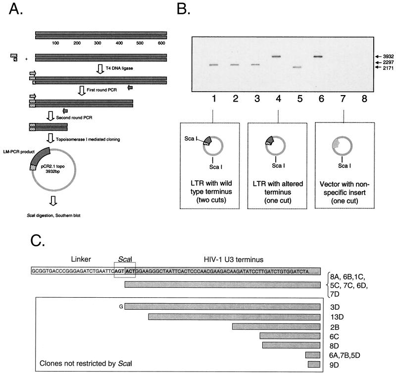
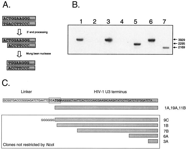
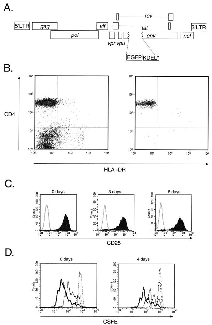
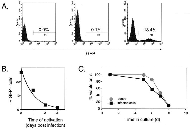
Most current evidence suggests that the infection of resting CD4(+) T cells by human immunodeficiency virus type 1 (HIV-1) is not productive due to partial or complete blocks in the viral life cycle at steps prior to integration of the viral genome into the host cell chromosome. However, stimulation of an infected resting T cell by antigen, cytokines, or microenvironmental factors can overcome these blocks and allow for the production of progeny virions. In this study, we sought to understand the structure and fate of the virus in unstimulated resting CD4(+) T cells. Using a novel linker-mediated PCR assay designed to detect and characterize linear unintegrated forms of the HIV-1 genome, we demonstrate that reverse transcription can proceed to completion following the infection of resting T cells, generating the substrate for the retroviral integration reaction. However, reverse transcription in resting T cells is far slower than in activated T cells, requiring 2 to 3 days to complete. The delay in completing reverse transcription may make the viral DNA genome more susceptible to competing decay processes. To explore the relationship between the formation of the linear viral genome and the stability of the preintegration state, we employed a recombinant HIV-1 virus expressing the enhanced green fluorescent protein to measure the rate at which HIV-1 decays in the preintegration state. Our results demonstrate that the preintegration state is labile and decays rapidly (half-life = 1 day) following the entry of HIV-1 into a resting T cell, with significant decay occurring during the slow process of reverse transcription.


Figures

FIG. 1.
FIG. 1.
Detection of the linear form of HIV-1 DNA using LM-PCR. The final product of the reverse transcription reaction is a linear genome with blunt ends. To detect linear products of the reverse transcription reaction, genomic DNA was isolated from infected cells and loaded into a ligation reaction mixture containing a vast molar excess of an asymmetric blunt linker. The ligation of linker onto the end of the genome was detected by PCR with an HIV-1-specific primer and one complementary to the linker. In some experiments, a heminested PCR approach was employed. Production of an HIV-1-specific amplicon was universally T4 ligase dependent.
FIG. 2.
FIG. 2.
Specificity of LM-PCR for molecules with blunt ends. (A) The specificity of LM-PCR for blunt targets representing completed reverse transcripts (top) or asymmetric targets representing the processed form of the HIV-1 genome (bottom) was evaluated with both blunt linkers and linkers with complementary overhangs. (B) Ten thousand copies of a linear molecule with blunt (StuI restricted) or asymmetric ends (AccI restricted) were loaded into a LM-PCR mixture. Ligation of the linker onto the terminus of the plasmid was evaluated with a 28-cycle PCR employing oligonucleotide 25t and LPCR-L. Numbers refer to the reactions shown in panel A.
FIG. 3.
FIG. 3.
Reverse transcription proceeds with protracted kinetics in highly purified resting T cells but can yield double-stranded HIV-1 DNA molecules of the appropriate length. Highly purified resting T cells were obtained from HIV-1-negative donors by cell sorting. Control MT-2 cells and resting CD4+ T cells were infected with HIV-1 IIIb at a multiplicity of infection of 1. At the indicated times, DNA was isolated and nested LM-PCR was performed to detect the U3 end of the linear HIV-1 genome. The LM-PCR involved an initial reaction of 15 cycles with outer LPCR-L and oligonucleotide 25t. Reaction products were diluted 1:4 in water, and a second 25-cycle PCR was performed using oligonucleotide 25t and LPCR-L. Reaction products were run on a 2% gel and Southern blotted with a U3-specific probe.
FIG. 4.
FIG. 4.
Characterization of complete reverse transcripts cloned from resting T lymphocytes. (A) LM-PCR assay for complete reverse transcripts. The ligation of an asymmetric linker containing one-half of a ScaI site onto the terminus of a full-length HIV-1 genome creates a novel ScaI site, allowing rapid screening of cloned LM-PCR products. (B) Restriction analysis of cloned LM-PCR products obtained from resting CD4+ T cells. Highly purified resting CD4+ T cells were infected with HIV-1 IIIB. Three days after infection, cells were lysed and DNA was isolated for LM-PCR. Cloned LM-PCR products were restricted with ScaI and analyzed by Southern blotting using an LTR-specific probe. The analysis allowed the identification of blunt linear molecules with either the predicted U3 end (lanes 1, 2, 3, and 5) or clones bearing alterations that result in a change in the sequence of the most-terminal U3 nucleotides (lanes 4 and 6). Because the insert was cloned in either a forward or reverse orientation, inserts of two different sizes were predicted for clones containing the novel ScaI site (lanes 1, 2, 3, and 5). Some clones did not contain an insert that contained a sequence from the HIV-1 LTR and were not characterized further (lanes 7 and 8). (C) Sequence analysis of LM-PCR clones obtained from infected resting CD4+ T cells.
FIG. 5.
FIG. 5.
Infected resting CD4+ T cells contain linear HIV-1 DNA molecules that have 3′ processing by integrase. (A) LM-PCR assay for complete reverse transcripts that have undergone 3′ processing by integrase. As an initial step in the integration reaction, integrase removes the terminal 2 nucleotides from the 3′ end of each strand of the full-length, linear HIV-1 DNA. The product of this reaction can be detected by removing the 5′ overhang with single-strand-specific MBN, generating a blunt-ended molecule whose terminal nucleotides form one-half of an NcoI site. The linker used in these experiments creates a novel NcoI site when ligated to the linear genome of HIV-1 that had been processed by integrase and cleaved by MBN. LM-PCR clones were restricted with NcoI, run on a 1% gel, and probed by Southern blotting with an LTR-specific probe. The vector used in these studies has an NcoI site; therefore, clones with inserts derived from linear DNA that has been processed by integrase will be cleaved twice in a manner analogous to that for the ScaI screen described in the legend for Fig. 4. (B) LM-PCR analysis of HIV-1 DNA from infected resting CD4+ T cells. Highly purified resting CD4+ T cells were infected with HIV-1 IIIB. Three days after infection, cells were lysed and DNA was isolated for LM-PCR. Restriction analysis of cloned LM-PCR products identified cloned inserts that were cleaved by NcoI, indicating prior processing by integrase (lanes 5 and 7) and inserts that were not cleaved (lanes 1, 3, and 6). Because the insert was cloned in either a forward or reverse orientation, inserts of two different sizes were predicted for clones containing the novel NcoI site. Some clones did not contain an HIV-1-derived insert (lanes 2 and 4). (C) Sequence analysis of LM-PCR clones obtained from MBN-treated DNA isolated from infected resting T lymphocytes.
FIG. 6.
FIG. 6.
Analysis of the decay of the preintegration state of HIV-1 latency. (A) Map of reporter virus NL4-3-GFP used to monitor the fate of HIV-1 in infected resting CD4+ T cells. NL4-3-GFP contains the EGFP coding sequence inserted in frame in the env gene, followed by codons for the KDEL sequence to retain GFP in the ER and a stop codon. Highly purified resting CD4+ T cells were infected with VSV-G-pseudotyped NL4-3-GFP. Following infection, the cells were extensively washed and cultured for various times before being subjected to activation with mitogen PHA and irradiated allogeneic PBMC. Expression of GFP by the infected cells was monitored by flow cytometry. (B) Purity of resting CD4+ T-cell preparations. Resting CD4+ T cells were isolated from the peripheral blood of HIV-negative donors by magnetic bead depletion and cell sorting. Cells stained with antibodies to CD4 and activation marker HLA-DR before (left) and after (right) magnetic bead depletion and cell sorting were analyzed by flow cytometry. (C) Efficiency of in vitro activation of CD4+ T cells. Uninfected resting CD4+ T cells were activated with PHA and irradiated feeders immediately after isolation (0 days) or after 3 or 6 days of culture in MM. Cells were stained with an anti-CD25 antibody 72 h after activation. Solid histogram, CD25 staining; open histogram, isotype control. (D) Proliferative responses of resting CD4+ T cells following activation with PHA and irradiated allogeneic PBMC. Resting CD4+ T cells were stained by CFSE and activated immediately after isolation (left) and after 4 days of culture (right). CFSE intensity was measured at 24 (dotted line), 48 (broken line), 72 (thin line), and 96 h (thick line) after activation.
FIG. 7.
FIG. 7.
Analysis of preintegration latency. (A) Dependence of viral gene expression on cellular activation. Resting CD4+ T cells were left uninfected (left) or were infected with pseudotyped NL4-3-GFP virus at a multiplicity of infection (MOI) of 1 (center and right). Four days after infection, GFP expression was analyzed by flow cytometry. In the absence of an activating stimulus (center), the percentage of cells with green fluorescence 4 days after infection was very low (0.1%), only slightly higher than the background observed in uninfected cells (left). However, if the resting cells were activated with PHA and irradiated allogeneic PBMC on day 1 postinfection, a significant portion of infected CD4+ T cells expressed GFP on day 4 postinfection (right). (B) Decay of HIV-1 in the preintegration state. Resting CD4+ T cells were infected with pseudotyped NL4-3-GFP at a MOI of 7, washed, and maintained in RPMI 1640 supplied with 10% fetal calf serum (FCS) for the indicated times before activation with PHA and allogeneic PBMC. The decay of virus in the preintegration state was measured as a decrease in the percentage of GFP-positive cells determined by FACS analysis 48 to 72 h after activation. The experiment shown is representative of six independent experiments, which are summarized in Table 1. (C) Viability of resting CD4+ T cells infected with pseudotyped NL4-3-GFP. Highly purified resting CD4+ T lymphocytes were isolated and infected with pseudotyped NL4-3-GFP at a MOI of 2. Both HIV-1-infected cells and noninfected resting T cells were maintained in RPMI 1640 supplemented in 10% FCS. Cell viability was measured over time by exclusion of vital dye trypan blue.
FIG. 8.
FIG. 8.
Contribution of completely reverse transcribed DNA. (A) Decay of HIV-1 in the preintegration state was measured as described for Fig. 7 in the presence (triangles) or absence (circles) of reverse transcription inhibitor 3TC at 10 μM. Resting CD4+ T cells were infected at a multiplicity of infection of 10 and then incubated for the indicated times before the addition of 3TC and activation with PHA and allogeneic feeder cells. 3TC was added 4 h prior to activation. (B) Relative contribution of completely reverse transcribed DNA to the rescue of virus from the preintegration state. The relative contribution was determined by expressing the number of cells with inducible GFP expression in the presence of 3TC as a percentage of the cells with induced GFP expression in the absence of 3TC.

References

    1. Blankson, J. N., D. Finzi, T. C. Pierson, B. P. Sabundayo, K. Chadwick, J. B. Margolick, T. C. Quinn, and R. F. Siliciano. 2000. Biphasic decay of latently infected CD4+ T cells in acute human immunodeficiency virus type 1 infection. J. Infect. Dis. 182:1636-1642. - PubMed
    1. Bukrinsky, M. I., T. L. Stanwick, M. P. Dempsey, and M. Stevenson. 1991. Quiescent T lymphocytes as an inducible virus reservoir in HIV-1 infection. Science 254:423-427. - PMC - PubMed
    1. Chun, T. W., L. Carruth, D. Finzi, X. Shen, J. A. DiGiuseppe, H. Taylor, M. Hermankova, K. Chadwick, J. Margolick, T. C. Quinn, Y. H. Kuo, R. Brookmeyer, M. A. Zeiger, P. Barditch-Crovo, and R. F. Siliciano. 1997. Quantification of latent tissue reservoirs and total body viral load in HIV-1 infection. Nature 387:183-188. - PubMed
    1. Chun, T. W., K. Chadwick, J. Margolick, and R. F. Siliciano. 1997. Differential susceptibility of naive and memory CD4+ T cells to the cytopathic effects of infection with human immunodeficiency virus type 1 strain LAI. J. Virol. 71:4436-4444. - PMC - PubMed
    1. Chun, T. W., D. Finzi, J. Margolick, K. Chadwick, D. Schwartz, and R. F. Siliciano. 1995. In vivo fate of HIV-1-infected T cells: quantitative analysis of the transition to stable latency. Nat. Med. 1:1284-1290. - PubMed

Publication types