. 2002 Sep;76(17):8560-71.
doi: 10.1128/jvi.76.17.8560-8571.2002.

mRNA degradation by the virion host shutoff (Vhs) protein of herpes simplex virus: genetic and biochemical evidence that Vhs is a nuclease

Affiliations

mRNA degradation by the virion host shutoff (Vhs) protein of herpes simplex virus: genetic and biochemical evidence that Vhs is a nuclease

David N Everly Jr et al. J Virol. 2002 Sep.

Abstract

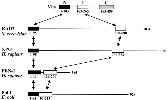
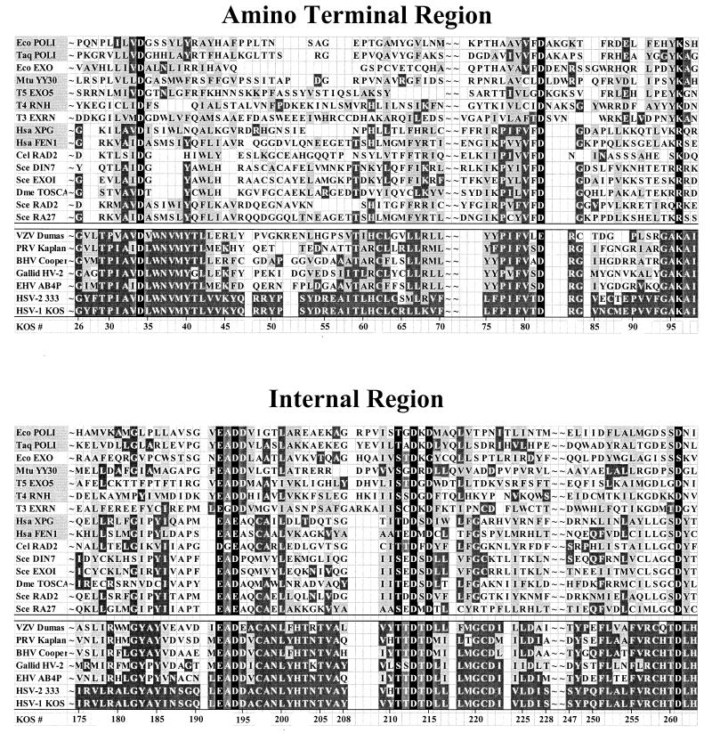
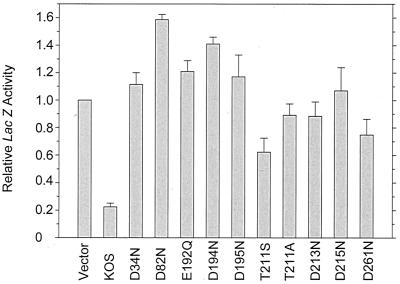
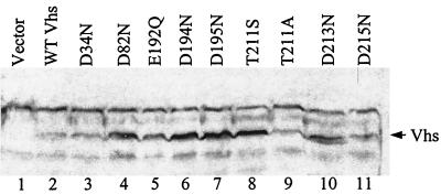
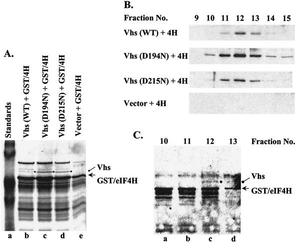
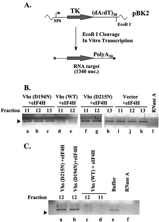
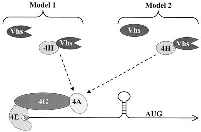
During lytic infections, the virion host shutoff (Vhs) protein (UL41) of herpes simplex virus destabilizes both host and viral mRNAs. By accelerating the decay of all mRNAs, it helps redirect the cell from host to viral gene expression and facilitates the sequential expression of different classes of viral genes. While it is clear that Vhs induces mRNA degradation, it is uncertain whether it is itself an RNase or somehow activates a cellular enzyme. This question was addressed by using a combination of genetic and biochemical approaches. The Vhs homologues of alphaherpesviruses share sequence similarities with a family of mammalian, yeast, bacterial, and phage nucleases. To test the functional significance of these similarities, Vhs was mutated to alter residues corresponding to amino acids known to be critical to the nuclease activity of cellular homologues. In every instance, mutations that inactivated the nuclease activity of cellular homologues also abolished Vhs activity. Recent experiments showed that Vhs interacts with the cellular translation initiation factor eIF4H. In this study, the coexpression of Vhs and a glutathione S-transferase (GST)-eIF4H fusion protein in bacteria resulted in the formation of a complex of the proteins. The wild-type Vhs/GST-eIF4H complex was isolated and shown to have RNase activity. In contrast, Vhs mutations that altered key residues in the nuclease motif abolished the nuclease activity of the recombinant Vhs/GST-eIF4H complex. The results provide genetic and biochemical evidence that Vhs is an RNase, either alone or as a complex with eIF4H.


Figures

FIG. 1.
FIG. 1.
Primary structures of Vhs and selected human, yeast, and bacterial nucleases. The primary structure of the Vhs polypeptides is depicted in the first line, with the three homology regions shared by the UL41 homologues from various alphaherpesviruses depicted by the black, white, and grey rectangles labeled N, I (internal), and C. The coordinates of the residues encompassing each of the homology regions differ slightly among the UL41 homologues, as do the distances separating them in the primary sequence. The coordinates shown below the first line refer to the Vhs polypeptide of HSV-1. The primary structures of the RAD2 protein from S. cerevisiae, the human XPG and FEN-1 polypeptides, and DNA polymerase I from E. coli are diagrammed in the second through fifth lines (26, 68). Each of these polypeptides has an amino-terminal domain (depicted by a black rectangle) that is similar to the amino-terminal domain of Vhs and an internal domain (depicted by a white rectangle) that is similar to the internal domain of Vhs. The amino acids that comprise the domains of each of the proteins are shown below the corresponding rectangles, and the overall length of each polypeptide is shown at the right. The distances separating the amino-terminal and internal domains of each polypeptide differ considerably from nuclease to nuclease. Arrows connect homologous domains in the different proteins.
FIG. 2.
FIG. 2.
Sequences of the amino-terminal and internal domains of Vhs and selected cellular nucleases. The amino acid sequences of the amino-terminal and internal domains of the Vhs polypeptides from seven alphaherpesviruses are shown below the double line; the sequences of the corresponding domains of selected cellular and phage nucleases (listed in Table 1) are shown above the double line. The amino acid coordinates of the Vhs polypeptide from HSV-1 strain KOS are shown at the bottom of each panel. Amino acids that are identical to the residues in HSV-1 strain KOS are shown in white lettering on a dark grey background. Amino acids that represent a conservative change from the residues in HSV-1 strain KOS are shown in black lettering on a light grey background. The names of cellular and phage nucleases for which structural and/or genetic data have identified key residues that are in the active site are highlighted by a light grey background in the leftmost column. The active-site residues from these nucleases that are conserved in other cellular and phage nucleases and in the Vhs polypeptides are shown in white lettering on a black background. These residues were the focus of site-directed mutagenesis of the Vhs polypeptide (see Fig. 3 and 4). VZV, varicella-zoster virus; PRV, pseudorabies virus; BHV, bovine herpesvirus; Galid HV-2, Gallid herpesvirus 2; EHV, equine herpesvirus.
FIG. 3.
FIG. 3.
Ability of wild-type Vhs and site-directed Vhs mutants to inhibit the expression of a cotransfected reporter gene. Triplicate cultures of Vero cells were transfected with 3 μg of the reporter plasmid pSV-β-galactosidase plus 0.73 pmol of pcDNA1.1amp (Vector) or UL41-containing effector plasmids encoding wild-type Vhs or the indicated mutant forms of Vhs. Transfection mixtures also contained enough salmon sperm carrier DNA to bring the total amount of DNA to 12 μg. Cell extracts were prepared 40 to 48 h after transfection and assayed for β-galactosidase activity as described in the text. For each transfection, the amount of β-galactosidase activity was expressed as a fraction of that observed in the transfection involving the vector pcDNA1.1amp. Errors bars indicate the standard error of the mean.
FIG. 4.
FIG. 4.
Expression of mutant and wild-type Vhs polypeptides in transfected cells. Vero cells were transfected with 0.73 pmol of pcDNA1.1amp (Vector) or plasmids encoding wild-type Vhs (WT Vhs) or the indicated mutant forms of Vhs. Whole-cell lysates were prepared 48 h after transfection and analyzed by sodium dodecyl sulfate-polyacrylamide gel electrophoresis and Western blotting with polyclonal rabbit antiserum raised against a Vhs-LacZ fusion protein. The arrow to the right of lane 11 indicates the position of the Vhs (UL41) polypeptide.
FIG. 5.
FIG. 5.
Expression of recombinant Vhs and GST-eIF4H. (A) Coomassie blue-stained gel of recombinant proteins bound to glutathione-Sepharose and eluted with 10 mM glutathione. Proteins are from E. coli expressing GST-eIF4H (GST/4H) (lanes b to e) and wild-type (WT) Vhs (lane b), D194N (lane c), or D215N (lane d). Material in lane e is from bacteria expressing GST-eIF4H and no Vhs. Vhs is indicated by a closed circle to the right of lanes b to d, and GST-eIF4H is indicated by an arrow. (B) Material that eluted from glutathione-Sepharose was applied to a column of HiTrap SP Sepharose and eluted with a gradient of 0 to 1 M salt. Fractions were analyzed for Vhs by Western blotting. 4H, eIF4H. (C) Coomassie blue-stained gel of HiTrap SP Sepharose fractions 10 to 13 from cells cells expressing wild-type Vhs and GST-eIF4H. Vhs is indicated by a closed circle, and GST-eIF4H is indicated by an arrow.
FIG. 6.
FIG. 6.
RNase activity of peak fractions containing Vhs and GST-eIF4H. (A) EcoRI cleavage of pBK2 followed by in vitro transcription with SP6 RNA polymerase in the presence of a 5′ cap analogue resulted in the production of a capped, 1,340 nucleotide (nuc.) target RNA with a 35-nucleotide poly(A) tail encoded by the plasmid. TK, thymidine kinase. (B and C) Target RNAs were incubated for 3 h (B) or 16 h (C) with degradation buffer (C, lane e), with RNase A (B, lane l, and C, lane f), or with the indicated HiTrap SP Sepharose fractions from bacteria expressing just GST-eIF4H (B, lanes i to k), GST-eIF4H plus Vhs D194N (B, lanes a to c, and C, lane b), GST-eIF4H plus Vhs D215N (B, lanes f to h, and C, lane A), or GST-eIF4H plus wild-type (WT) Vhs (B, lanes d and e, and C, lanes c and d). RNAs were extracted with phenol-chloroform, precipitated from ethanol, electrophoresed through 1.2% agarose gels, and visualized by staining with ethidium bromide.
FIG. 7.
FIG. 7.
Models for Vhs targeting. In model 1, Vhs is itself an RNase. It is targeted to mRNAs and to regions of translation initiation through its interaction with eIF4H. The functional, and perhaps physical, interaction of eIF4H with eIF4A is depicted by the broken arrow. In model 2, Vhs is an essential component of an RNase. However, it does not become active until after binding to eIF4H. The active nuclease is targeted by virtue of its eIF4H component.

References

    1. Allmang, C., E. Petfalski, A. Podtelejnikov, M. Mann, D. Tollervey, and P. Mitchell. 1999. The yeast exosome and human PM-Scl are related complexes of 3′ → 5′ exonucleases. Genes Dev. 13:2148-2158. - PMC - PubMed
    1. Altschul, S. F., T. L. Madden, A. A. Schaffer, J. Zhang, Z. Zhang, W. Miller, and D. J. Lipman. 1997. Gapped BLAST and PSI-BLAST: a new generation of protein database search programs. Nucleic Acids Res. 25:3389-3402. - PMC - PubMed
    1. Baldi, P., Y. Chauvin, T. Hunkapiller, and M. A. McClure. 1994. Hidden Markov models of biological primary sequence information. Proc. Natl. Acad. Sci. USA 91:1059-1063. - PMC - PubMed
    1. Becker, Y., E. Tavor, Y. Asher, C. Berkowiltz, and M. Moyal. 1993. Effect of herpes simplex virus type-1 UL41 gene on the stability of mRNA from the cellular genes: beta-actin, fibronectin, glucose transporter-1, and docking protein, and on virus intraperitoneal pathogenicity of newborn mice. Virus Genes 7:133-143. - PubMed
    1. Berthomme, H., B. Jacquemont, and A. Epstein. 1993. The pseudorabies virus host-shutoff homolog gene: nucleotide sequence and comparison with alphaherpesvirus protein counterparts. Virology 193:1028-1032. - PubMed

Publication types

MeSH terms

LinkOut - more resources