. 2002 Sep;76(17):8572-81.
doi: 10.1128/jvi.76.17.8572-8581.2002.

Mechanisms of inhibition of nuclear hormone receptor-dependent hepatitis B virus replication by hepatocyte nuclear factor 3beta

Affiliations

Mechanisms of inhibition of nuclear hormone receptor-dependent hepatitis B virus replication by hepatocyte nuclear factor 3beta

Hong Tang et al. J Virol. 2002 Sep.

Abstract

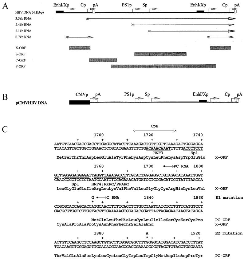
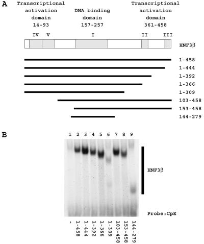
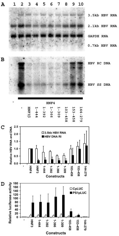
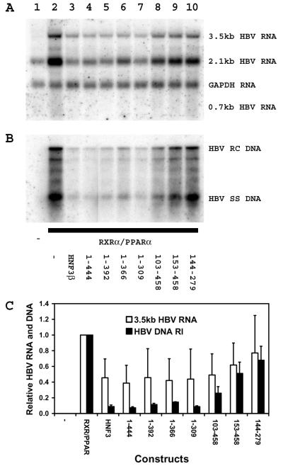
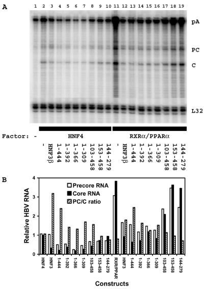
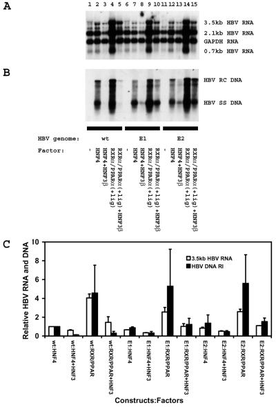
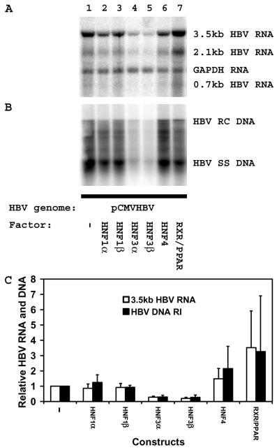
The nuclear hormone receptors hepatocyte nuclear factor 4 (HNF4) and the retinoid X alpha (RXRalpha) plus the peroxisome proliferator-activated receptor alpha (PPARalpha) heterodimer support hepatitis B virus (HBV) replication in nonhepatoma cells. Hepatocyte nuclear factor 3 (HNF3) inhibits nuclear hormone receptor-mediated viral replication. Inhibition of HBV replication by HNF3beta is associated with the preferential reduction in the level of the pregenomic RNA compared with that of precore RNA. Hepatitis B e antigen (HBeAg), encoded by the precore RNA, mediates part of the inhibition of viral replication by HNF3beta. The amino-terminal transcriptional activation domain of HNF3beta is essential for the inhibition of HBV replication. The activation of transcription by HNF3 from HBV promoters downstream from the nucleocapsid promoter appears to contribute indirectly to the reduction in the steady-state level of 3.5-kb HBV RNA, possibly by interfering with the elongation rate of these transcripts. Therefore, transcriptional interference mediated by HNF3 may also regulate HBV RNA synthesis and viral replication.


Figures

FIG. 1.
FIG. 1.
Structures and sequences of the HBV constructs supporting viral transcription and replication in mouse fibroblasts. (A) Structure of the HBV DNA (4.1-kbp) construct used in transient-transfection analysis. The 4.1-kbp greater-than-genome length HBV DNA sequence in this construct spans coordinates 1072 to 3182 plus 1 to 1990 of the HBV genome (subtype ayw). The locations of the 3.5-, 2.4-, 2.1-, and 0.7-kb HBV transcripts are indicated. EnhI/Xp, enhancer I/X-gene promoter region; Cp, nucleocapsid or core promoter; pA, polyadenylation site; PS1p, presurface antigen promoter; Sp, major surface antigen promoter; X, X gene; S, surface antigen gene; C, core gene; P, polymerase gene; ORF, open reading frame. (B) Structure of the pCMVHBV DNA construct used in transient-transfection analysis. The CMV immediate-early promoter (region from coordinates −522 to −1) directs the expression of the 3.5-kb HBV pregenomic RNA from the greater-than-genome length HBV DNA sequence in this construct that spans coordinates 1821 to 3182 plus 1 to 1990 of the HBV genome (subtype ayw). The locations of the 3.5-, 2.4-, 2.1-, and 0.7-kb HBV transcripts are the same as indicated for the HBV DNA (4.1-kbp) construct. (C) Sequence of the HBV core promoter region. The E1 (A1816G) and E2 (G1898A) mutations in the precore open reading frame (PC-ORF) prevent the expression of HBeAg from the HBV DNA (4.1-kbp) E1 and E2 mutant constructs. The sequence of the X-gene-encoded polypeptide is not changed by the E1 mutation in the X-gene open reading frame (X-ORF). The location of the CpE double-stranded oligonucleotide (HNF3 recognition site) used for electrophoretic mobility shift analysis is indicated. The HNF3, Sp1, and nuclear hormone receptor (HNF4 and RXRα-PPARα) binding sites are also indicated. PC RNA, precore 3.5-kb RNA; C RNA, pregenomic 3.5-kb RNA.
FIG. 2.
FIG. 2.
Expression of truncated HNF3β polypeptides in mouse NIH 3T3 fibroblasts. (A) Schematic representations of the HNF3β polypeptides showing the locations of the transcriptional activation domains and the DNA binding domain (32, 33) The amino-terminal transcriptional activation domain includes conserved sequence regions IV and V (33). The carboxyl-terminal transcriptional activation domain includes conserved sequence regions II and III (32). The winged helix DNA binding domain spans the conserved sequence region I (8, 32, 33). The amino acids of the truncated HNF3β polypeptides are shown to the right of the schematic representations. (B) Electrophoretic mobility shift analysis of a HBV nucleocapsid promoter HNF3 recognition site with truncated HNF3β polypeptides. The 32P-labeled, double-stranded oligonucleotide CpE (20) and whole-cell extracts prepared from mouse fibroblasts transfected with an empty vector control (−) (lane 1), an expression vector encoding the full-length HNF3β polypeptide (amino acid residues 1 to 458) (lane 2), and expression vectors encoding the truncated HNF3β polypeptides spanning amino acid residues 1 to 444 (lane 3), 1 to 392 (lane 4), 1 to 366 (lane 5), 1 to 309 (lane 6), 103 to 458 (lane 7), 153 to 458 (lane 8), and 144 to 279 (lane 9) were used for this analysis.
FIG. 3.
FIG. 3.
Modulation of HNF4-dependent HBV transcription and replication by truncated HNF3β polypeptides. Mouse NIH 3T3 fibroblasts were transiently transfected with the HBV DNA (4.1-kbp) construct (lanes 1 to 10) plus the HNF4 expression vector (lanes 2 to 10) and the HNF3β expression vectors (lanes 3 to 10). (A) RNA (Northern) filter hybridization analysis of HBV transcripts. The glyceraldehyde 3-phosphate dehydrogenase (GAPDH) transcript was used as an internal control for RNA loading per lane. (B) DNA (Southern) filter hybridization analysis of HBV replication intermediates. HBV RC DNA, HBV relaxed circular DNA; HBV SS DNA, HBV single-stranded DNA. The amino acids of the HNF3β polypeptides for panels A and B are shown below the gel in panel B. (C) Quantitative analysis of the 3.5-kb HBV RNA and DNA replication intermediates. The levels of the 3.5-kb HBV RNA and HBV DNA replication intermediates (HBV DNA RI) are reported relative to the levels of the HBV DNA (4.1-kbp) construct in the presence of HNF4 expression (lane 2), which are set at 1.0. The mean RNA and DNA levels plus standard deviations (indicated by the error bars) from two independent analyses are shown. (D) The effects of truncated HNF3β polypeptides on transcription from the nucleocapsid and large surface antigen promoter constructs CpLUC and PS1pLUC, respectively, were examined. Relative activities of the constructs in mouse fibroblast in the absence orpresence of ectopically expressed truncated HNF3β polypeptides are indicated. The amino acids of the HNF3β polypeptides are indicated below the graph. The transcriptional activities are reported relative to those of the CpLUC and PS1pLUC constructs in the absence of HNF3β expression (−), with a relative activity set at 1.0. The internal control used to correct for transfection efficiencies was pCMVβ. The mean luciferase activities plus standard deviations (indicated by the error bars) from three independent analyses are shown.
FIG. 4.
FIG. 4.
Modulation of RXRα-PPARα-dependent HBV transcription and replication by truncated HNF3β polypeptides. Mouse NIH 3T3 fibroblasts were transiently transfected with the HBV DNA (4.1-kbp) construct (lanes 1 to 10) plus the RXRα-PPARα expression vector (lanes 2 to 10) and the HNF3β expression vectors (lanes 3 to 10). (A) RNA (Northern) filter hybridization analysis of HBV transcripts. The glyceraldehyde 3-phosphate dehydrogenase (GAPDH) transcript was used as an internal control for RNA loading per lane. (B) DNA (Southern) filter hybridization analysis of HBV replication intermediates. HBV RC DNA, HBV relaxed circular DNA; HBV SS DNA, HBV single-stranded DNA. The amino acids of the HNF3β polypeptides in panels A and B are shown below the gel in panel B. (C) Quantitative analysis of the 3.5-kb HBV RNA and HBV DNA replication intermediates. The levels of the 3.5-kb HBV RNA and HBV DNA replication intermediates (HBV DNA RI) are reported relative to those of the HBV DNA (4.1-kbp) construct in the presence of RXRα-PPARα expression (lane 2), which are set at 1.0. All-trans retinoic acid and clofibric acid at 1 μM and 1 mM, respectively, were used to activate the nuclear hormone receptors RXRα and PPARα. The mean RNA and DNA levels plus standard deviations (indicated by the error bars) from two independent analyses are shown. The amino acids of the HNF3β polypeptides are shown below the graph.
FIG. 5.
FIG. 5.
Effects of truncated HNF3β polypeptides on the relative levels of precore and pregenomic RNA synthesis. Mouse NIH 3T3 fibroblasts were transiently transfected with the HBV DNA (4.1-kbp) construct (lanes 1 to 19), the HNF4 expression vector (lanes 2 to 10), the RXRα-PPARα expression vectors (RXRα/PPARα) (lanes 11 to 19), and the truncated HNF3β expression vectors (lanes 3 to 10 and 12 to 19) as indicated. The amino acids of the truncated HNF3β expression vectors are shown below the gel. (A) RNase protection analysis was performed to map the transcription initiation sites of the HBV precore (PC) and pregenomic or core (C) transcripts. The HBV probe also protected a fragment (pA) derived from the 3′ ends of all the HBV RNAs that terminated at the HBV polyadenylation site. A riboprobe detecting the ribosomal gene L32 transcripts was included as an internal control. (B) Quantitative analysis of the 3.5-kb HBV precore (PC) and core (C) RNA levels. The levels of the 3.5-kb HBV PC and C RNAs are reported relative to those of the C RNA transcribed from the HBV DNA (4.1-kbp) construct in the presence of HNF4 expression (lane 2), which are set at 1.0. The quantitative analyses of lanes 2 to 19 in panel A are shown.
FIG. 6.
FIG. 6.
Transcription and replication of wild-type and HBeAg-minus HBV DNA (4.1-kbp) constructs in mouse NIH 3T3 fibroblasts. Cells were transiently transfected with the wild-type (wt) HBV DNA (4.1-kbp) construct (lanes 1 to 5) and the HBeAg-minus HBV DNA (4.1-kbp) constructs (E1 [lanes 6 to 10] and E2 [lanes 11 to 15]) and liver-enriched transcription factors as indicated. (A) RNA (Northern) filter hybridization analysis of HBV transcripts. The glyceraldehyde 3-phosphate dehydrogenase (GAPDH) transcript was used as an internal control for RNA loading per lane. (B) DNA (Southern) filter hybridization analysis of HBV replication intermediates. HBV RC DNA, HBV relaxed circular DNA; HBV SS DNA, HBV single-stranded DNA. All-trans retinoic acid and clofibric acid at 1 μM and 1 mM, respectively, were used to activate the nuclear hormone receptors RXRα and PPARα. (C) Quantitative analysis of the 3.5-kb HBV RNA and HBV DNA replication intermediates. The levels of the 3.5-kb HBV RNA and HBV DNA replication intermediates (HBV DNA RI) are reported relative to those of the HBV DNA (4.1-kbp) construct in the presence of HNF4 expression (lane 2), which are set at 1.0. The mean RNA and DNA levels plus standard deviations (indicated by the error bars) from two independent analyses are shown.
FIG. 7.
FIG. 7.
HNF3β inhibits HBV pregenomic RNA synthesis and viral replication from regulatory elements downstream from the nucleocapsid promoter in the mouse NIH 3T3 fibroblasts. Cells were transiently transfected with the pCMVHBV DNA construct (lanes 1 to 7) and liver-enriched transcription factors (lanes 2 to 7) as indicated. (A) RNA (Northern) filter hybridization analysis of HBV transcripts. The glyceraldehyde 3-phosphate dehydrogenase (GAPDH) transcript was used as an internal control for RNA loading per lane. (B) DNA (Southern) filter hybridization analysis of HBV replication intermediates. HBV RC DNA, HBV relaxed circular DNA; HBV SS DNA, HBV single-stranded DNA. All-trans retinoic acid and clofibric acid at 1 μM and 1 mM, respectively, were used to activate the nuclear hormone receptors RXRα and PPARα. (C) Quantitative analysis of the 3.5-kb HBV RNA and DNA replication intermediates. The levels of the 3.5-kb HBV RNA and HBV DNA replication intermediates (HBV DNA RI) are reported relative to those of the pCMVHBV DNA construct in the absence of ectopic transcription factor expression (lane 1), which are set at 1.0. The mean RNA and DNA levels plus standard deviations (indicated by the error bars) from three independent analyses are shown.

Similar articles

Cited by

References

    1. Akahane, Y., T. Yamanaka, H. Suzuki, Y. Sugai, F. Tsuda, S. Yotsumoto, S. Omi, H. Okamoto, Y. Miyakawa, and M. Mayumi. 1990. Chronic active hepatitis with hepatitis B virus DNA and antibody against e antigen in the serum. Disturbed synthesis and secretion of e antigen from hepatocytes due to a point mutation in the precore region. Gastroenterology 99:1113-1119. - PubMed
    1. Andrews, N. C., and D. V. Faller. 1991. A rapid micropreparation technique for extraction of DNA-binding proteins from limiting numbers of mammalian cells. Nucleic Acids Res. 19:2499.. - PMC - PubMed
    1. Boshart, M., F. Weber, G. Jahn, K. Dorsch-Hasler, B. Fleckenstein, and W. Schaffner. 1985. A very strong enhancer is located upstream of an immediate early gene of human cytomegalovirus. Cell 41:521-530. - PubMed
    1. Buckwold, V. E., M. Chen, and J. H. Ou. 1997. Interaction of transcription factors RFX1 and MIBP1 with the gamma motif of the negative regulatory element of the hepatitis B virus core promoter. Virology 227:515-518. - PubMed
    1. Buckwold, V. E., Z. C. Xu, M. Chen, T. S. B. Yen, and J. H. Ou. 1996. Effects of a naturally occurring mutation in the hepatitis B virus basal core promoter on precore gene expression and viral replication. J. Virol. 70:5845-5851. - PMC - PubMed

Publication types

MeSH terms

LinkOut - more resources