. 2002 Sep;76(17):8596-608.
doi: 10.1128/jvi.76.17.8596-8608.2002.

Identification and expression of human cytomegalovirus transcription units coding for two distinct Fcgamma receptor homologs

Affiliations

Identification and expression of human cytomegalovirus transcription units coding for two distinct Fcgamma receptor homologs

Ramazan Atalay et al. J Virol. 2002 Sep.

Abstract

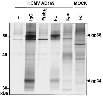
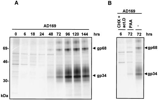
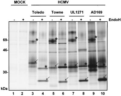
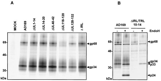
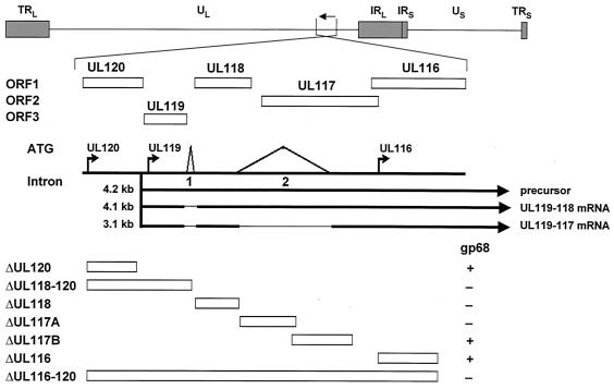
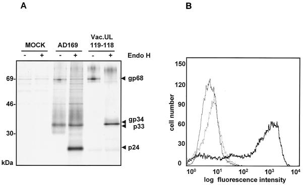
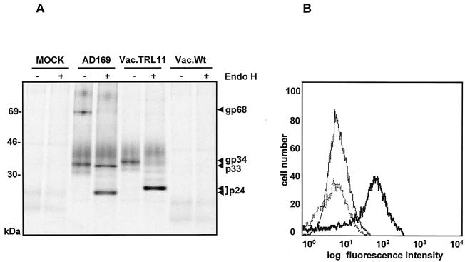
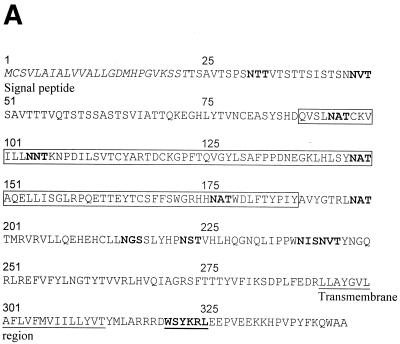
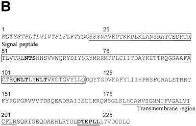
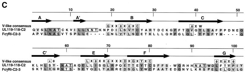
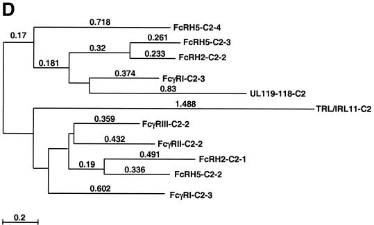
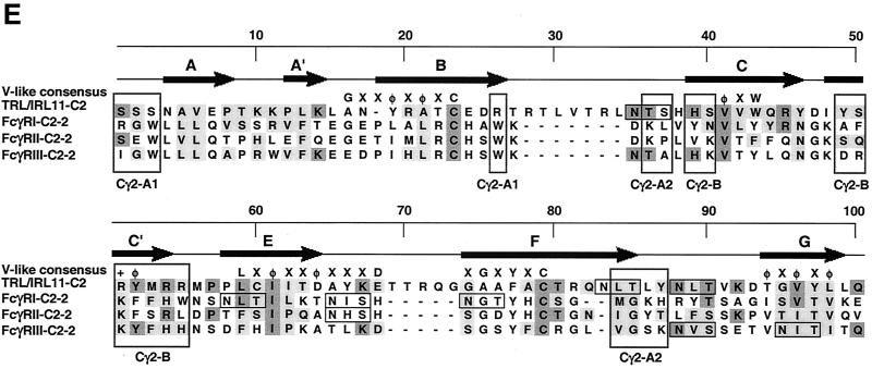
Cellular receptors for the Fc domain of immunoglobulin G (IgG) (FcgammaRs) comprise a family of surface receptors on immune cells connecting humoral and cellular immune responses. Several herpesviruses induce FcgammaR activities in infected cells. Here we identify two distinct human cytomegalovirus (HCMV)-encoded vFcgammaR glycoproteins of 34 and 68 kDa. A panel of HCMV strains exhibited a slight molecular microheterogeneity between Fcgamma-binding proteins, suggesting their viral origin. To locate the responsible genes within the HCMV genome, a large set of targeted HCMV deletion mutants was constructed. The mutant analysis allowed the identification of a spliced UL119-UL118 mRNA to encode vFcgammaR gp68 and TRL11/IRL11 to encode vFcgammaR gp34. Both vFcgammaRs are surface resident type I transmembrane glycoproteins. Significant relatedness of sequences in the extracellular chain of gpUL119-118 and gpTRL11 with particular immunoglobulin supergene family domains present in FcgammaR I and FcgammaRs II/III, respectively, indicates a different ancestry and function of gpUL119-118 and gpTRL11. The HCMV-encoded vFcgammaRs highlight an impressive diversification and redundancy of FcgammaR structures.


Figures

FIG. 1.
FIG. 1.
Precipitation of FcBPs from HCMV AD169-infected cells. Cells were labeled with [35S]methionine at 72 h p.i. for 1 h. The precipitation from lysates was done with Sepharose covalently coupled with complete nonimmune huIgG, Fc fragments of huIgG, F(ab)2 fragments of huIgG, and β2m. Proteins were separated by SDS-11.5 to 15% PAGE. FcBPs gp34 and gp68 are indicated by arrowheads.
FIG. 2.
FIG. 2.
Expression kinetics of FcBPs during the HCMV replication cycle. (A) MRC5 cells were infected with HCMV strain AD169 at an MOI of 3 for the times indicated. Cell lysates were precipitated with Fc fragments of huIgG, and precipitates were separated by 11.5-to-15%-gradient SDS-PAGE. FcBPs gp34 and gp68 are indicated by arrowheads. (B) Selective and enhanced synthesis of HCMV IE proteins was used by infecting cells in the presence of CHX, which was replaced by ActD (act.D), as described in Materials and Methods. PAA was used as an inhibitor of late phase gene expression.
FIG. 3.
FIG. 3.
Expression of FcBPs in HCMV strains and clinical isolates. MRC5 cells were infected at an MOI of 3 for 72 h with HCMV laboratory strains and the clinical isolate UL1271. Cells were metabolically labeled with [35S]methionine, and proteins from lysates were precipitated with Fc fragments and protein A Sepharose. Before separation of proteins by SDS-11.5 to 15% PAGE, an aliquot of each precipitate was deglycosylated with Endo H. FcBPs and their deglycosylated products are indicated (gp68 and p33 by asterisks; gp34 and p24 by arrows).
FIG. 4.
FIG. 4.
Analysis of FcBPs in HCMV pHB5-derived deletions mutants. (A) MRC5 fibroblasts were infected with HCMV deletion mutants for 72 h, labeled with [35S]methionine, and lysed. Lysates were precipitated with Fc fragments and protein A Sepharose before analysis by SDS-11.5 to 15% PAGE. (B) MRC5 cells were infected with ΔTRL10-14/IRL for 72 h, labeled with [35S]methionine, and lysed. Precipitation of lysates was performed with Fc fragments and protein A Sepharose. Precipitates were digested with Endo H before analysis by SDS-11.5 to 15% PAGE.
FIG. 5.
FIG. 5.
Identification of the transcription unit coding for FcBP gp68 within the HCMV UL120-UL116 gene region. The prototype arrangement of the viral genome consisting of long (UL) and short (US) unique components bounded by terminal repeats (TR) and inverted internal repeats (IR) as well as the position and the direction of transcription of the UL120-UL116 genes are indicated at the top panel. Below, open boxes designate the UL120-UL116 ORFs. The region contains three possible start ATGs indicated by arrows and two possible intron sequences. The mRNAs derived from the 4.2-kb mRNA precursor transcribed from the UL119 promoter are shown as lines interrupted by thin lines indicating splicing events. The positions of deletion mutants within the UL120-UL116 genomic region is given by boxes at the bottom. +, presence of vFcγR gp68 synthesis in cells infected with HCMV deletion mutants; −, absence of vFcγR gp68 synthesis.
FIG. 6.
FIG. 6.
Expression of vFcγR gp68 by vacUL119-118. CV1 cells were infected at an MOI of 3 for 14 h. (A) After labeling with [35S]methionine was done, precipitation of proteins was performed with Fc fragments and protein A Sepharose. Precipitates were digested with Endo H and analyzed by SDS-11.5 to 15% PAGE. (B) Cells were stained with huIgGFc-FITC, harvested using cell dissociation solution, and analyzed by flow cytometry. Thin line, CV1 cells mock infected; dotted line, wild-type vaccinia-infected cells, bold line, cells infected with vacUL119-118.
FIG. 7.
FIG. 7.
Expression of vFcγR gp34 by vacTRL11. CV1 cells were infected at an MOI of 3 for 14 h. (A) After labeling with [35S]methionine was done, precipitation of proteins was performed with Fc fragments and protein A Sepharose. Precipitates were digested with Endo H digestion before analysis by SDS-11.5 to 15% PAGE. (B) Cells were stained with huIgGFc-FITC, harvested using cell dissociation solution, and analyzed by flow cytometry. Thin line, CV1 cells mock infected; dotted line, wild-type vaccinia-infected cells; bold line, cells infected with vacTRL11.
FIG. 8.
FIG. 8.
Sequence analysis and alignment of gpUL119-118 and gpTRL11/IRL11. The deduced amino acid sequences of the UL119-118 ORF (A) and the TRL11/IRL11 ORF (B) are shown. The putative signal peptides are shown in italics, the putative transmembrane regions are underlined, and N-glycosylation sites are given in bold. The boxes indicate the putative IgSF domains of gpUL119-118 and gpTRL11. Putative cytoplasmic signaling motifs are in bold and underlined, i.e., a potential tyrosine-based motif in gpUL119-118 and an internalization motif for gpTRL11. Sequence alignments were performed by the program MacVector 7.02r using the ClustalW method with a PAM350 similarity matrix. For detailed analysis, the gpUL119-118 IgSF-like domain (aa 91 to 190) was aligned with the third domain of human FcγRI (aa 191 to 282; GenBank accession number X14356). (C) Amino acid residues are represented by the one-letter code, with dashes indicating gaps in the alignments. Identical residues are shaded in dark grey; residues showing similarity, defined as acidic (D or E), basic (H, K, or R), hydrophobic (A, F, I, L, M, P, V, or W) or polar (C, G, N, Q, S, T, or Y) are shaded in light grey. The N-glycosylation sites are boxed in black. The V-like consensus sequence (56) is shown above the alignment (+ represents a basic residue; φ represents a hydrophobic residue). Residues fitting the consensus are boxed in dark grey. The arrows above the sequence indicate the putative β-sheets deduced from the crystal structure of FcγRII (60). (D) A phylogenetic tree was generated by alignment of the IgSF-like domains of gpUL119-118 (aa 91 to 190) and gpTRL (aa 24 to 122) with the Fc-binding second domains of human FcγRI (aa 102 to 187; GenBank accession no. X14356), FcγRII (aa 121 to 207; accession no. M31932), and FcγRIII (aa 106 to 192; accession no. X16863), the third domain of human FcγRI (aa 191 to 282), and different domains of two human cellular Fc receptor homologs (12), FcRH2 (GenBank accession no. AY043465) domains 1 (aa 17 to 103) and 2 (aa 110 to 198), as well as FcRH5 (accession no. AF397453) domains 2 (aa 102 to 187), 3 (aa 193 to 282), and 4 (aa 288 to 377). Alignment of the TRL11 IgSF-like domain (aa 24 to 122) with the Fc-binding second domains of human FcγRI (aa 102 to 187; GenBank accession number X14356), FcγRII (aa 121 to 207; accession number M31932), and FcγRIII (aa 106 to 192; accession number X16863) is shown (E). The arrows above the sequence indicate the β-sheet structure of FcγRII/III based on crystal structure analysis (60, 68). The boxes indicate the contact regions for the two Fc-chains of huIgG (Cγ2-A and Cγ2-B) as determined by crystal structure analysis (61).
FIG. 8.
FIG. 8.
Sequence analysis and alignment of gpUL119-118 and gpTRL11/IRL11. The deduced amino acid sequences of the UL119-118 ORF (A) and the TRL11/IRL11 ORF (B) are shown. The putative signal peptides are shown in italics, the putative transmembrane regions are underlined, and N-glycosylation sites are given in bold. The boxes indicate the putative IgSF domains of gpUL119-118 and gpTRL11. Putative cytoplasmic signaling motifs are in bold and underlined, i.e., a potential tyrosine-based motif in gpUL119-118 and an internalization motif for gpTRL11. Sequence alignments were performed by the program MacVector 7.02r using the ClustalW method with a PAM350 similarity matrix. For detailed analysis, the gpUL119-118 IgSF-like domain (aa 91 to 190) was aligned with the third domain of human FcγRI (aa 191 to 282; GenBank accession number X14356). (C) Amino acid residues are represented by the one-letter code, with dashes indicating gaps in the alignments. Identical residues are shaded in dark grey; residues showing similarity, defined as acidic (D or E), basic (H, K, or R), hydrophobic (A, F, I, L, M, P, V, or W) or polar (C, G, N, Q, S, T, or Y) are shaded in light grey. The N-glycosylation sites are boxed in black. The V-like consensus sequence (56) is shown above the alignment (+ represents a basic residue; φ represents a hydrophobic residue). Residues fitting the consensus are boxed in dark grey. The arrows above the sequence indicate the putative β-sheets deduced from the crystal structure of FcγRII (60). (D) A phylogenetic tree was generated by alignment of the IgSF-like domains of gpUL119-118 (aa 91 to 190) and gpTRL (aa 24 to 122) with the Fc-binding second domains of human FcγRI (aa 102 to 187; GenBank accession no. X14356), FcγRII (aa 121 to 207; accession no. M31932), and FcγRIII (aa 106 to 192; accession no. X16863), the third domain of human FcγRI (aa 191 to 282), and different domains of two human cellular Fc receptor homologs (12), FcRH2 (GenBank accession no. AY043465) domains 1 (aa 17 to 103) and 2 (aa 110 to 198), as well as FcRH5 (accession no. AF397453) domains 2 (aa 102 to 187), 3 (aa 193 to 282), and 4 (aa 288 to 377). Alignment of the TRL11 IgSF-like domain (aa 24 to 122) with the Fc-binding second domains of human FcγRI (aa 102 to 187; GenBank accession number X14356), FcγRII (aa 121 to 207; accession number M31932), and FcγRIII (aa 106 to 192; accession number X16863) is shown (E). The arrows above the sequence indicate the β-sheet structure of FcγRII/III based on crystal structure analysis (60, 68). The boxes indicate the contact regions for the two Fc-chains of huIgG (Cγ2-A and Cγ2-B) as determined by crystal structure analysis (61).

References

    1. Amigorena, S., and C. Bonnerot. 1999. Fc receptor signaling and trafficking: a connection for antigen processing. Immunol. Rev. 172:279-284. - PubMed
    1. Antonsson, A., and P. J. Johansson. 2001. Binding of human and animal immunoglobulins to the IgG Fc receptor induced by human cytomegalovirus. J. Gen. Virol. 82:1137-1145. - PubMed
    1. Baucke, R. B., and P. G. Spear. 1979. Membrane proteins specified by herpes simplex viruses. V. Identification of an Fc-binding glycoprotein. J. Virol. 32:779-789. - PMC - PubMed
    1. Borst, E. M., G. Hahn, U. H. Koszinowski, and M. Messerle. 1999. Cloning of the human cytomegalovirus (HCMV) genome as an infectious bacterial artificial chromosome in Escherichia coli: a new approach for construction of HCMV mutants. J. Virol. 73:8320-8329. - PMC - PubMed
    1. Burmeister, W. P., L. N. Gastinel, N. E. Simister, M. L. Blum, and P. J. Bjorkman. 1994. Crystal structure at 2.2 A resolution of the MHC-related neonatal Fc receptor. Nature 372:336-343. - PubMed

Publication types

MeSH terms

Associated data

LinkOut - more resources