. 2002 Sep;76(17):8729-36.
doi: 10.1128/jvi.76.17.8729-8736.2002.

Hemagglutinin protein of wild-type measles virus activates toll-like receptor 2 signaling

Affiliations

Hemagglutinin protein of wild-type measles virus activates toll-like receptor 2 signaling

Karen Bieback et al. J Virol. 2002 Sep.

Abstract

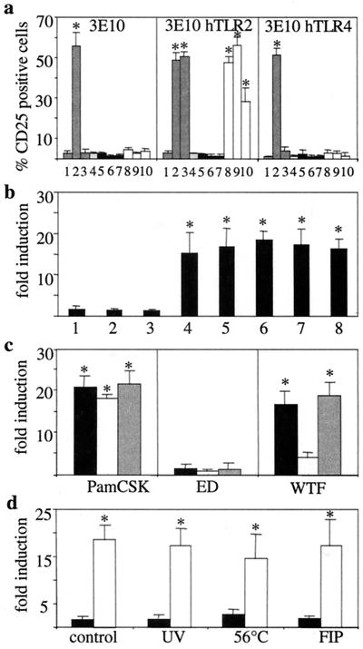
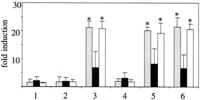
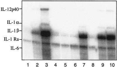
Pattern recognition via Toll-like receptors (TLR) by antigen-presenting cells is an important element of innate immunity. We report that wild-type measles virus but not vaccine strains activate cells via both human and murine TLR2, and this is a property of the hemagglutinin (H) protein. The ability to activate cells via TLR2 by wild-type MV H protein is abolished by mutation of a single amino acid, asparagine at position 481 to tyrosine, as is found in attenuated strains, which is important for interaction with CD46, the receptor for these strains. TLR2 activation by MV wild-type H protein stimulates induction of proinflammatory cytokines such as interleukin-6 (IL-6) in human monocytic cells and surface expression of CD150, the receptor for all MV strains. Confirming the specificity of this interaction, wild-type H protein did not induce IL-6 release in macrophages from TLR2-/- mice. Thus, the unique property of MV wild-type strains to activate TLR2-dependent signals might essentially contribute not only to immune activation but also to viral spread and pathogenicity by upregulating the MV receptor on monocytes.


Figures

FIG. 1.
FIG. 1.
TLR2-mediated cell activation by MV. (a) CHO cells stably expressing an NF-κB-dependent reporter gene (ELAM.TAC) together with CD14 alone (3E10) (left panel) or with human TLR2 (3E10 hTLR2) (middle panel) or TLR4 (3E10 hTLR4) (right panel) were analyzed for CD25 expression after incubation with medium (lane 1), LPS (lane 2), PamCSK (lane 3), mock supernatant (lane 4), ED (black bars) (lane 5, MOI = 10; lane 6, MOI = 1; lane 7, MOI = 0.1), or WTF (white bars) (lane 8, MOI = 10; lane 9, MOI = 1; lane 10, MOI = 0.1). (b) 3E10 hTLR2 cells were incubated with MV vaccine strains ED (lane 1), AIK-C (lane 2), Moraten (lane 3), or lymphotropic wild-type strains WTF (lane 4), Bilthoven (lane 5), Wü5404 (lane 6), Wü5679 (lane 7), and Wü4797 (lane 8) (MOI = 1 for each). (c) 3E10 hTLR2 cells (black bars), EL1 mTLR2 cells (white bars), or 3E10 mTLR2 cells (grey bars) were incubated with ED or WTF (MOI = 1 for each), or with PamCSK. (d) 3E10 hTLR2 cells were incubated with ED (black bars) or WTF (white bars) (MOI = 1 for each) as live virus (control), inactivated by UV irradiation or heating at 56°C, or as live virus in the presence of FIP. Data shown were obtained in three (a, b, and d) or five (c) independent experiments, with P values of <0.01 (a to c) and <0.05 (d) with respect to the mock control. Statistically significant differences from the control are indicated by asterisks.
FIG. 2.
FIG. 2.
A single amino acid exchange within the H protein discriminates whether TLR2 signaling is induced by MV. 3E10 hTLR2 cells (grey bars), EL1 mTLR2 cells (black bars), or 3E10 mTLR2 cells (white bars) were incubated with purified, UV-inactivated ED (lane 1), ED(WTF-F) (lane 2), ED(WTF-H) (lane 3), ED(WTF-H; N→Y) (lane 4), ED(WTF-F/H) (lane 5), or WTF (lane 6) and analyzed for CD25 induction. Values shown were obtained in three independent experiments (P < 0.01 with respect to the mock control). Statistically significant differences from the control are indicated by asterisks.
FIG. 3.
FIG. 3.
Induction of monokine-specific transcripts by MV expressing the WTF-H protein. THP-1 cells matured by vitamin D3 were incubated with medium (lane 1), LPS (lane 2), PamCSK (lane 3), mock supernatant (lane 4), ED (lane 5), ED(WTF-F) (lane 6), ED(WTF-H) (lane 7), ED(WTF-H; N→Y) (lane 8), ED(WTF-F/H) (lane 9), or WTF (lane 10), and RNAs were harvested 12 h later and analyzed by an RNase protection assay with 32P-labeled probes.
FIG. 4.
FIG. 4.
Activation of TLR2 by viruses expressing the WTF-H protein triggers release of IL-6 and expression of CD150 in human monocytes. (a and b) Cells were left untreated (lane 1) or were stimulated with LPS (lane 2), PamCSK (lane 3), mock supernatant (lane 4), ED (lane 5), ED(WTF-F) (lane 6), ED(WTF-H) (lane 7), ED(WTF-H; N→Y) (lane 8), ED(WTF-F/H) (lane 9), or WTF (lane 10). After 12 h, supernatants were harvested for determination of IL-6 (a), while cells were stained for CD150 surface expression (b). Values represent means of seven (a) or four (b) individual donors and were normalized to the IL-6 content of the inoculum (determined on the basis of the medium control; P ≤ 0.01) (a) or to the medium control (P ≤ 0.01) (b). (c and d) Cells (three donors) were pretreated with TLR2-, TLR4-, or CD14-specific antibodies and incubated with PamCSK (2.5, 0.5, and 0.1 μg/ml) (black bars) or WTF (10, 2 or 0.4 μg/ml of purified virus) (white bars). Alternatively, PamCSK or WTF (same concentrations as above) were treated with MV N- or MV H-specific antibodies prior to incubation with monocytes. Release of IL-6 (c) or induction of CD150 (d) was determined, and inhibition by the antibodies used (percent) was determined with respect to the control in the absence of antibody (P ≤ 0.01). Statistically significant differences from the control are indicated by asterisks.
FIG. 5.
FIG. 5.
IL-6 release from TLR2−/− murine macrophages cannot be stimulated by MV WTF. IL-6 concentrations (in nanograms per milliliter) were determined in supernatants of peritoneal macrophages from wild-type CH3/HeN (grey bars), C3H/TLR2−/− (black bars), or C3H/HeJ (white bars) (expressing nonfunctional TLR4) cells stimulated for 12 h with medium (lane 1), LPS (lane 2), PamCSK (lane 3), mock (lane 4), ED (lane 5), or WTF (lane 6). Values shown are means of two independent experiments (three animals of each genotype were used per experiment) (P ≤ 0.01). Statistically significant differences from the control are indicated by asterisks.

References

    1. Aarden, L. A., E. R. De Groot, O. L. Schaap, and P. M. Lansdorp. 1987. Production of hybridoma growth factor by human monocytes. Eur. J. Immunol. 17:1411-1417. - PubMed
    1. Akira, S., K. Takeda, and T. Kaisho. 2001. Toll-like receptors: critical proteins linking innate and acquired immunity. Nat. Immunol. 2:675-680. - PubMed
    1. Alexopoulou, L., A. Holt, R. Medzhitov, and R. A. Flavell. 2001. Recognition of double-stranded RNA and activation of NF-κB by Toll-like receptor 3. Nature 413:732-738. - PubMed
    1. Arbibe, L., J. P. Mira, N. Teusch, L. Kline, M. Guha, N. Mackman, P. J. Godowski, R. J. Ulevitch, and U. G. Knaus. 2000. Toll-like receptor 2-mediated NF-κB activation requires a RacI-dependent pathway. Nat. Immunol. 1:533-540. - PubMed
    1. Astier, A., M. C. Trescol-Biemont, O. Azocar, B. Lamouille, and C. Rabourdin-Combe. 2000. CD46, a new costimulatory molecule for T cells, that induces p120CBL and LAT phosphorylation. J. Immunol. 164:6091-6095. - PubMed

Publication types

MeSH terms