. 2002 Sep;76(17):8820-33.
doi: 10.1128/jvi.76.17.8820-8833.2002.

Identification and characterization of the pseudorabies virus tegument proteins UL46 and UL47: role for UL47 in virion morphogenesis in the cytoplasm

Affiliations

Identification and characterization of the pseudorabies virus tegument proteins UL46 and UL47: role for UL47 in virion morphogenesis in the cytoplasm

Martina Kopp et al. J Virol. 2002 Sep.

Abstract

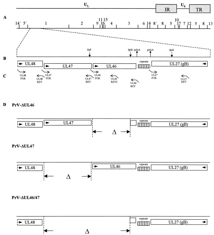
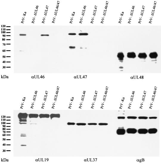
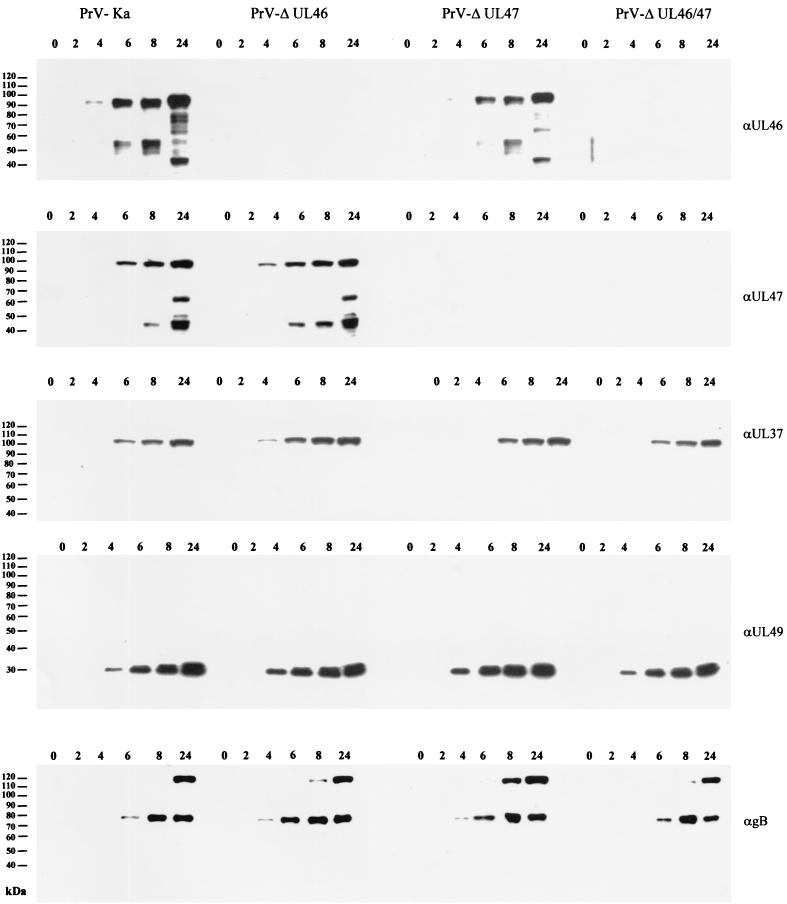
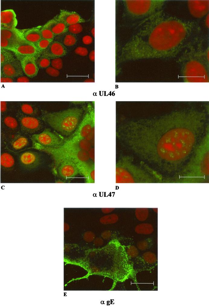
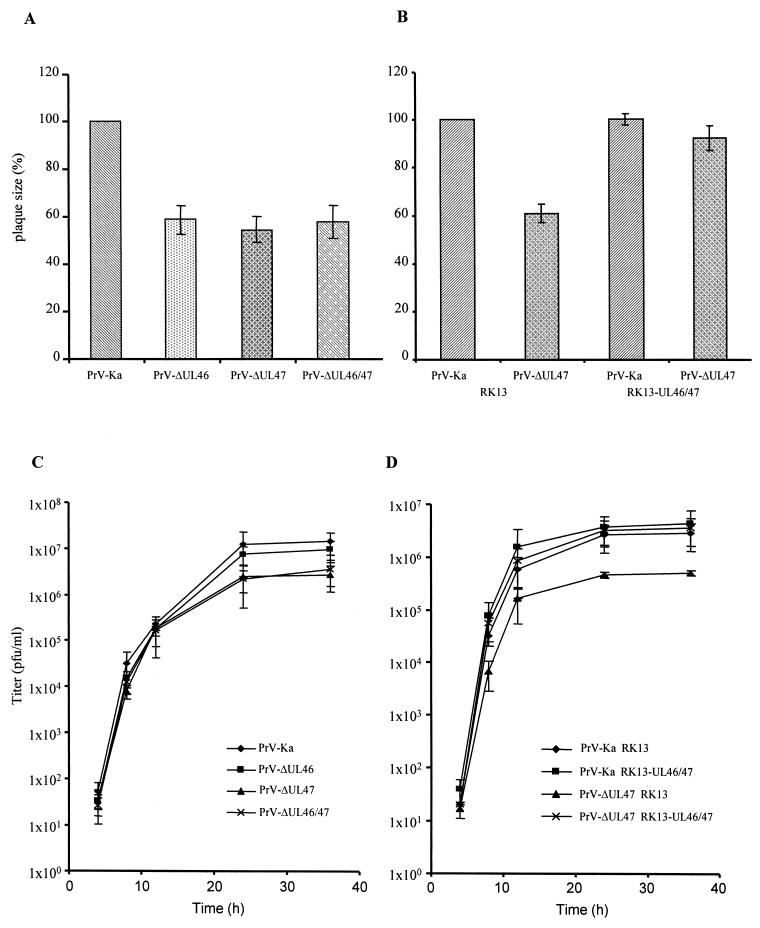
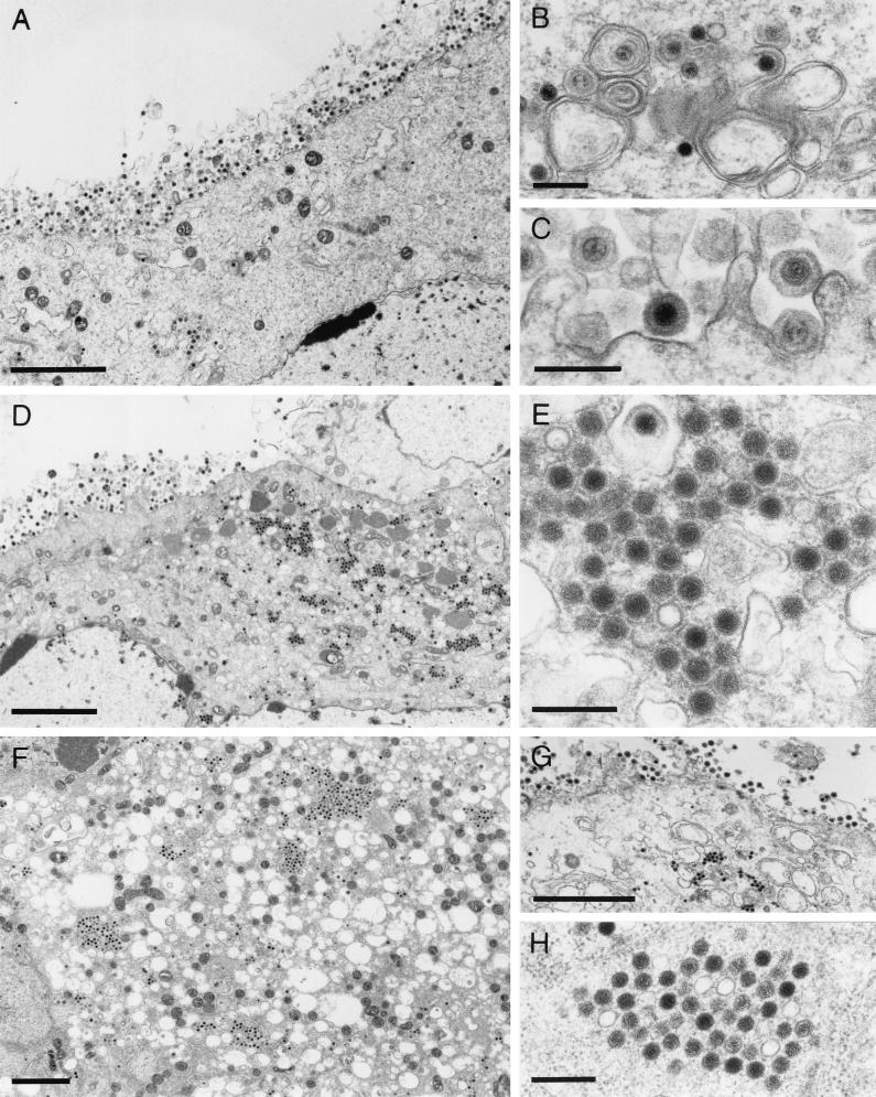
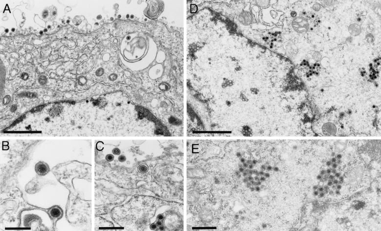
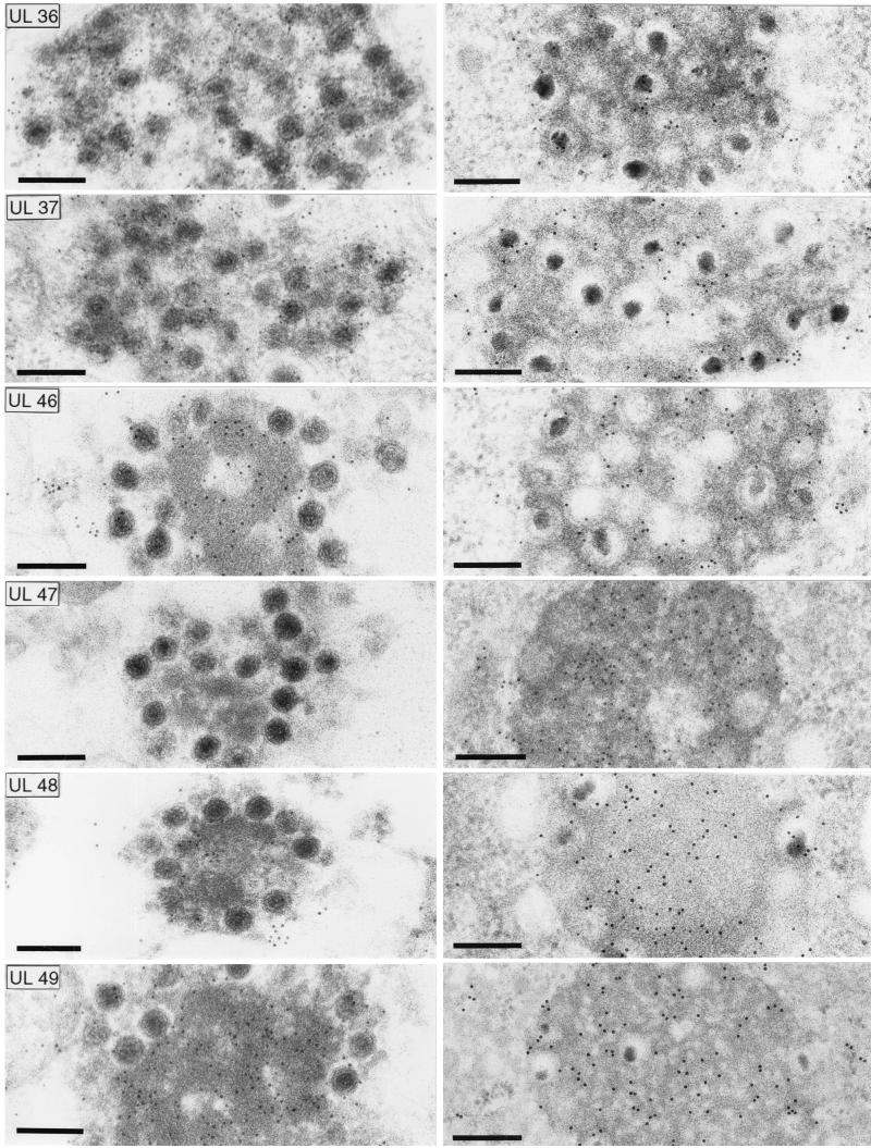
Proteins encoded by the UL46 and UL47 genes of herpes simplex virus type 1 (HSV-1) constitute major components of the viral tegument. However, their functions have so far not been elucidated in detail. By use of monospecific antisera directed against bacterially expressed glutathione-S-transferase fusion proteins, the homologous UL46 and UL47 proteins of the alphaherpesvirus pseudorabies virus (PrV) were identified in virus-infected cells and in virions. The PrV UL46 gene product of 693 amino acids (aa) exhibits an apparent molecular mass of 95 kDa, whereas the UL47 product of 750 aa was identified as a 97-kDa protein. Both are present in purified virions, correlating with their role as tegument proteins. Immunofluorescence analysis by confocal laser scan microscopy showed that late in infection the UL46 product is detectable in the cytoplasm, whereas the UL47 product was observed to be diffuse in the cytoplasm and speckled in the nucleus. Virus mutants lacking either the UL46 or the UL47 gene or both were isolated on noncomplementing cells, demonstrating that these genes either singly or in combination are not required for productive viral replication. However, plaque sizes were decreased. Interestingly, in one-step growth analysis, UL47 deletion mutants exhibited an approximately 10-fold decrease in final titers, whereas the UL46 deletion mutant was not affected. This finding correlated with ultrastructural observations which showed unimpaired virion morphogenesis in the absence of the UL46 protein, whereas in the absence of the UL47 protein intracytoplasmic aggregates of partially tegumented capsids were observed. In summary, we identified the PrV UL46 and UL47 proteins and show that the UL47 protein plays an important role in virion assembly in the cytoplasm.


Figures

FIG. 1.
FIG. 1.
Construction of PrV UL46 and UL47 deletion mutants. (A) A schematic map of the PrV genome shows the long (UL) and short (US) unique regions, the inverted repeat sequences (IR, TR), and the positions of BamHI restriction sites. Numbers indicate BamHI restriction fragments. (B) Coding regions of proteins in the genomic region relevant for this study. Arrows indicate transcriptional orientations. Also indicated is a region of repeated sequences between the UL27 and UL46 genes, as well as location of consensus sequences for polyadenylation (polyA) and relevant restriction sites. (C) Location of primers used for PCR amplification (see Table 1). (D) Genotypes of the constructed viral mutants.
FIG. 2.
FIG. 2.
Identification and virion localization of PrV UL46 and UL47 proteins. Purified virions of PrV-Ka, PrV-ΔUL46, PrV-ΔUL47, and PrV-ΔUL46/47 were analyzed by immunoblotting using monospecific antisera against the UL46, UL47, UL48, UL19, and UL37 proteins or a monoclonal antibody against gB.
FIG. 3.
FIG. 3.
Expression kinetics of PrV UL46 and UL47 proteins. RK13 cells were infected at an MOI of 5 with PrV-Ka, PrV-ΔUL46, PrV-ΔUL47, or PrV-ΔUL46/47 and harvested and analyzed at the indicated hours after infection. Blots were probed with monospecific antisera against the UL46, UL47, UL37, and UL49 proteins or a monoclonal antibody against gB.
FIG. 4.
FIG. 4.
Intracellular localization of PrV UL46 and UL47 proteins. Immunofluorescence analysis by confocal laser scan microscopy of RK13 cells infected with PrV-Ka under plaque assay conditions was performed by using monospecific antisera against the UL46 (upper row) or UL47 proteins (middle row) or a monoclonal antibody against gE (bottom). For UL46 and UL47 detection, two different magnifications are shown (bars represent 25 μm in left panels and 50 μm in right panels). Red, propidium iodide stain; green, reactivity of specific antibodies.
FIG. 5.
FIG. 5.
Plaque size and one-step growth kinetics of PrV UL46 and UL47 deletion mutants. Growth properties of deletion mutants in UL46 (PrV-ΔUL46) or UL47 (PrV-ΔUL47) or both (PrV-ΔUL46/47) were assessed by analyzing relative plaque size on noncomplementing (A) or RK13-UL46/47 (B) cells compared to that of PrV-Ka, which was taken as 100%. (C and D) One-step replication kinetics on noncomplementing and RK13-UL46/47 cells.
FIG. 6.
FIG. 6.
Electron microscopy of mutant virus-infected cells. RK13 cells were infected with PrV-ΔUL46 (A to C), PrV-ΔUL47 (D and E), or PrV-ΔUL46/47 (F to H) and analyzed by electron microscopy 16 h after infection. (A to C) Unimpaired virion morphogenesis in the absence of the UL46 protein including secondary envelopment in the cytoplasm (B) as well as numerous extracellular virions (A and C) are shown. (D) Overview of a PrV-ΔUL47-infected cell demonstrating that besides all stages of normal virion morphogenesis, intracytoplasmic aggregations of capsids can be observed (shown at higher magnification in panel E). Similar aggregations were also observed in PrV-ΔUL46/47-infected cells (F to H). Bars, 3 μm in panels A, D, F, and G and 300 nm in panels B, C, E, and H.
FIG. 7.
FIG. 7.
Electron microscopy of infected RK13-UL46/47 cells. Transcomplementing RK13-UL46/47 and normal RK13 cells were infected with PrV-ΔUL47 and analyzed by electron microscopy 16 h after infection. Panels A to C show unimpaired virion formation on complementing cells, whereas panels D and E demonstrate, in a parallel assay, the formation of capsid aggregates in the cytoplasm of noncomplementing cells. Bars, 1.5 μm in panels A and D, 100 nm in panel B, and 500 nm in panels C and E.
FIG. 8.
FIG. 8.
Immunoelectron microscopy. Intracytoplasmic aggregates formed in the absence of the UL47 protein (left panel) or in the absence of glycoproteins gE/I and gM (right panel) (7) were analyzed by immunoelectron microscopy using monospecific antisera against the tegument proteins UL36, UL37, UL46, UL47, UL48, and UL49. Bar, 250 nm.

Similar articles

Cited by

References

    1. Baer, R., A. T. Bankier, M. D. Biggin, P. L. Deininger, P. J. Farrell, T. J. Gibson, G. F. Hatfull, G. S. Hudson, S. C. Satchwell, C. Seguin, P. Tuffnell, and B. G. Barrell. 1984. DNA sequence and expression of the B95-8 Epstein-Barr virus genome. Nature (London) 310:207-211. - PubMed
    1. Barker, D., and B. Roizman. 1990. Identification of three genes nonessential for growth in cell culture near the right terminus of the unique sequences of long component of herpes simplex virus 1. Virology 177:684-692. - PubMed
    1. Batterson, W., and B. Roizman. 1983. Characterization of the herpes simplex virion-associated factor responsible for the induction of α genes. J. Virol. 46:371-377. - PMC - PubMed
    1. Bechtel, J., and T. Shenk. 2002. Human cytomegalovirus UL47 tegument protein functions after entry and before immediate-early gene expression. J. Virol. 76:1043-1050. - PMC - PubMed
    1. Blaho, J. A., C. Mitchell, and B. Roizman. 1994. An amino acid sequence shared by the herpes simplex virus 1 alpha regulatory proteins 0, 4, 22, and 27 predicts the nucleotidylylation of the UL21, UL31, UL47 and UL49 gene products. J. Biol. Chem. 269:17401-17410. - PubMed

Publication types

MeSH terms

Substances

LinkOut - more resources