

Long-term effect of interferon on keratinocytes that maintain human papillomavirus type 31
- PMID: 12163606
- PMCID: PMC136980
- DOI: 10.1128/jvi.76.17.8864-8874.2002
Long-term effect of interferon on keratinocytes that maintain human papillomavirus type 31
Abstract
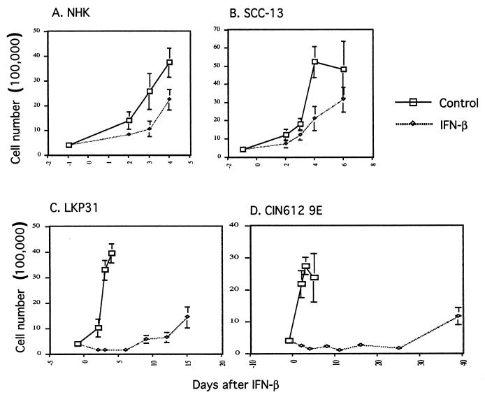
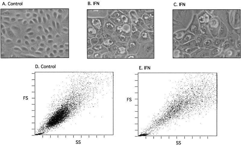
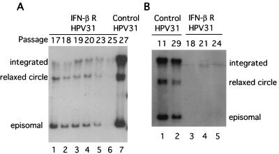
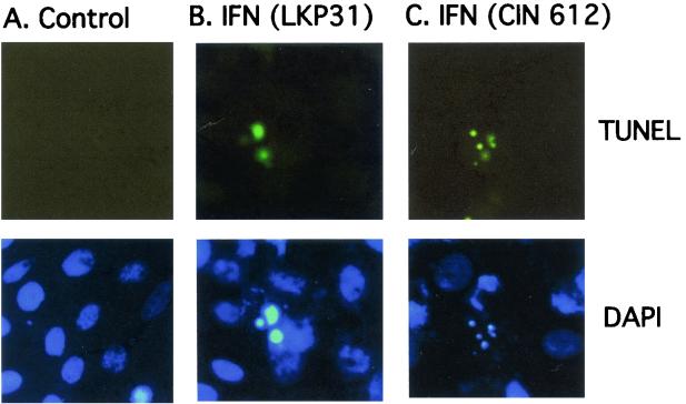
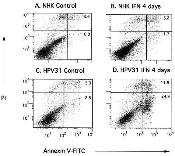
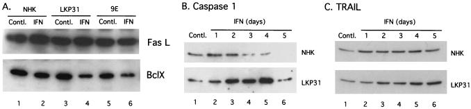
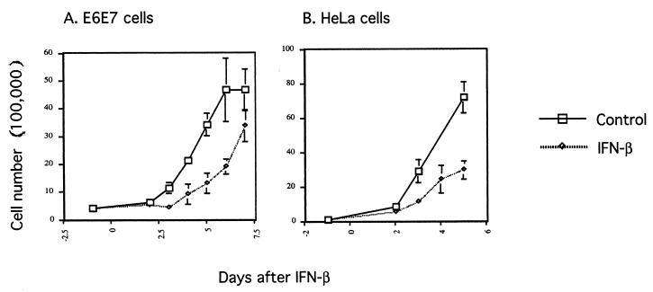
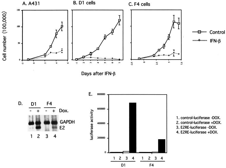
The long-term effects of interferon treatment on cell lines that maintain human papillomavirus type 31 (HPV-31) episomes have been examined. High doses and prolonged interferon treatment resulted in growth arrest of HPV-positive cells, with a high percentage of cells undergoing apoptosis. These effects were not seen with interferon treatment of either normal human keratinocytes or cells derived from HPV-negative squamous carcinomas, which exhibited only slight decreases in their rates of growth. Within 2 weeks of the initiation of treatment, a population of HPV-31-positive cells that were resistant to interferon appeared consistently and reproducibly. The resistant cells had growth and morphological characteristics similar to those of untreated cells. Long-term interferon treatment of HPV-positive cells also resulted in a reduction in HPV episome levels but did not significantly decrease the number of integrated copies of HPV. Cells that maintained HPV genomes lacking E5 were sensitive to interferon, while cells expressing only the E6/E7 genes were resistant. In contrast, cells that expressed E2 from a tetracycline-inducible promoter were found to be significantly more sensitive to interferon treatment than parental cells. This suggests that at least a portion of the sensitivity to interferon could be mediated through the E2 protein. These studies indicate that cells maintaining HPV episomes are highly sensitive to interferon treatment but that resistant populations arise quickly.
Figures
Similar articles
-
Interferon-beta treatment of cervical keratinocytes naturally infected with human papillomavirus 16 episomes promotes rapid reduction in episome numbers and emergence of latent integrants.Carcinogenesis. 2006 Nov;27(11):2341-53. doi: 10.1093/carcin/bgl172. Epub 2006 Sep 14. Carcinogenesis. 2006. PMID: 16973673
-
Papillomavirus type 16 oncogenes downregulate expression of interferon-responsive genes and upregulate proliferation-associated and NF-kappaB-responsive genes in cervical keratinocytes.J Virol. 2001 May;75(9):4283-96. doi: 10.1128/JVI.75.9.4283-4296.2001. J Virol. 2001. PMID: 11287578 Free PMC article.
-
Human papillomavirus type 31 oncoproteins E6 and E7 are required for the maintenance of episomes during the viral life cycle in normal human keratinocytes.Proc Natl Acad Sci U S A. 1999 Jul 20;96(15):8449-54. doi: 10.1073/pnas.96.15.8449. Proc Natl Acad Sci U S A. 1999. PMID: 10411895 Free PMC article.
-
Human papillomaviruses and the interferon response.J Interferon Cytokine Res. 2009 Sep;29(9):629-35. doi: 10.1089/jir.2009.0075. J Interferon Cytokine Res. 2009. PMID: 19715460 Free PMC article. Review.
-
[Genomic organization and proteins of human papillomavirus].Mikrobiyol Bul. 2012 Jul;46(3):507-15. Mikrobiyol Bul. 2012. PMID: 22951665 Review. Turkish.
Cited by
-
Interferon Kappa Inhibits Human Papillomavirus 31 Transcription by Inducing Sp100 Proteins.J Virol. 2015 Oct 21;90(2):694-704. doi: 10.1128/JVI.02137-15. Print 2016 Jan 15. J Virol. 2015. PMID: 26491169 Free PMC article.
-
High-risk human papillomaviruses repress constitutive kappa interferon transcription via E6 to prevent pathogen recognition receptor and antiviral-gene expression.J Virol. 2011 Nov;85(21):11372-80. doi: 10.1128/JVI.05279-11. Epub 2011 Aug 17. J Virol. 2011. PMID: 21849431 Free PMC article.
-
Degradation of p53, not telomerase activation, by E6 is required for bypass of crisis and immortalization by human papillomavirus type 16 E6/E7.J Virol. 2004 Jun;78(11):5698-706. doi: 10.1128/JVI.78.11.5698-5706.2004. J Virol. 2004. PMID: 15140967 Free PMC article.
-
The Interaction Between Human Papillomaviruses and the Stromal Microenvironment.Prog Mol Biol Transl Sci. 2016;144:169-238. doi: 10.1016/bs.pmbts.2016.09.003. Epub 2016 Oct 11. Prog Mol Biol Transl Sci. 2016. PMID: 27865458 Free PMC article. Review.
-
Vesicular trafficking permits evasion of cGAS/STING surveillance during initial human papillomavirus infection.PLoS Pathog. 2020 Nov 30;16(11):e1009028. doi: 10.1371/journal.ppat.1009028. eCollection 2020 Nov. PLoS Pathog. 2020. PMID: 33253291 Free PMC article.
References
-
- Barber, G. N. 2001. Host defense, viruses and apoptosis. Cell Death Differ. 8:113-126. - PubMed
-
- Barnard, P., and N. A. J. McMillan. 1999. The human papillomavirus E7 oncoprotein abrogates signaling mediated by interferon-α. Virology 259:305-313. - PubMed
-
- Boehm, U., T. Klamp, M. Groot, and J. C. Howard. 1997. Cellular responses to interferon-gamma. Annu. Rev. Immunol. 15:749-795. - PubMed
-
- Borden, E. C., T. F. Hogan, and J. G. Voelkel. 1982. Comparative antiproliferative activity in vitro of natural interferons alpha and beta for diploid and transformed human cells. Cancer Res. 42:4948-4953. - PubMed
Publication types
MeSH terms
Substances
Grants and funding
LinkOut - more resources
Full Text Sources
Other Literature Sources
