. 2002 Sep;76(17):8875-89.
doi: 10.1128/jvi.76.17.8875-8889.2002.

Stabilization of the soluble, cleaved, trimeric form of the envelope glycoprotein complex of human immunodeficiency virus type 1

Affiliations

Stabilization of the soluble, cleaved, trimeric form of the envelope glycoprotein complex of human immunodeficiency virus type 1

Rogier W Sanders et al. J Virol. 2002 Sep.

Abstract

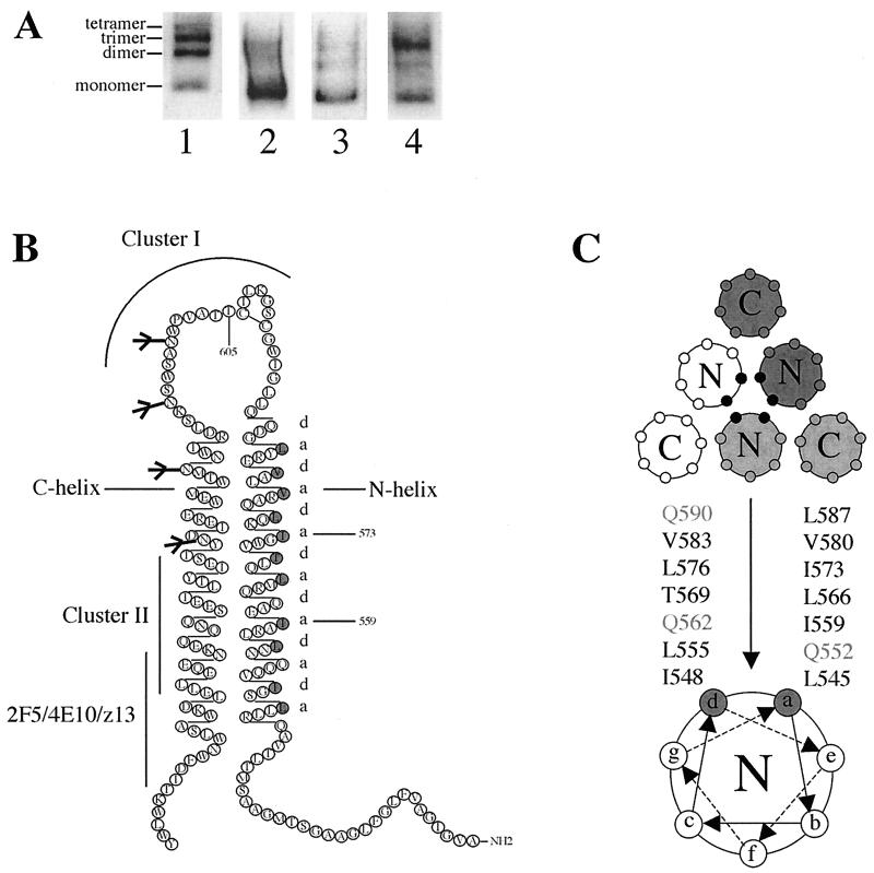
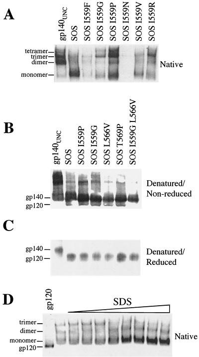
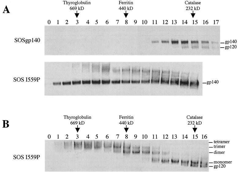
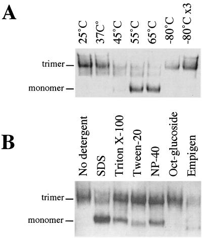
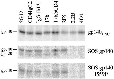
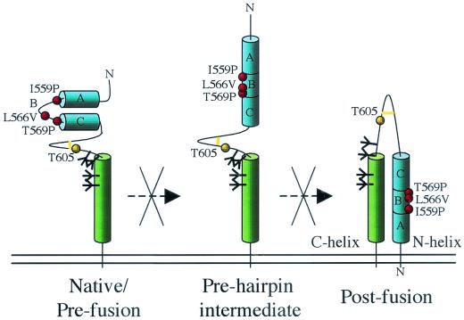
The envelope glycoprotein (Env) complex of human immunodeficiency virus type 1 has evolved a structure that is minimally immunogenic while retaining its natural function of receptor-mediated virus-cell fusion. The Env complex is trimeric; its six individual subunits (three gp120 and three gp41 subunits) are associated by relatively weak, noncovalent interactions. The induction of neutralizing antibodies after vaccination with individual Env subunits has proven very difficult, probably because they are inadequate mimics of the native complex. Our hypothesis is that a stable form of the Env complex, perhaps with additional modifications to rationally alter its antigenic structure, may be a better immunogen than the individual subunits. A soluble form of Env, SOS gp140, can be made that has gp120 stably linked to the gp41 ectodomain by an intermolecular disulfide bond. This protein is fully cleaved at the proteolysis site between gp120 and gp41. However, the gp41-gp41 interactions in SOS gp140 are too weak to maintain the protein in a trimeric configuration. Consequently, purified SOS gp140 is a monomer (N. Schülke, M. S. Vesanen, R. W. Sanders, P. Zhu, D. J. Anselma, A. R. Villa, P. W. H. I. Parren, J. M. Binley, K. H. Roux, P. J. Maddon, J. P. Moore, and W. C. Olson, J. Virol. 76:7760-7776, 2002). Here we describe modifications of SOS gp140 that increase its trimer stability. A variant SOS gp140, designated SOSIP gp140, contains an isoleucine-to-proline substitution at position 559 in the N-terminal heptad repeat region of gp41. This protein is fully cleaved, has favorable antigenic properties, and is predominantly trimeric. SOSIP gp140 trimers are noncovalently associated and can be partially purified by gel filtration chromatography. These gp140 trimers are dissociated into monomers by anionic detergents or heat but are relatively resistant to nonionic detergents, high salt concentrations, or exposure to a mildly acidic pH. SOSIP gp140 should be a useful reagent for structural and immunogenicity studies.


Figures

FIG. 1.
FIG. 1.
Instability of SOS gp140 and a mutagenesis strategy for gp41ECTO. (A) BN-PAGE analysis of SOS gp140 present in culture supernatants derived from four individual experiments each involving transient transfection of 293T cells with the same Env and furin expression plasmids. (B) The residues at positions a and d in the N-terminal heptad repeat of gp41ECTO, depicted in gray in the secondary structure, were substituted in this study. Major MAb epitopes are indicated (26, 68, 100, 106). (C) Schematic representation of a cross-section of the six-helix bundle, postfusion form of gp41ECTO and a helical-wheel representation of one N-terminal helix. The residues at positions a and d of the N-terminal heptad repeat (black symbols) form the trimer interface.
FIG. 2.
FIG. 2.
PAGE analysis of SOS gp140. (A) BN-PAGE of SOS gp140 containing changes at position 559. gp140UNC was included for comparison. (B and C) SDS-PAGE of SOS gp140 variants under nonreducing (B) or reducing (C) conditions. (D) BN-PAGE analysis after treatment with increasing SDS concentrations. SOSIG gp140 was treated with SDS for 1 h at 25°C. The SDS concentrations used were 0, 0.005, 0.01, 0.015, 0.02, 0.025, 0.03, 0.035, and 0.04%, increasing from left to right. BN-PAGE analysis was then performed. Monomeric gp120 (20 ng) served as a molecular weight standard.
FIG. 3.
FIG. 3.
Gel filtration analysis of SOSIP gp140. (A) SOS gp140 and SOSIP gp140 (SOS I559P) were fractionated on a Superdex 200 column. The individual fractions were analyzed by SDS-PAGE and Western blotting. (B) The SOSIP gp140 fractions from panel A were analyzed by BN-PAGE and Western blotting. The elution positions (peak fractions) of standard proteins are indicated.
FIG. 4.
FIG. 4.
Stability of SOSIP gp140 trimers. Fractions 6 and 7 from a gel filtration profile similar to that shown in Fig. 3 were pooled as a source of SOSIP gp140 trimers. The trimers were incubated for 1 h at the temperatures indicated (A) or exposed to a 0.1% concentration of various detergents for 1 h at 25°C (B). The proteins were then analyzed by BN-PAGE and Western blotting. −80°C ×3, three freeze-thaw cycles at −80°C; NP-40, Nonidet P-40; Oct-glucoside, n-octyl β-d-glucopyranoside.
FIG. 5.
FIG. 5.
Antigenic structure analysis of gp140. Radioimmunoprecipitation assays were performed with gp140UNC, SOS gp140, and SOSIP gp140 (SOS gp140 I559P) and various neutralizing and nonneutralizing MAbs as previously described (5). sCD4, soluble CD4.
FIG. 6.
FIG. 6.
HIV-1 gp41ECTO core structure and mutant peptides. (A) The N36-C34 crystal structure is shown with one N36 helix and one C34 helix labeled at the amino termini. Three C34 helices (red) pack against the N36 trimeric coiled coil (blue). The van der Waal surfaces of residues at positions a (red) and d (green) are superimposed on the helix backbone of the N36 coiled coil. Amino acids substituted in this study are indicated above the a and d layers. The diagram was prepared by using the program GRASP (64). (B) Thermal melting transition curves of the N36(L6)C34 (open circles), I559G (closed circles), I559P (open squares), L566V (open diamonds), and T569P (open triangles) peptides were determined by CD spectroscopy at 222 nm and at a peptide concentration of 10 μM in PBS. The increase in the fraction of unfolded molecules is shown as a function of temperature. All melts were reversible. Superimposable folding and unfolding curves were observed, and >90% of the signal was regained upon cooling. (C) Equilibrium sedimentation analysis of the T569P peptide.Representative data for this peptide were collected at 20°C, 20,000 rpm, and a peptide concentration of ∼30 μM in PBS. The data fit best to a trimer model (curve 3). Curves for a dimer (curve 2) and a tetramer (curve 4) are depicted for comparison. Analyses of residual differences from curve 3 did not reveal a systematic error.
FIG. 7.
FIG. 7.
Model of gp41ECTO and its transitions during fusion. (Left panel) Hypothetical, native prefusion configuration of gp41 (39). (Middle panel) Prehairpin intermediate form. (Right panel) Postfusion state. In the prefusion configuration, the N-terminal helix is not present, and the region around positions 559 to 569 is not helical. The I559P and related substitutions are proposed to disrupt either the formation of the N-terminal helix in the prehairpin intermediate form or the formation of the six-helix bundle. As a result, modified SOS gp140 is maintained in the prefusion configuration. The position of the T605C substitution that creates SOS gp140 is also specified, as is the adjacent intermolecular disulfide bond (yellow bar). The positions of N-linked glycans are represented as black branching structures. Only two helices from one gp41 molecule are shown, for clarity. The letters A, B, and C are added to assist with orientation of the helices.

Similar articles

Cited by

References

    1. Allaway, G. P., K. L. Davis-Bruno, G. A. Beaudry, E. B. Garcia, E. L. Wong, A. M. Ryder, K. W. Hasel, M. C. Gauduin, R. A. Koup, J. S. McDougal, and P. J. Maddon. 1995. Expression and characterization of CD4-IgG2, a novel heterotetramer that neutralizes primary HIV type 1 isolates. AIDS Res. Hum. Retrovir. 11:533-539. - PubMed
    1. Barnett, S. W., S. Lu, I. Srivastava, S. Cherpelis, A. Gettie, J. Blanchard, S. Wang, I. Mboudjeka, L. Leung, Y. Lian, A. Fong, C. Buckner, A. Ly, S. Hilt, J. Ulmer, C. T. Wild, J. R. Mascola, and L. Stamatatos. 2001. The ability of an oligomeric human immunodeficiency virus type 1 (HIV-1) envelope antigen to elicit neutralizing antibodies against primary HIV-1 isolates is improved following partial deletion of the second hypervariable region. J. Virol. 75:5526-5540. - PMC - PubMed
    1. Bernstein, H. B., S. P. Tucker, S. R. Kar, S. A. McPherson, D. T. McPherson, J. W. Dubay, J. Lebowitz, R. W. Compans, and E. Hunter. 1995. Oligomerization of the hydrophobic heptad repeat of gp41. J. Virol. 69:2745-2750. - PMC - PubMed
    1. Bewley, C. A., J. M. Louis, R. Ghirlando, and G. M. Clore. 2002. Design of a novel peptide inhibitor of HIV fusion that disrupts the internal trimeric coiled-coil of gp41. J. Biol. Chem. 277:14238-14245. - PubMed
    1. Binley, J. M., R. W. Sanders, B. Clas, N. Schuelke, A. Master, Y. Guo, F. Kajumo, D. J. Anselma, P. J. Maddon, W. C. Olson, and J. P. Moore. 2000. A recombinant human immunodeficiency virus envelope glycoprotein complex stabilized by an intermolecular disulfide bond between the gp120 and gp41 subunits is an antigenic mimic of the trimeric virion-associated structure. J. Virol. 74:627-643. - PMC - PubMed

Publication types

MeSH terms

Substances

LinkOut - more resources