

Hantaan virus infection causes an acute neurological disease that is fatal in adult laboratory mice
- PMID: 12163608
- PMCID: PMC137000
- DOI: 10.1128/jvi.76.17.8890-8899.2002
Hantaan virus infection causes an acute neurological disease that is fatal in adult laboratory mice
Abstract
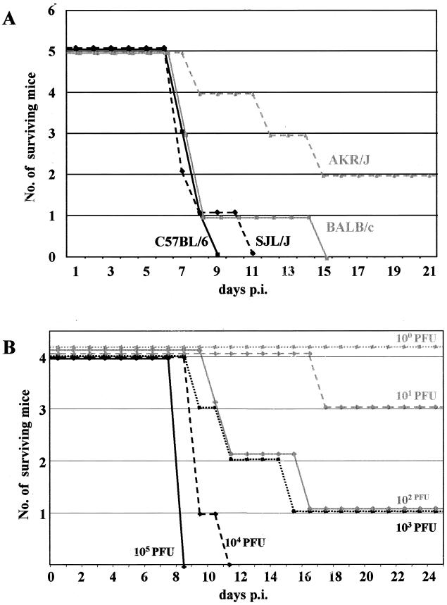
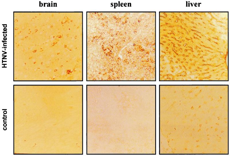
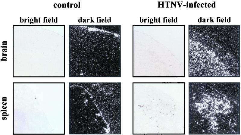
Hantaan virus, the etiological agent of Korean hemorrhagic fever, is transmitted to humans from persistently infected mice (Apodemus agrarius), which serve as the primary reservoir. Here we demonstrate that several strains of adult Mus musculus domesticus (C57BL/6, BALB/c, AKR/J, and SJL/J) were susceptible to Hantaan virus infection when infected intraperitoneally. First clinical signs were loss of weight, ruffled fur, and reduced activity, which were followed by neurological symptoms, such as paralyses and convulsions. Within 2 days of disease onset, the animals died of acute encephalitis. PCR analysis indicated a systemic infection with viral RNA present in all major organs. Immunohistochemical and in situ hybridization analyses of postmortem material detected viral antigen and RNA in the central nervous system (predominantly brain), liver, and spleen. In the central nervous system, viral antigen and RNA colocalized with perivascular infiltrations, the predominant pathological finding. To investigate the involvement of the interferon system in Hantaan virus pathogenesis, we infected alpha/beta interferon receptor knockout mice. These animals were more susceptible to Hantaan virus infection, indicating an important role of interferon-induced antiviral defense mechanisms in Hantaan virus pathogenesis. The present model may help to overcome shortcomings in the development of therapeutic and prophylactic measurements against hantavirus infections.
Figures
Similar articles
-
Isolation of Hantaan virus, the etiologic agent of Korean hemorrhagic fever, from wild urban rats.J Infect Dis. 1982 Nov;146(5):638-44. doi: 10.1093/infdis/146.5.638. J Infect Dis. 1982. PMID: 6127366
-
Dobrava-Belgrade hantavirus from Germany shows receptor usage and innate immunity induction consistent with the pathogenicity of the virus in humans.PLoS One. 2012;7(4):e35587. doi: 10.1371/journal.pone.0035587. Epub 2012 Apr 24. PLoS One. 2012. PMID: 22545121 Free PMC article.
-
Dobrava hantavirus causes hemorrhagic fever with renal syndrome in central Europe and is carried by two different Apodemus mice species.J Med Virol. 2001 Feb;63(2):158-67. J Med Virol. 2001. PMID: 11170053
-
Hantaan virus infection in suckling mice: virologic and pathologic correlates.J Med Virol. 1985 Oct;17(2):107-17. doi: 10.1002/jmv.1890170203. J Med Virol. 1985. PMID: 2865331
-
[Detection of HFRS Hantaan virus RNA sequences by PCR].Nihon Rinsho. 1992 Jul;50 Suppl:305-9. Nihon Rinsho. 1992. PMID: 1357208 Review. Japanese. No abstract available.
Cited by
-
Hantaviruses induce antiviral and pro-inflammatory innate immune responses in astrocytic cells and the brain.Viral Immunol. 2014 Aug;27(6):256-66. doi: 10.1089/vim.2014.0019. Epub 2014 Jun 17. Viral Immunol. 2014. PMID: 24937036 Free PMC article.
-
Emerging infectious diseases: the Bunyaviridae.J Neurovirol. 2005 Oct;11(5):412-23. doi: 10.1080/13550280591002496. J Neurovirol. 2005. PMID: 16287682 Review.
-
The Syrian hamster model of hantavirus pulmonary syndrome.Antiviral Res. 2012 Sep;95(3):282-92. doi: 10.1016/j.antiviral.2012.06.002. Epub 2012 Jun 15. Antiviral Res. 2012. PMID: 22705798 Free PMC article. Review.
-
Hantavirus infection in type I interferon receptor-deficient (A129) mice.J Gen Virol. 2020 Oct;101(10):1047-1055. doi: 10.1099/jgv.0.001470. J Gen Virol. 2020. PMID: 32667279 Free PMC article.
-
Bunyaviruses and the type I interferon system.Viruses. 2009 Dec;1(3):1003-21. doi: 10.3390/v1031003. Epub 2009 Nov 23. Viruses. 2009. PMID: 21994579 Free PMC article.
References
-
- Duchin, J. S., F. T. Koster, C. J. Peters, G. L. Simpson, B. Tempest, S. R. Zaki, T. G. Ksiazek, P. E. Rollin, S. T. Nichol, E. T. Umland, R. L. Moolenaar, S. E. Reef, K. B. Nolte, M. M. Gallaher, J. C. Butler, R. F. Breiman, and The Hantavirus Study Group. 1994. Hantavirus pulmonary syndrome: a clinical description of 17 patients with a newly recognized disease. N. Engl. J. Med. 330:949-955. - PubMed
-
- Elliott, R. M. 1990. Molecular biology of Bunyaviridae. J. Gen. Virol. 71:501-522. - PubMed
-
- Feldmann, H., A. Sanchez, S. Morzunov, C. F. Spiropoulou, P. E. Rollin, T. G. Ksiazek, C. J. Peters, and S. T. Nichol. 1993. Utilization of autopsy RNA for the synthesis of the nucleoprotein antigen of a newly recognized virus associated with hantavirus pulmonary syndrome. Virus Res. 30:351-367. - PubMed
-
- Feldmann, H. March 2000, posting date. Hantaviruses, p. 1-8. In R. M. Atlas, M. M. Cox, S. F. Gilbert, J. W. Roberts, and B. A. Wood (ed.), Encyclopedia of life sciences. [Online.] Nature Publishing Group, London, England. http://www.els.net.
Publication types
MeSH terms
Substances
LinkOut - more resources
Full Text Sources
Molecular Biology Databases
