. 2002 Sep;76(17):8920-30.
doi: 10.1128/jvi.76.17.8920-8930.2002.

Molecular analysis of three Ljungan virus isolates reveals a new, close-to-root lineage of the Picornaviridae with a cluster of two unrelated 2A proteins

Affiliations

Molecular analysis of three Ljungan virus isolates reveals a new, close-to-root lineage of the Picornaviridae with a cluster of two unrelated 2A proteins

Susanne Johansson et al. J Virol. 2002 Sep.

Abstract

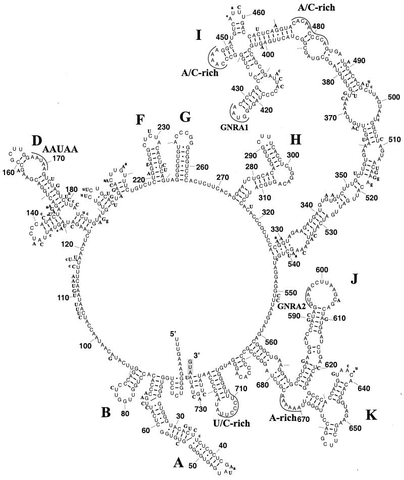
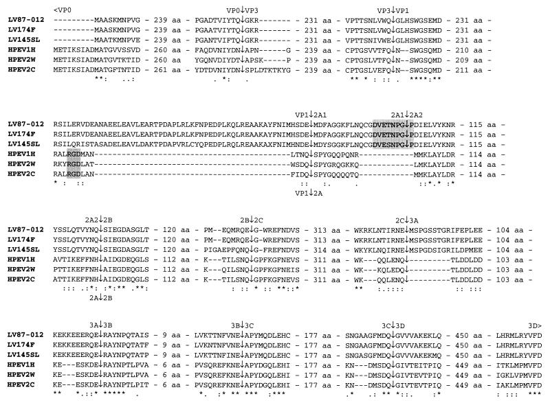
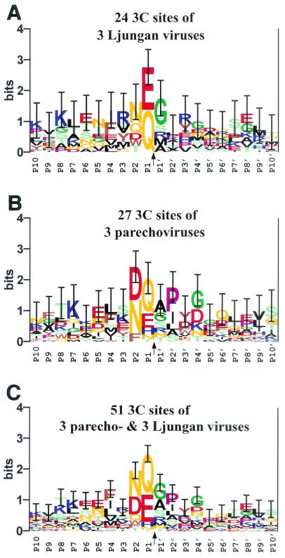
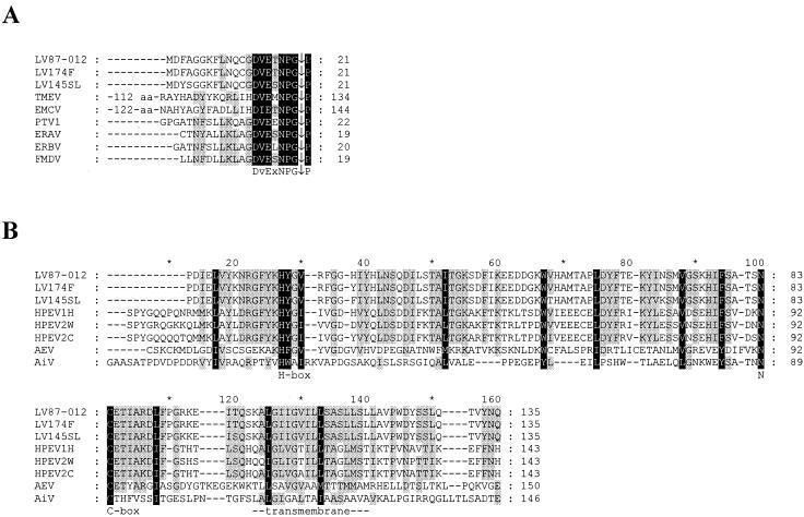
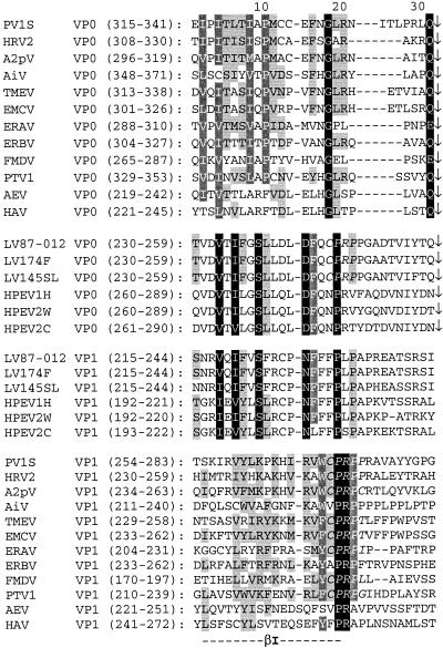
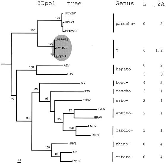
Ljungan virus (LV) is a suspected human pathogen recently isolated from bank voles (Clethrionomys glareolus). In the present study, it is revealed through comparative sequence analysis that three newly determined Swedish LV genomes are closely related and possess a deviant picornavirus-like organization: 5' untranslated region-VP0-VP3-VP1-2A1-2A2-2B-2C-3A-3B-3C-3D-3' untranslated region. The LV genomes and the polyproteins encoded by them exhibit several exceptional features, such as the absence of a predicted maturation cleavage of VP0, a conserved sequence determinant in VP0 that is typically found in VP1 of other picornaviruses, and a cluster of two unrelated 2A proteins. The 2A1 protein is related to the 2A protein of cardio-, erbo-, tescho-, and aphthoviruses, and the 2A2 protein is related to the 2A protein of parechoviruses, kobuviruses, and avian encephalomyelitis virus. The unprecedented association of two structurally different 2A proteins is a feature never previously observed among picornaviruses and implies that their functions are not mutually exclusive. Secondary polyprotein processing of the LV polyprotein is mediated by proteinase 3C (3C(pro)) possessing canonical affinity to Glu and Gln at the P1 position and small amino acid residues at the P1' position. In addition, LV 3C(pro) appears to have unique substrate specificity to Asn, Gln, and Asp and to bulky hydrophobic residues at the P2 and P4 positions, respectively. Phylogenetic analysis suggests that LVs form a separate division, which, together with the Parechovirus genus, has branched off the picornavirus tree most closely to its root. The presence of two 2A proteins indicates that some contemporary picornaviruses with a single 2A may have evolved from the ancestral multi-2A picornavirus.


Figures

FIG. 1.
FIG. 1.
Predicted stem-loop structures of the LV 5′ UTR. Stem-loop structures in the 5′ UTR were predicted for three LV isolates as described in Materials and Methods. Illustrated is the predicted organization of the 5′ UTR of strain LV 87-012 drawn with the XRNA program (B. Wasei and H. F. Noller, unpublished data). The secondary-structure elements recognized in the 5′ UTRs harboring group II IRESs are labeled. Also highlighted are various conserved sequence elements discussed in the text. Stem-loop structures A through H were derived from the RNAGA consensus program (5) which compared LV strains and three parechoviruses. The folding of the I and J-K stem-loop regions was supported by the consensus analysis and was predicted using the MFOLD program (67). Replacements in strains 145SL and 174F are indicated in boldface uppercase and lowercase letters, respectively, and insertions and deletions are marked with asterisks and arrows, respectively. Base pairings predicted for LV 87-012 are indicated with continuous lines, and base pairings that are possible in either of the two other isolates are indicated with broken lines. The initiator codon is indicated with a shaded box.
FIG. 2.
FIG. 2.
Comparison of the terminal regions and the predicted cleavage sites of the polyproteins of LV strains 87-012, 174F, and 145SL with those of parechoviruses (HPEV1H, HPEV2W, and HPEV2C). ∗, identical residues; : and ·, strong and weak conserved groups, respectively, of amino acid residues as defined by the Clustal X program (59, 60). The RGD motif in the C-terminal end of VP1 in HPEV is highlighted in the smaller gray box and is located in the last conserved region between HPEV and LV in VP1. The conserved DvExNPG|P motif putatively responsible for the LV 2A1 C-terminal polyprotein processing is highlighted in the larger gray box. Two of the previously predicted cleavage sites for parechoviruses, 2A|2B and 3B|3C, are revised here based on the now available LV amino acid sequences. The numbers of amino acids separating the amino acids shown in detail around the proposed scissile bonds are indicated in the alignments.
FIG. 3.
FIG. 3.
Conservation of 3Cpro sites in LV and parechovirus polyproteins. Three separate multiple, gap-free 20-aa-long alignments around the P10-P10′ positions of the sites (presumably) cleaved by 3Cpro domains of LVs (A), parechoviruses (B), or both groups of viruses (C) were converted into sequence logos presentations (4, 55). Due to a greater uncertainty of the prediction for the LV VP1|2A1 sites (see the text), they were not included in the analysis. In the logos, the height of each letter (amino acid residue) is proportional to its frequency at the specific position, and the letters are sorted so that the most common residue is on the top of the stack. The height of each stack is proportional to the information content (measured in bits) of the sequences at this position. The upper limit of information at any position (4.32 bits) is determined by the natural diversity of the 20 aa, which is expressed as a logarithm of 20. The most conserved and important positions are relatively high and easily recognized. Vertical bars, whose sizes are reversibly proportional to the sampling size, indicate 1 standard deviation of the information content at each position (55). The letters indicating amino acid residues are colored as follows: light green for S, T, and C; orange for N and Q; red for D and E; blue for K, R, and H; brown for W, F, and Y; black for A, L, I, V, and M; pink for P; and green for G.
FIG. 4.
FIG. 4.
Multiple sequence alignments of 2A1 and 2A2 of LVs and selected picornaviruses. Black and gray backgrounds highlight alignment columns with 100 and 60% conserved residues, respectively, as defined in the GeneDoc default similarity groups (37). (A) Alignment of the LV 2A1 and the C-terminal regions of the picornavirus NPGP 2A protein family. (B) Alignment of the LV 2A2 and the picornavirus H-NC 2A protein family. The C-terminal border of parechovirus 2As is depicted as it was predicted (Fig. 2). The actual boundaries of 2A proteins from AiV and AEV were not defined. The conserved H-box and NC-box and a putative transmembrane domain made up of an approximately 20-aa-long hydrophobic region are labeled. ∗, each noneven count of 10.
FIG. 5.
FIG. 5.
Conservation between the C-terminal regions of VP0 and VP1 of picornaviruses. Multiple alignments of VP0 and VP1 of a set of picornaviruses representing all genera and LVs were generated as described in Materials and Methods. Presented is an alignment of sequences in the vicinity of the β-strands I of VP0 and VP1 split into four virus-protein groups separated by horizontal spaces. The top group comprises VP0 of all picornavirus genera except those of LVs and parechoviruses, which form the second group; the bottom group comprises VP1 of all picornavirus genera except those of LVs and parechoviruses, which form the third group. The amino acid conservation is highlighted separately for the top, bottom, and two middle groups. Black, dark gray (with white letters) and light gray (with black letters) backgrounds highlight alignment columns with 100, 80, and 60% conserved residues, respectively, as defined in the GeneDoc default similarity groups (37). The CysProArgPro (CPRP) tetrapeptide is indicated by italic type. Arrows indicate the position of the scissile bond at the VP0-VP3 junction.
FIG. 6.
FIG. 6.
Phylogenetic relationships among picornaviruses. A phylogenetic tree of picornavirus 3Dpol protein sequences (left) representing all genera and LVs was generated by using the Proml program in the PHYLIP 3.6a2 package as described in Materials and Methods. Prior to the tree inference, a likelihood mapping analysis of the 3Dpol alignment showed that the phylogenetic signal could be inferred by using bifurcating trees (data not shown). The tree was rooted by using two 3D-like sequences encoded by InFV and SBV as the out-group. All bifurcations supported in more than 70 of 100 bootstrap trials are labeled. Bar, 0.1 substitutions per site. The LV division is highlighted in the gray oval. The picornavirus genera are listed to the right of the tree. To illustrate the diversity of picornaviruses outside 3Dpol, structural variants of 2A and L proteins were arbitrarily assigned unique numbers, and these are listed along with their respective virus genera (right). The number 0 indicates the lack of L protein. Note that AEV and HAV of hepatoviruses encode different combinations of L and 2A proteins.

Similar articles

Cited by

References

    1. Auvinen, P., and T. Hyypiä. 1990. Echoviruses include genetically distinct serotypes. J. Gen. Virol. 71:2133-2139. - PubMed
    1. Beard, C. W., and P. W. Mason. 2000. Genetic determinants of altered virulence of Taiwanese foot-and-mouth disease virus. J. Virol. 74:987-991. - PMC - PubMed
    1. Bertioli, D. 1997. Rapid amplification of cDNA ends, p. 233-238. In B. A. White (ed.), PCR cloning protocols: from molecular cloning to genetic engineering, vol. 67. Humana Press Inc., Totowa, N.J.
    1. Blom, N., J. Hansen, D. Blaas, and S. Brunak. 1996. Cleavage site analysis in picornaviral polyproteins: discovering cellular targets by neural networks. Protein Sci. 5:2203-2216. - PMC - PubMed
    1. Chen, J. H., S. Y. Le, and J. V. Maizel. 2000. Prediction of common secondary structures of RNAs: a genetic algorithm approach. Nucleic Acids Res. 28:991-999. - PMC - PubMed

Publication types

MeSH terms

Associated data

LinkOut - more resources