. 2002 Sep;76(17):8966-72.
doi: 10.1128/jvi.76.17.8966-8972.2002.

Genetically stable picornavirus expression vectors with recombinant internal ribosomal entry sites

Affiliations

Genetically stable picornavirus expression vectors with recombinant internal ribosomal entry sites

Andrew T Dufresne et al. J Virol. 2002 Sep.

Abstract

In many respects, picornaviruses are well suited for their proposed use as immunization vectors. However, their inherent genetic instability hinders application for prophylactic purposes. We demonstrate the improved expression and stability of a heterologous insert through a novel vector design strategy that partially replaces noncoding regulatory sequences with coding sequences for foreign gene products.


Figures

FIG. 1.
FIG. 1.
Genetic structure of proposed picornavirus-based expression vectors. (A) Wild-type (Wt) poliovirus. (B) Capsid display vectors with foreign peptides (hatched box) incorporated into the viral capsid (6, 14). (C) Dicistronic vectors expressing a foreign insert (hatched box) driven by a secondary encephalomyocarditis virus (EMCV) IRES (2). The intercistronic EMCV IRES may be placed between P1 and P2 or between the insert and P1. (D) Polyprotein fusion vectors (5, 8). A foreign insert is fused to the viral polyprotein either in between P1 and P2 or at the N terminus. Proteolytic processing of the fusion polyprotein occurs through artificial cleavage sites for the viral 2Apro or 3Cpro protease, respectively (5, 8).
FIG. 2.
FIG. 2.
Construction of recombinant IRES expression vectors. Both vector types feature artificial cleavage sites for 2Apro for the release of foreign polypeptides from the fusion polyprotein. Inserted SIVp17 sequences are indicated by hatched boxes. (A) Genetic structure of a conventional fusion polyprotein expression vector based on PVS-RIPO. The rhinoviral IRES element is shown. The enlarged sequence detail depicts HRV2 IRES stem-loop domain VI and the genetic structure of the insert region. Initiation of translation of the fusion polyprotein occurs from the authentic HRV2 initiation codon, preceding the N-terminal 4 amino acids of the poliovirus polyprotein and the ORF for SIVp17 (boldface letters). (B) Sequence and proposed secondary structure of the SIV AUG loop (7). The initiating AUG of SIVgag (boldface letters) is in a position similar to that of Y(n)X(m)AUG in the HRV2 IRES, forming the base of stem-loop domain VI [for the position of the Y(n)X(m)AUG motif, compare with Fig. 3A]. (C) Genetic structure of RPδ6/SIVp17. Sequences shown indicate the SIV AUG loop which has been used to replace HRV2 IRES stem-loop domain VI. Initiation occurs at Y(n)X(m)AUG, which had been placed in Kozak context (ACCAUGG). X(m) has been altered to contain a BglII restriction site. SIVp17 sequences are shown in boldface.
FIG. 3.
FIG. 3.
Position and structure of the Y(n)X(m)AUG motif within type 1 (enterovirus and rhinovirus) IRES elements. The polypyrimidine tract [Y(n)], spacer [X(m)], and cryptic AUG (asterisks) in the intact HRV2 IRES (A) and in a stem-loop domain VI deletion mutant (B) are indicated. The sequence of X(m) was altered in panel B to put the adjacent cryptic AUG into Kozak context (CUUAUGG to ACCAUGG). (C) Growth characteristics of PVS-RIPO (open squares) and RPδ6 (filled diamonds) in HeLa cells. The IRES deletion construct gave rise to viable virus that grew only slightly less efficiently than did PVS-RIPO in HeLa cells. p.i., postinfection.
FIG.4.
FIG.4.
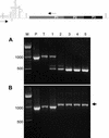
RT-PCR analysis of total RNA preparations obtained from serial passages of RIPO/SIVp17 and RPδ6/SIVp17 expression vectors. Lanes are marked according to the origin of template used in the diagnostic PCR: M, molecular mass marker; P, plasmid DNA; T, lysate of transfected cells; 1 through 5, individual passages. The schematic above indicates the positions of annealing sequences for primers used in the PCR. (A) Transfection of RIPO/SIVp17 RNA and subsequent passaging in HeLa cells resulted in the rapid deletion of inserted foreign sequences. (B) Transfection of RPδ6/SIVp17 RNA and subsequent passaging of virus resulted in a genetic adaptation event characterized by an insert enlarged by 114 nt (arrowhead). The enlarged insert was retained after five passages, without evidence for the occurrence of deletion variants.
FIG. 5.
FIG. 5.
Sequence and growth characteristics of variant RPδ6/SIVp17(21). (A) The amplified PCR product obtained through RT-PCR from total cellular RNA prepared from the fifth consecutive passage of RPδ6/SIVp17 in HeLa cells (Fig. 4B) was subjected to sequence analysis. The variant virus had acquired a duplication of IRES and insert sequences (duplicated sequences are boxed in gray). The duplication was in frame, containing two tandem AUG codons in Kozak context (capital letters). (B) RPδ6/SIVp17(21) virus (filled diamonds) exhibited propagation kinetics in HeLa cells that resembled those of PVS-RIPO (open squares). p.i., postinfection.
FIG. 5.
FIG. 5.
Sequence and growth characteristics of variant RPδ6/SIVp17(21). (A) The amplified PCR product obtained through RT-PCR from total cellular RNA prepared from the fifth consecutive passage of RPδ6/SIVp17 in HeLa cells (Fig. 4B) was subjected to sequence analysis. The variant virus had acquired a duplication of IRES and insert sequences (duplicated sequences are boxed in gray). The duplication was in frame, containing two tandem AUG codons in Kozak context (capital letters). (B) RPδ6/SIVp17(21) virus (filled diamonds) exhibited propagation kinetics in HeLa cells that resembled those of PVS-RIPO (open squares). p.i., postinfection.
FIG. 6.
FIG. 6.
Western blot analysis of the kinetics of viral and foreign gene expression in HeLa cells infected with RPδ6/SIVp17(21). Lane M, molecular mass marker. (A) A monoclonal antibody against the poliovirus gene products 2C and its precursor 2BC was used in Western blot assays of cell lysates obtained at the indicated intervals postinfection (p.i.). Initial viral gene expression could be detected at 3 h p.i. and reached its peak at 6 h p.i. (B) Serum from an SHIV-infected rhesus macaque was used to sample infected cell lysates assayed in panel A for expression of SIVp17(21). In parallel with native viral gene expression, SIVp17(21) could be detected at 3 h p.i. Synthesis greatly increased until 6 h p.i., synchronous with viral gene expression. The gel migration rate of SIVp17(21) is in accordance with that of an enlarged gene product produced through initiation at the first Y(n)X(m)AUG motif in variant RPδ6/SIVp17(21).
FIG. 7.
FIG. 7.
Genetic stability of variant RPδ6/SIVp17(21) after extended passages. (A) RT-PCR analysis of 20 passages of variant RPδ6/SIVp17(21) in HeLa cells. Methods and primers are those employed for Fig. 4A and B. The source of the template cDNA is indicated by the following lane designations: M, molecular mass marker; P, original RPδ6/SIVp17 plasmid DNA; T, lysate of cells transfected with RPδ6/SIVp17 transcript RNA; 1 through 20, individual passages; P−, RPδ6 plasmid DNA). Two distinct deletion variants emerged after the ninth passage, slowly replacing the full-length RPδ6/SIVp17(21) variant. Deletions were comparatively minor [fragments corresponding to partially deleted insert sequences are labeled (i) and (ii)]. The amplicon from the P− template represents the expected size of the fragment obtained from virus genomes devoid of any insert (compare with Fig. 4A). (B) Comparative analysis of SIVp17(21) expression by variant RPδ6/SIVp17(21) after transfection of transcript RNA and 20 subsequent passages in HeLa cells. No expression of SIVp17 was observed in transfected HeLa cells and the first passage under experimental conditions that readily revealed SIVp17(21) expression in later passages. (C) Genetic structure and sequence of two deletion variants (i and ii from panel A) isolated from variant RPδ6/SIVp17(21) after 20 passages in HeLa cells. Internal deletions of 114 and 240 nt, respectively, were flanked by identical complementary sequences. Duplicated sequences and engineered stem-loop structures were not affected by deletion events.

Similar articles

Cited by

References

    1. Agol, V. I., E. V. Pilipenko, and O. R. Slobodskaya. 1996. Modification of translational control elements as a new approach to design of attenuated picornavirus strains. J. Biotechnol. 44:119-128. - PubMed
    1. Alexander, L., H. H. Lu, and E. Wimmer. 1994. Polioviruses containing picornavirus type 1 and/or type 2 internal ribosomal entry site elements: genetic hybrids and the expression of a foreign gene. Proc. Natl. Acad. Sci. USA 91:1406-1410. - PMC - PubMed
    1. Almond, J. W., D. Stone, K. Burke, M. A. Skinner, A. J. Macadam, D. Wood, M. Ferguson, and P. D. Minor. 1993. Approaches to the construction of new candidate poliovirus type 3 vaccine strains. Dev. Biol. Stand. 78:161-169. - PubMed
    1. Altmeyer, R., N. Escriou, M. Girard, A. Palmenberg, and S. van der Werf. 1994. Attenuated Mengo virus as a vector for immunogenic human immunodeficiency virus type 1 glycoprotein 120. Proc. Natl. Acad. Sci. USA 91:9775-9779. - PMC - PubMed
    1. Andino, R., D. S. D. Silvera, P. L. Suggett, C. J. Achacoso, C. J. Miller, D. Baltimore, and M. B. Feinberg. 1994. Engineering poliovirus as a vaccine vector for the expression of diverse antigens. Science 265:1448-1451. - PubMed

Publication types

Substances

LinkOut - more resources