. 2002 Aug 20;99(17):11103-8.
doi: 10.1073/pnas.162039399. Epub 2002 Aug 5.

Transition to the open state of the TolC periplasmic tunnel entrance

Affiliations

Transition to the open state of the TolC periplasmic tunnel entrance

Christian Andersen et al. Proc Natl Acad Sci U S A. .

Abstract

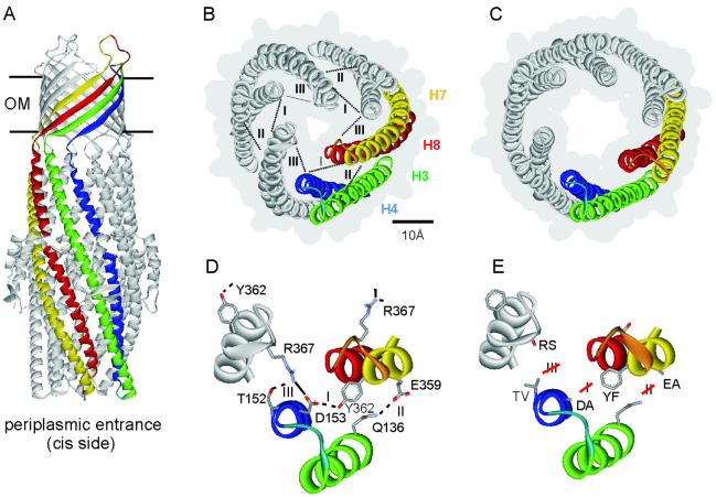
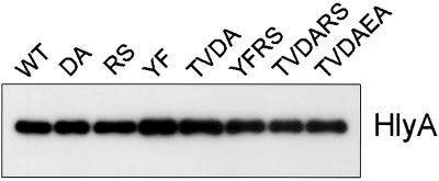
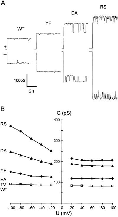
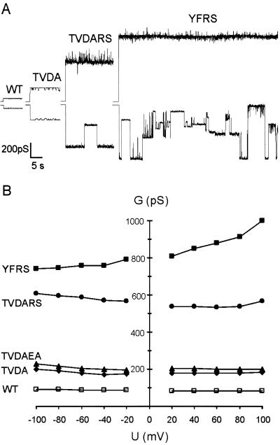
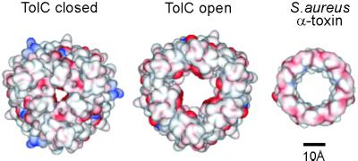
The TolC channel-tunnel spans the bacterial outer membrane and periplasm, providing a large exit duct for protein export and multidrug efflux when recruited by substrate-engaged inner membrane complexes. The sole constriction in the single pore of the homotrimeric TolC is the periplasmic tunnel entrance, which in its resting configuration is closed by dense packing of the 12 tunnel-forming alpha-helices. Recruitment of TolC must trigger opening for substrate transit to occur, but the mechanism underlying transition from the closed to the open state is not known. The high resolution structure of TolC indicates that the tunnel helices are constrained at the entrance by a circular network of intra- and intermonomer hydrogen bonds and salt bridges. To assess how opening is achieved, we disrupted these connections and monitored changes in the aperture size by measuring the single channel conductance of TolC derivatives in black lipid bilayers. Elimination of individual connections caused incremental weakening of the circular network, accompanied by gradual relaxation from the closed state and increased flexibility of the entrance. Simultaneous abolition of the key links caused a substantial increase in conductance, generating an aperture that corresponds to the modeled open state, with the capacity to allow access and passage of diverse substrates. The results support a model in which transition to the open state of TolC is achieved by an iris-like realignment of the tunnel entrance helices.


Figures

Fig 1.
Fig 1.
The structure of TolC and its periplasmic entrance. (A) The TolC structure (9) is 140 Å in length. The 40 Å long β-barrel domain spanning the outer membrane (OM) is assembled from 12 β-sheets (four per monomer) and is constitutively open. The 100 Å long α-helical domain is formed by 12 α-helices and projects across the periplasmic space. The entrance is located at the bottom of this domain as indicated. One monomer is highlighted in the same color scheme as in B. (B) The closed state of the entrance seen from the periplasmic side. Helices of one of the three monomers are colored green (H3, outer coiled coil), blue (H4, outer), yellow (H7, inner coiled coil), and red (H8, inner). The numbering of the helices is taken from the high resolution structure (9). The connecting turns between H3/H4 and between H7/H8 are shown respectively in light blue and orange. Intramonomer (I and II) and intermonomer connections (III) are shown as dotted lines. The gray background outlines the surface representation, showing the small periplasmic entrance. (C) The modeled open state of the entrance (9), derived by realigning the inner coiled coils (H7/H8) to the same configuration as the outer coiled coils (H3/H4). (D) Magnification of the circular network at the tunnel entrance, showing the residues central to the intramonomer (I and II) and intermonomer (III) links. The salt bridge is shown in full, the hydrogen bonds are indicated by broken lines. (E) Substitutions made to disrupt the hydrogen bonds and salt bridge shown in D. Depictions were generated by using weblab viewerlite (Molecular Simulations, Cambridge, U.K.).
Fig 2.
Fig 2.
TolC-dependent protein export from E. coli. Cell-free supernatants (100 μl) of exponentially growing cultures of E. coli BL923 expressing either WT or mutant TolC protein were subjected to SDS/PAGE and blotted with anti-HlyA antiserum.
Fig 3.
Fig 3.
Single channel conductance of TolC variants with single substitutions. (A) At +80 mV (upper trace) and −80 mV (lower trace). (B) Dependence on applied membrane potential. Each point represents the main (maximum) conductance achieved at the voltage. Proteins were added to the cis side of the membrane bathed in 1 M KCl, pH 7.5.
Fig 4.
Fig 4.
Single channel conductance of TolC variants with multiple substitutions. (A) At +80 mV (upper trace) and −80 mV (lower trace). (B) Dependence of the main conductance on applied membrane potential. Conditions as in Fig. 3.
Fig 5.
Fig 5.
Putative size of the opened entrance. Comparison of the modeled open state of the TolC entrance (Center) with the actual closed state (Left) (9) and the Staphylococcus aureus α-toxin membrane domain (Right) (22). The surface representations show electronegative and electropositive surfaces colored red and blue, respectively (generated by using weblab viewerlite, Molecular Simulations).

References

    1. Thanassi D. G. & Hultgren, S. J. (2000) Curr. Opin. Cell. Biol. 12, 420-430. - PubMed
    1. Andersen C., Hughes, C. & Koronakis, V. (2000) EMBO Rep. 1, 313-318. - PMC - PubMed
    1. Thanabalu T., Koronakis, E., Hughes, C. & Koronakis, V. (1998) EMBO J. 17, 6487-6496. - PMC - PubMed
    1. Koronakis V., Li, J., Koronakis, E. & Stauffer, K. (1997) Mol. Microbiol. 23, 617-626. - PubMed
    1. Nikaido H. (1994) Science 264, 382-388. - PubMed

Publication types

MeSH terms

LinkOut - more resources