

Massive cross-modal cortical plasticity and the emergence of a new cortical area in developmentally blind mammals
- PMID: 12163645
- PMCID: PMC123273
- DOI: 10.1073/pnas.162342799
Massive cross-modal cortical plasticity and the emergence of a new cortical area in developmentally blind mammals
Abstract
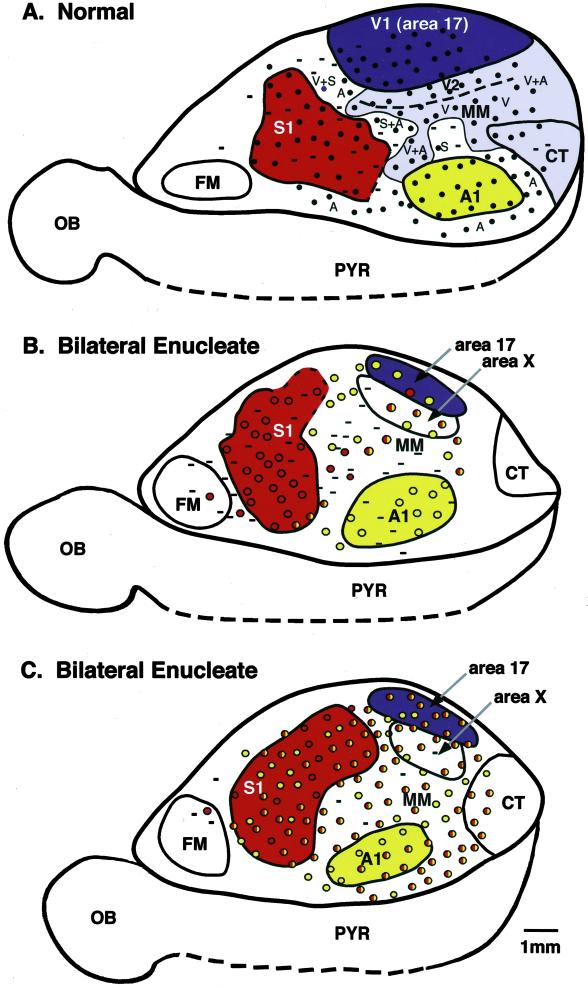
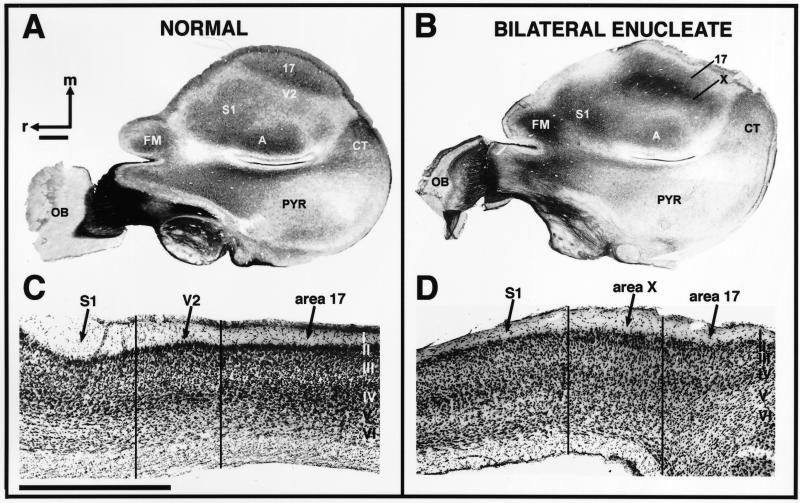
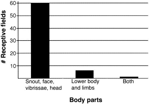
In the current investigation, the neurophysiological organization of the neocortex was examined in adult animals that were bilaterally enucleated very early in life, before the retino-geniculo-cortical pathway was established. Our results indicate that some aspects of development of cortical fields are not mediated by specific sensory inputs. However, the current study also demonstrates that peripheral innervation plays a large role in the organization of the neocortex, as cortical territories normally involved in visual processing are completely captured by the auditory and somatosensory system. Thus, a large degree of phenotypic variability in cortical organization can be accomplished solely by removing or modifying sensory inputs.
Figures
Similar articles
-
Alterations in cortical and thalamic connections of somatosensory cortex following early loss of vision.J Comp Neurol. 2019 Jul 1;527(10):1675-1688. doi: 10.1002/cne.24582. Epub 2018 Dec 9. J Comp Neurol. 2019. PMID: 30444542 Free PMC article.
-
Cross-modal plasticity for the spatial processing of sounds in visually deprived subjects.Exp Brain Res. 2009 Jan;192(3):343-58. doi: 10.1007/s00221-008-1553-z. Epub 2008 Sep 2. Exp Brain Res. 2009. PMID: 18762928 Review.
-
Mental Imagery Follows Similar Cortical Reorganization as Perception: Intra-Modal and Cross-Modal Plasticity in Congenitally Blind.Cereb Cortex. 2019 Jul 5;29(7):2859-2875. doi: 10.1093/cercor/bhy151. Cereb Cortex. 2019. PMID: 30060011
-
Early blindness results in abnormal corticocortical and thalamocortical connections.Neuroscience. 2006 Oct 27;142(3):843-58. doi: 10.1016/j.neuroscience.2006.06.055. Epub 2006 Aug 24. Neuroscience. 2006. PMID: 16934941
-
Human brain plasticity: evidence from sensory deprivation and altered language experience.Prog Brain Res. 2002;138:177-88. doi: 10.1016/S0079-6123(02)38078-6. Prog Brain Res. 2002. PMID: 12432770 Review.
Cited by
-
Auditory attention activates peripheral visual cortex.PLoS One. 2009;4(2):e4645. doi: 10.1371/journal.pone.0004645. Epub 2009 Feb 27. PLoS One. 2009. PMID: 19247451 Free PMC article.
-
Metabolic Changes in the Bilateral Visual Cortex of the Monocular Blind Macaque: A Multi-Voxel Proton Magnetic Resonance Spectroscopy Study.Neurochem Res. 2017 Feb;42(2):697-708. doi: 10.1007/s11064-016-2126-3. Epub 2016 Dec 1. Neurochem Res. 2017. PMID: 27909856
-
Evolution of mammalian sensorimotor cortex: thalamic projections to parietal cortical areas in Monodelphis domestica.Front Neuroanat. 2015 Jan 7;8:163. doi: 10.3389/fnana.2014.00163. eCollection 2014. Front Neuroanat. 2015. PMID: 25620915 Free PMC article.
-
Alterations in cortical and thalamic connections of somatosensory cortex following early loss of vision.J Comp Neurol. 2019 Jul 1;527(10):1675-1688. doi: 10.1002/cne.24582. Epub 2018 Dec 9. J Comp Neurol. 2019. PMID: 30444542 Free PMC article.
-
Shaping Diversity Into the Brain's Form and Function.Front Neural Circuits. 2018 Oct 10;12:83. doi: 10.3389/fncir.2018.00083. eCollection 2018. Front Neural Circuits. 2018. PMID: 30364100 Free PMC article. Review.
References
-
- Röder B., Rosler, F. & Neville, H. J. (1999) Neurosci. Lett. 264, 53-56. - PubMed
-
- Röder B., Rosler, F. & Neville, H. J. (2000) Neuropsychologia 38, 1482-1502. - PubMed
-
- Sadato N., Pascualleone, A., Grafman, J., Ibanez, V., Deiber, M. P., Dold, G. & Hallett, M. (1996) Nature (London) 380, 526-528. - PubMed
-
- Cohen L. G., Celnik, P., PascualLeone, A., Corwell, B., Faiz, L., Dambrosia, J., Honda, M., Sadato, N., Gerloff, C., Catala, M. D. & Hallett, M. (1997) Nature (London) 389, 180-183. - PubMed
MeSH terms
LinkOut - more resources
Full Text Sources
