. 2002 Aug;129(2):198-207.
doi: 10.1046/j.1365-2249.2002.01924.x.

Coupling complement regulators to immunoglobulin domains generates effective anti-complement reagents with extended half-life in vivo

Affiliations

Coupling complement regulators to immunoglobulin domains generates effective anti-complement reagents with extended half-life in vivo

C L Harris et al. Clin Exp Immunol. 2002 Aug.

Abstract

Complement activation and subsequent generation of inflammatory molecules and membrane attack complex contributes to the pathology of a number of inflammatory and degenerative diseases, including arthritis, glomerulonephritis and demyelination. Agents that specifically inhibit complement activation might prove beneficial in the treatment of these diseases. Soluble recombinant forms of the naturally occurring membrane complement regulatory proteins (CRP) have been exploited for this purpose. We have undertaken to design better therapeutics based on CRP. Here we describe the generation of soluble, recombinant CRP comprising rat decay accelerating factor (DAF) or rat CD59 expressed as Fc fusion proteins, antibody-like molecules comprising two CRP moieties in place of the antibody Fab arms (CRP-Ig). Reagents bearing DAF on each arm (DAF-Ig), CD59 on each arm (CD59-Ig) and a hybrid reagent containing both DAF and CD59 were generated. All three reagents inhibited C activation in vitro. Compared with soluble CRP lacking Fc domains, activity was reduced, but was fully restored by enzymatic release of the regulator from the Ig moiety, implicating steric constraints in reducing functional activity. In vivo studies showed that DAF-Ig, when compared to soluble DAF, had a much extended half-life in the circulation in rats and concomitantly caused a sustained reduction in plasma complement activity. When given intra-articularly to rats in a model of arthritis, DAF-Ig significantly reduced severity of disease. The data demonstrate the potential of CRP-Ig as reagents for sustained therapy of inflammatory disorders, including arthritis, but emphasize the need for careful design of fusion proteins to retain function.


Figures
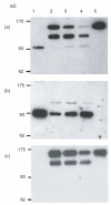
Fig. 1
Fig. 1
Western blot analysis of supernatant from transfected cells. Supernatant from cells transfected with plasmid encoding CD59-spacer-Ig (lane 1), DAF-Ig (lane 5) or both plasmids together (lanes 2–4) was subjected to SDS-PAGE on a 7·5% nonreducing gel and Western blot analysis. Lanes 2–4 represent cotransfection with 2 μg total DNA with the ratio of DAF-Ig: CD59-Ig plasmid being 2 : 1, 1 : 1 and 1 : 2, respectively. Blots were probed with HRPO-conjugated polyclonal anti-human Fc (a), monoclonal anti-rat CD59 (b) or monoclonal anti-rat DAF (c). Monoclonal antibodies were detected using HRPO conjugated secondary antibody and bands were visualized using ECL.
Fig. 2
Fig. 2
Purification of CRP-Ig and sDAF. DAF-Ig, CD59-Ig and CD59-spacer-Ig were purified from culture supernatant by protein A affinity chromatography. Purified proteins were subjected to SDS-PAGE on a (a) 7·5% or (b) 10% nonreducing gel and stained with Coomassie Blue. Human IgG1 was run for comparison. (c) Soluble, recombinant rat DAF was purified from culture supernatant by affinity chromatography on a monoclonal anti-rat DAF column followed by gel filtration. The pure protein was subjected to nonreducing (NR) or reducing (R) SDS-PAGE on a 12·5% gel. Proteins were visualized by silver staining.
Fig. 3
Fig. 3
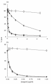
In vitro complement regulatory function of DAF-Ig. (a) Sensitized erythrocytes were incubated in GVB with rat serum and different concentrations of sCR1 (□), rat DAF-Ig (▴), sDAF (▾) or a nonregulatory Ig fusion protein (○). Haemolysis was assessed by release of haemoglobin to the supernatant and percent lysis was determined. Results represent the mean value ± SD of three determinations. (b) Inhibition of lysis by sDAF (▾) is compared to that achieved with soluble DAF released from DAF-Ig using papain (◊) and a nonregulatory Ig fusion protein (○).
Fig. 4
Fig. 4
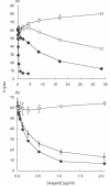
In vitro complement regulatory function of CD59-Ig. Guinea pig erythrocytes bearing C5b-7 sites were incubated in PBS/EDTA with rat serum and different concentrations of test protein. Haemolysis was assessed by release of haemoglobin to the supernatant and percent lysis was determined. Results represent the mean value ± SD of three determinations. (a) Functional comparison of soluble, recombinant CD59 (•), CD59-Ig (□), CD59-spacer-Ig (◊) and a nonregulatory Ig fusion protein (○). (b) Inhibition of lysis by a nonregulatory Ig fusion protein (○), soluble, recombinant CD59 (•) and soluble CD59 released from CD59-Ig using papain (▴).
Fig. 5
Fig. 5
In vivo half-life of DAF-Ig and sDAF. Radiolabelled DAF-Ig (•) and sDAF (○) were administered to rats, blood was removed at specific timepoints and protein bound radioactivity was determined. Results are expressed as percent of levels at 3 min and represent the mean of five animals ± SD.
Fig. 6
Fig. 6
Effect of circulating DAF-Ig on plasma CH50. Rats were dosed intravenously with 10 mg/kg DAF-Ig. (a) Plasma was removed at specific timepoints and levels of DAF-Ig in plasma were determined by ELISA, data are expressed as percent of levels at 1 h; results represent the mean of five animals ± SD. (b) Haemolytic activity (CH50) was also determined, results are expressed as percent of haemolytic activity prior to reagent administration; results represent the mean of five animals ± SEM.
Fig. 7
Fig. 7
Therapeutic effect of DAF-Ig on AIA. Methylated BSA was introduced into the right knee of immune rats. DAF-Ig (○) or saline (•, control) was administered to the joint at the same time. Swelling of the joint was measured daily and compared to that of the left knee. Results represent the mean of five animals ± SEM (* P <0·01; ** P <0·001).

Similar articles

Cited by

References

    1. Morgan BP, Meri S. Membrane proteins that protect against complement lysis. Springer Semin Immunopathol. 1994;15:369–96. - PubMed
    1. Morgan BP, Harris CL. London: Academic Press; 1999. Complement Regulatory Proteins.
    1. Kinoshita T, Lavoie S, Nussenzweig V. Regulatory proteins for the activated third and fourth components of complement (C3b and C4b) in mice. II. Identification and properties of complement receptor type 1 (CR1) J Immunol. 1985;134:2564–70. - PubMed
    1. Reid KBM, Bentley DR, Campbell RD, Chung LP, Sim RB, Kristensen T, Tack BF. Complement proteins which interact with C3b or C4b. A superfamily of structurally related proteins. Immunol Today. 1986;7:230–4. - PubMed
    1. Morgan BP, Daniels RH, Williams BD. Measurement of Terminal Complement Complexes in Rheumatoid-Arthritis. Clin Exp Immunol. 1988;73:473–8. - PMC - PubMed

Publication types

MeSH terms