. 2002 Aug;129(2):272-80.
doi: 10.1046/j.1365-2249.2002.01890.x.

Profiles of cell-to-cell interaction of Mycobacterium intracellulare-induced immunosuppressive macrophages with target T cells in terms of suppressor signal transmission

Affiliations

Profiles of cell-to-cell interaction of Mycobacterium intracellulare-induced immunosuppressive macrophages with target T cells in terms of suppressor signal transmission

K Ogasawara et al. Clin Exp Immunol. 2002 Aug.

Abstract

Previously, we have found that immunosuppressive macrophages (M(phi)s) induced by Mycobacterium intracellulare-infection (MI-M(phi)s) required cell contact with target T cells to express their suppressor activity against concanavalin A (Con A)-induced T cell mitogenesis. In this study, we examined the profiles of cell-to-cell interaction of MI-M(phi)s with target T cells. First, MI-M(phi)s displayed suppressor activity in an H-2 allele-unrestricted manner, indicating that MHC molecules are not required for cell contact. The suppressor activity of MI-M(phi)s was reduced markedly by paraformaldehyde fixation or treatment with cytochalasin B or colchicine, indicating that vital membrane functions are required for their suppressor activity. Secondly, the suppressor activity of MI-M(phi)s was independent of cell-to-cell interaction via CD40 ligand/CD40 and M(phi)-derived indoleamine 2,3-dioxygenase, which causes rapid degradation of tryptophan in T cells. Thirdly, precultivation of splenocytes with MI-M(phi)s, allowing cell-to-cell contact, reduced Con A- or anti-CD3 antibody-induced mitogenesis but not phorbol myristate acetate/calcium ionophore A23187-elicited proliferation of T cells. In addition, co-cultivation of T cells with MI-M(phi)s caused marked changes in profiles of the tyrosine phosphorylation of 33 kDa, 34 kDa and 35-kDa proteins and, moreover, the activation of protein kinase C and its translocation to the cell membrane. It thus appears that suppressor signals of MI-M(phi)s, which are transmitted to the target T cells via cell contact, principally cross-talk with the early signalling events before the activation of PKC and/or intracellular calcium mobilization.


Figures
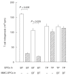
Fig. 1
Fig. 1
Cell-to-cell contact-mediated expression of the suppressor activity of M. intracellulare-induced macrophages (MI-Mφs) against Con A-induced T cell mitogenesis. Using a dual-chamber system, spleen cells (SPCs) (5 × 10⁵) were cultured in either the ‘bottom chamber' (BT), on which monolayer cultures of MI-Mφs (3 × 10⁵/well) were prepared (hatched bars) or not prepared (open bars), or the ‘top chamber' (TP) equipped with a 0·45 μm Millipore filter-bottom. In some cases, 5 × 10⁵ of SPCs pretreated with 100 μg/ml mitomycin C for 1 h (MMC-SPCs) were also added to the ‘bottom chamber' (BT). Each bar indicates the mean ± s.e.m. (n = 3).
Fig. 2
Fig. 2
MHC-unrestricted expression of the suppressor activity by M. intracellulare-induced macrophages (MI-Mφs) against target T cells. Con A-stimulated spleen cells (SPCs) (2·5 × 105) from BALB/c (H-2d), C57BL/6 (H-2b) or CBA/JN (H-2k) mice were co-cultured with MI-Mφs (2 × 10⁴/well) from BALB/c mice. Each bar indicates the mean ± s.e.m. (n = 4).
Fig. 3
Fig. 3
Deprivation of the suppressor activity from M. intracellulare-induced macrophages (MI-Mφs) by paraformaldehyde fixation. MI-Mφs prepared on microculture wells were fixed with 1% paraformaldehyde for 10 min and measured for their suppressor activity against Con A-induced T cell mitogenesis. Dotted bar: co-cultured with live MI-Mφs. Hatched bar: co-cultivated with fixed MI-Mφs. Each bar indicates the mea± s.e.m. (n = 8).
Fig. 4
Fig. 4
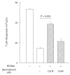
Effects of microfilament and microtuble inhibitors on the suppressor activity of M. intracellulare-induced macrophages (MI-Mφs). MI-Mφs (4 × 10⁴) were pretreated with cytochalasin B (CytB) or colchicine (Colch) at 2 μm each for 30 min, washed with 2% FBS-HBSS, and measured for the suppressor activity against Con A-induced T cell mitogenesis. Each bar indicates the mean ± s.e.m. (n = 3).
Fig. 5
Fig. 5
Effects of anti-CD40 monoclonal antibody (MoAb) blocking on cytokine mRNA expression by M. intracellulare-induced macrophages (MI-Mφs) with or without cocultivation with T cells in the presence of Con A (2 μg/ml). Levels of mRNA expression of test cytokines were measured by the RNase protection assay. Lane 1, MW marker; lane 2, MI-Mφs; lane 3, anti-CD40 MoAb treated (50 μg/ml at 4°C for 3 h) MI-Mφs; lane 4, MI-Mφs co-cultivated with T cells in the presence of Con A for 24 h; lane 5, MI-Mφs which were pretreated with anti-CD40 MoAb and then co-cultivated with T cells in the presence of Con A for 24 h; lane 6, positive control for each cytokine. The mRNA expression levels of a housekeeping gene (L32) of lanes 2–5 were essentially the same as each other (data not shown). In the right table, relative intensities of cytokine mRNA expression in terms of cytokine/L32 band ratio are indicated.
Fig. 6
Fig. 6
Profiles of Con A signal-associated tyrosine phosphorylation of cytoplasmic proteins in T cells co-cultivated with M. intracellulare-induced macrophages (MI-Mφs). The splenic lymphocytes which had been precultivated with or without MI-Mφs for 23 h were cultivated for 10 min after Con A stimulation, and then cytoplasmic proteins were extracted as mentioned in the Method section. The cell lysate preparation containing cytoplasmic proteins (35 μg each) was subjected to sodium dodecylsulphate (SDS)–12% polyacrylamide gel electrophoresis (SDS-PAGE) followed by Western blotting using antiphosphotyrosine antibody. In control experiments, the SDS-PAGE patterns of cytoplasmic proteins extracted from splenic T cells with or without co-cultivation with MI-Mφs were the same as each other, indicating that the profile of protein expression by T cells was not altered due to cell contact with MI-Mφs (data not shown). In the right table, the band intensities of the 15-kDa, 16-kDa, 33-kDa, 34-kDa and 35-kDa proteins measured using a densitometer are indicated.

Similar articles

Cited by

References

    1. Ellner JJ. Suppressor adherent cells in human tuberculosis. J Immunol. 1978;121:2573–9. - PubMed
    1. Edwards CK, Hedegaar HB, Zlotnik A, et al. Chronic infection due to Mycobacterium intracellulare in mice: association with macrophage release of prostaglandin E2 and reversal by injection of indomethacin, muramyl dipeptide, and interferon-γ. J Immunol. 1986;136:1820–7. - PubMed
    1. Tomioka H. Immunosuppressive macrophages. Clin Immunol. 2000;33:29–36.
    1. Tomioka H, Saito H, Yamada Y. Characteristics of immunosuppressive macrophages induced in spleen cells by Mycobacterium avium complex infections in mice. J Gen Microbiol. 1990;136:965–74. - PubMed
    1. Tomioka H, Saito H, Sato K. Characteristics of immunosuppressive macrophages induced in host spleen cells by Mycobacterium avium complex and Mycobacterium tuberculosis infections in mice. Microbiol Immunol. 1990;34:283–97. - PubMed

Publication types

Substances