. 2002 Aug 20;99(17):11055-60.
doi: 10.1073/pnas.162368399. Epub 2002 Aug 7.

Interactions between lipids and bacterial reaction centers determined by protein crystallography

Affiliations

Interactions between lipids and bacterial reaction centers determined by protein crystallography

A Camara-Artigas et al. Proc Natl Acad Sci U S A. .

Abstract

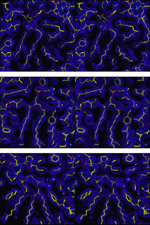
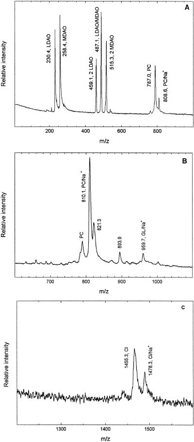
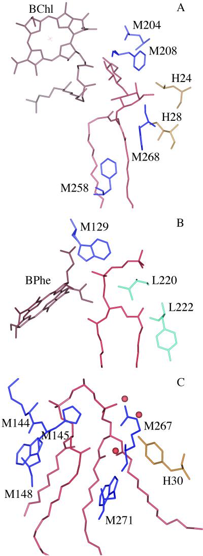
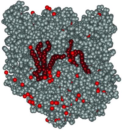
The structure of the reaction center from Rhodobacter sphaeroides has been solved by using x-ray diffraction at a 2.55-A resolution limit. Three lipid molecules that lie on the surface of the protein are resolved in the electron density maps. In addition to a cardiolipin that has previously been reported [McAuley, K. E., Fyfe, P. K., Ridge, J. P., Isaacs, N. W., Cogdell, R. J. & Jones, M. R. (1999) Proc. Natl. Acad. Sci. USA 96, 14706-14711], two other major lipids of the cell membrane are found, a phosphatidylcholine and a glucosylgalactosyl diacylglycerol. The presence of these three lipids has been confirmed by laser mass spectroscopy. The lipids are located in the hydrophobic region of the protein surface and interact predominately with hydrophobic amino acids, in particular aromatic residues. Although the cardiolipin is over 15 A from the cofactors, the other two lipids are in close contact with the cofactors and may contribute to the difference in energetics for the two branches of cofactors that is primarily responsible for the asymmetry of electron transfer. The glycolipid is 3.5 A from the active bacteriochlorophyll monomer and shields this cofactor from the solvent in contrast to a much greater exposed surface evident for the inactive bacteriochlorophyll monomer. The phosphate atom of phosphatidylcholine is 6.5 A from the inactive bacteriopheophytin, and the associated electrostatic interactions may contribute to electron transfer rates involving this cofactor. Overall, the lipids span a distance of approximately 30 A, which is consistent with a bilayer-like arrangement suggesting the presence of an "inner shell" of lipids around membrane proteins that is critical for membrane function.


Figures

Fig 1.
Fig 1.
Stereoview of electron density maps (2|FO| − |Fc|) for the three bound lipids (Top) glucosylgalactosyl diacylglycerol, (Middle) phosphatidylcholine, (Bottom) cardiolipin and the surrounding protein residues. The identification of the glycolipid and cardiolipin is clear based on the electron density. However, the phosphatidylcholine can also be substituted with the slightly smaller phosphatidylethanolamine. The contour level is 1 σ.
Fig 2.
Fig 2.
Representative spectra of purified RCs from laser mass spectroscopy using different matrices and ion modes. (A) Peaks at 787.0 and 808.6 Da correspond to the calculated molecular masses of phosphatidylcholine (PC), 786.1 Da, the sodium adduct (PC/Na+), 808.6 Da. The lower mass peaks at 230.4 and 258.4 Da are due to the detergent and show a mixture of lauryl and myrstyl dimethylamine oxide (LDAO and MDAO) that have calculated molecular masses of 230.4 and 258.4 Da, respectively. Also evident are dimers of lauryl and myrstyl dimethylamine oxide at 459.1 and 515.3 Da, as well as a lauryl/myrstyl dimethylamine oxide heterodimer at 487.1 Da. This spectrum was taken by using 4-hydroxybenzylidenemalononitritrile in positive ion mode. (B) A peak at 959.7 Da corresponding to the sodium adduct of the glycolipid (GL/Na+) with a calculated mass of 957.1 Da is evident using this 4-hydroxybenzylidenemalononitritrile matrix. The amplitude of this peak was found to track with the amount of NaI added to the matrix. Also seen in the spectrum is a major contribution from a sodium adduct of phosphatidylcholine at 810.1 Da. A minor contribution from the monoprotonated form is seen near 786 Da although the peak position is not precisely determined because of the presence of an overlapping peak. The peak at 893.9 Da arises from a contaminant. (C) Peaks at 1,455.3 and 1,478.3 Da corresponding to monoprotonated and monosodiated cardiolipin anions (CL and CL/Na+) with calculated molecular masses of 1,455.9 and 1,477.9 Da, respectively. This spectrum was taken by using a terthiophene matrix in negative ion mode.
Fig 3.
Fig 3.
Structure of the lipids with nearby cofactors and the surrounding protein. (A) The glycolipid (red) binds near the active bacteriochlorophyll monomer (brown) and nearby amino acid residues of the M subunit (blue) Leu M204, Phe M208, Phe M258, and Trp M268, and the H subunit residues (yellow) Leu H24 and Ile H28. (B) The phospholipid phosphatidylcholine (red) binds near the L subunit residues (light blue) Val L220 and Tyr L222, and the M subunit residue Trp M129 (dark blue), and the inactive bacteriopheophytin (brown). (C) The cardiolipin (red) binds on the M subunit (dark blue) and near the H subunit (yellow) with the phosphate atoms of the lipid interacting with His M145, Arg M267, Tyr H30, and several water molecules (orange spheres). Also shown are Lys M144, Trp M148, and Trp M271.
Fig 4.
Fig 4.
Surface representation of the RC structure showing the glycolipid and cardiolipid (dark red), the protein (gray), and bound water molecules (light red). Both of the lipids are tightly bound, with the glycolipid fitting between the M and H subunits whereas the cardiolipin is more exposed but held by electrostatic interactions involving the two phosphates and nearby charged residues. The two lipids are positioned with a separation of about 30 Å between the phosphate and saccharide moieties.

References

    1. Benning C. (1998) in Lipids in Photosynthesis: Structure, Function, and Genetics, eds. Siegenthaler, P. A. & Murata, N. (Kluwer, Dordrecht, The Netherlands), pp. 83–101.
    1. Michel H., (1991) Crystallization of Membrane Proteins (CRC, Boca Raton, FL).
    1. Feher G., Allen, J. P., Okamura, M. Y. & Rees, D. C. (1989) Nature (London) 339, 111-116.
    1. Blankenship R. E., Madigan, M. T. & Bauer, C. E., (1995) Anoxygenic Photosynthetic Bacteria (Kluwer, Dordrecht, The Netherlands).
    1. McAuley K. E., Fyfe, P. K., Ridge, J. P., Isaacs, N. W., Cogdell, R. J. & Jones, M. R. (1999) Proc. Natl. Acad. Sci. USA 96, 14706-14711. - PMC - PubMed

Publication types

MeSH terms

Substances

LinkOut - more resources