

DNA detection using water-soluble conjugated polymers and peptide nucleic acid probes
- PMID: 12167673
- PMCID: PMC123191
- DOI: 10.1073/pnas.162375999
DNA detection using water-soluble conjugated polymers and peptide nucleic acid probes
Abstract
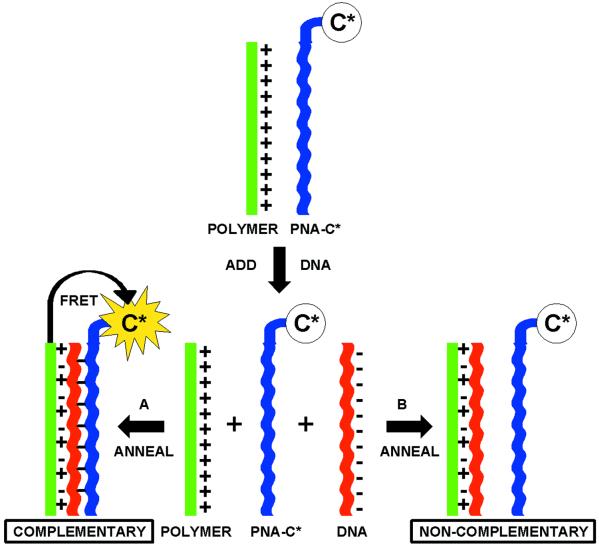
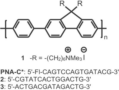
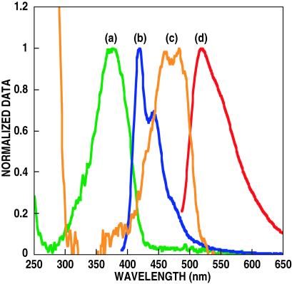
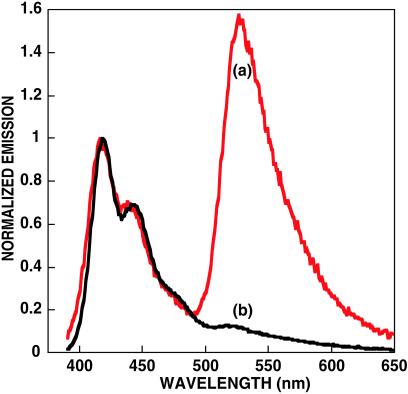
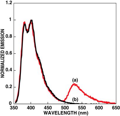
The light-harvesting properties of cationic conjugated polymers are used to sensitize the emission of a dye on a specific peptide nucleic acid (PNA) sequence for the purpose of homogeneous, "real-time" DNA detection. Signal transduction is controlled by hybridization of the neutral PNA probe and the negative DNA target. Electrostatic interactions bring the hybrid complex and cationic polymer within distances required for Förster energy transfer. Conjugated polymer excitation provides fluorescein emission >25 times higher than that obtained by exciting the dye, allowing detection of target DNA at concentrations of 10 pM with a standard fluorometer. A simple and highly sensitive assay with optical amplification that uses the improved hybridization behavior of PNA/DNA complexes is thus demonstrated.
Figures
References
-
- Schork N. J., Fallin, D. & Lanchbury, J. S. (2000) Clin. Genet. 58, 250-264. - PubMed
-
- Balakin K. V., Korshun, V. A., Mikhalev, I. I., Maleev, G. V., Malakhov, A. D., Prokhorenko, I. A. & Berlin, Y. A. (1998) Biosens. Bioelectron. 13, 771-778. - PubMed
-
- LePecq J. B. & Paoletti, C. (1967) J. Mol. Biol. 27, 87-106. - PubMed
Publication types
MeSH terms
Substances
Grants and funding
LinkOut - more resources
Full Text Sources
Other Literature Sources
