. 2002 Sep;22(17):6079-88.
doi: 10.1128/MCB.22.17.6079-6088.2002.

Inactivation of NF-kappaB-dependent cell survival, a novel mechanism for the proapoptotic function of c-Abl

Affiliations

Inactivation of NF-kappaB-dependent cell survival, a novel mechanism for the proapoptotic function of c-Abl

Hidehiko Kawai et al. Mol Cell Biol. 2002 Sep.

Abstract

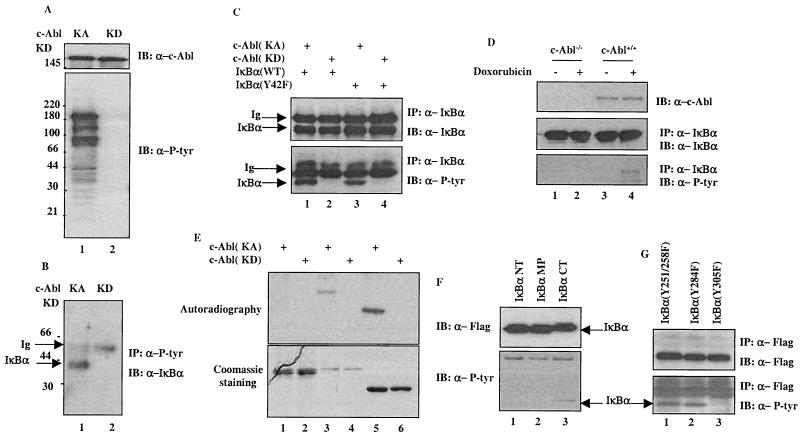
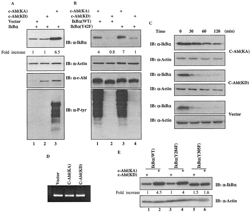
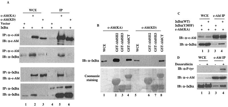
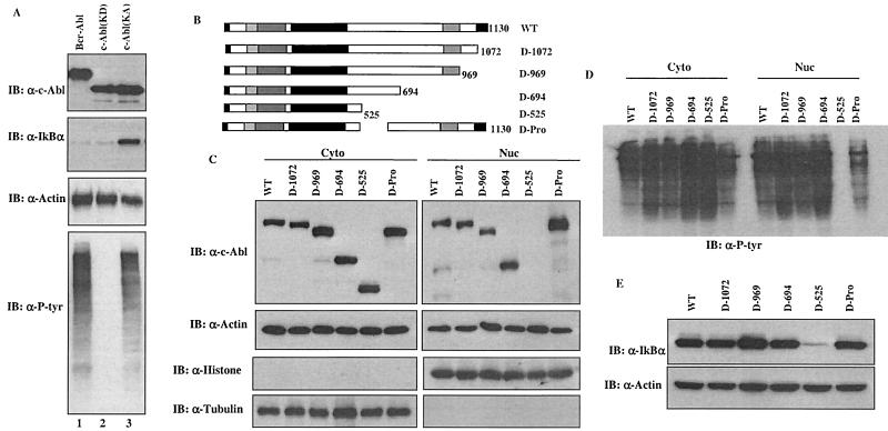
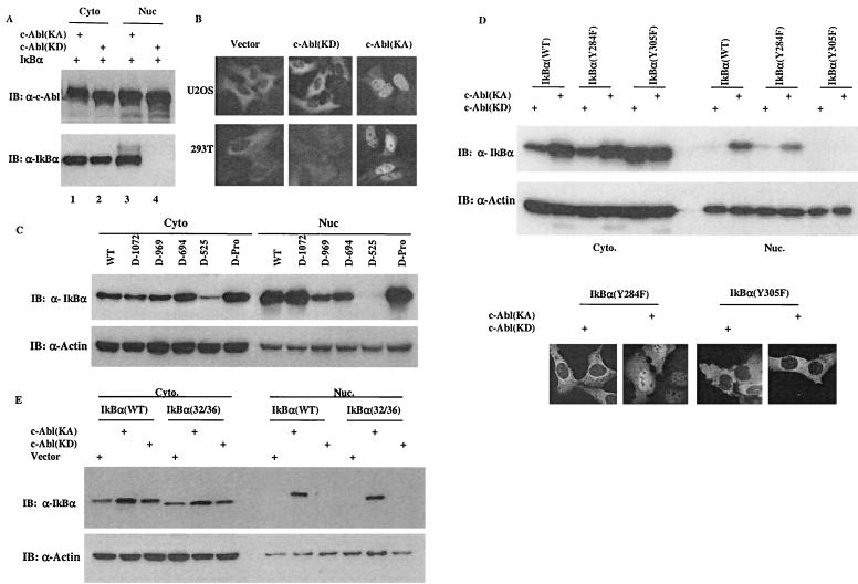
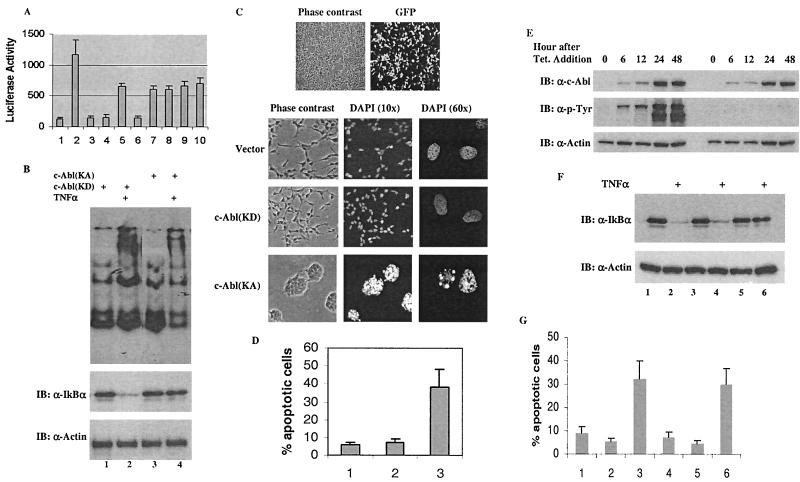
Using a system that expresses a constitutively kinase-active c-Abl protein [c-Abl(KA)], we identified the protein IkappaBalpha as a novel substrate of c-Abl. This kinase-substrate relationship is not only confirmed at the level of endogenous proteins but is also supported by a physical interaction between the two proteins. Interestingly, the association of c-Abl with IkappaBalpha, which is detectable in the form of nonphosphorylated proteins, is remarkably enhanced by an inducible binding of tyrosine-phosphorylated IkappaBalpha to the c-Abl SH2 domain. In contrast to the serine 32/34 phosphorylation that triggers ubiquitination and degradation of IkappaBalpha, c-Abl-mediated phosphorylation at tyrosine 305 is associated with an increase of the IkappaBalpha protein stability. Significantly, this activity is not shared by the oncogenic Bcr-Abl, because it is unique to the nuclear c-Abl. We also demonstrate that c-Abl targets the nuclear subpopulation of IkappaBalpha for phosphorylation and induces it to accumulate in the nucleus. As a consequence, NF-kappaB transcription activity is abolished, leading to an increased cellular sensitivity to the induction of apoptosis. The functional importance of c-Abl-mediated IkappaBalpha phosphorylation is highlighted by a loss of response of the IkappaBalpha(Y305F) protein to c-Abl-mediated regulation. Using cells expressing the c-Abl(KA) protein under the control of an inducible promoter, we demonstrate inactivation of the NF-kappaB-dependent cell survival pathway as one of the mechanisms for c-Abl-mediated apoptosis.


Figures

FIG. 1.
FIG. 1.
IκBα is a novel substrate of c-Abl. (A) A plasmid (2 μg) encoding c-Abl(KA) (lane 1) or c-Abl(KD) (lane 2) was transfected into 293T cells. The cells were harvested at 24 h posttransfection and subjected to Western analysis with anti-c-Abl (top panel) or anti-P-Tyr (bottom panel). (B) Cell lysates prepared from the transfectants were also analyzed by anti-P-tyr immunoprecipitation followed by anti-IκBα immunoblotting. (C) A vector expressing c-Abl(KA) (lanes 1 and 3) or c-Abl(KD) (lanes 2 and 4) was cotransfected with wild type (lanes 1 and 2) or Y42F (lanes 3 and 4) of IκBα into U2OS cells. The cells were subjected to anti-IκBα immunoprecipitation at 24 h posttransfection. The immunocomplexes were analyzed by immunoblotting using anti-IκBα (top panel) or anti-P-tyr (bottom panel). (D) c-Abl−/− or c-Abl+/+ MEFs were treated with doxorubicin (2 μM; lanes 2 and 4) or left untreated (lanes 1 and 3). The cells were harvested 12 h after the treatment and subjected to anti-IκBα immunoprecipitation. Proteasome inhibitor (MG132; 7.5 μM; Sigma) was added 6 h before harvesting. The IκBα immunocomplexes were analyzed with anti-IκBα (middle panel) or anti-P-tyr (bottom panel). (E) Purified recombinant c-Abl(KA) (lanes 1, 3, and 5) or c-Abl(KD) (lanes 2, 4, and 6) was incubated with GST-fusion proteins of p53DBD (lanes 1 and 2), IκBα (lanes 3 and 4) or Crk (lanes 5 and 6) in the presence of [32P]ATP for 15 min at 30°C. After washing, the GST-fusion proteins were resolved on SDS-PAGE and the gel was either stained with Coomassie dye (bottom panel) or dried followed by autoradiography (top panel). (F) Immunopurified Flag-tagged IκBα NT, middle portion (MP) or CT was incubated with purified recombinant c-Abl(KA) for 30 min at 30°C. The reaction products were analyzed by Western analysis using anti-IκBα (upper panel) or anti-P-tyr (lower panel). (G) A plasmid encoding IκBα(Y251/258F), (Y284F) or (Y305F) was cotransfected with c-Abl(KA). Lysates prepared 24 h posttransfection were immunoprecipitated with anti-Flag followed by Western analysis using anti-Flag (upper panel) or anti-P-tyr (lower panel).
FIG. 2.
FIG. 2.
Upregulation of IκBα by c-Abl in a kinase-dependent manner. (A) A vector expressing IκBα was cotransfected with a plasmid encoding control vector (lane 1), c-Abl(KD) (lane 2) or c-Abl(KA) (lane 3) into 293T cells. Cell lysates prepared at 24 h after transfection were subjected to Western analysis using the indicated antibody. The densitometric intensity of the IκBα band was scanned and numbers represent fold increase over that of vector control that was set as 1. (B) A plasmid containing cDNA of wild type (lanes 1 and 2) or Y42F (lanes 3 and 4) mutant of IκBα was coexpressed with c-Abl(KA) (lanes 1 and 3) or c-Abl(KD) (lanes 2 and 4) in U2OS cells and the cells were analyzed as described for panel A. (C) Half-life of IκBα in the vector, c-Abl(KD)- or c-Abl(KA)-expressing cells was determined by monitoring the disappearance of IκBα after addition of cycloheximide (20 μg/ml). (D) mRNA was isolated and analyzed by RT-PCR using a kit according to the manufacturer's protocol (Promega). (E) A plasmid encoding wild-type IκBα (lanes 1 and 2), IκBα(Y284F) (lanes 3 and 4) or IκBα(Y305F) (lanes 5 and 6) was cotransfected with c-Abl(KA) (lanes 2, 4 and 6) or c-Abl(KD) (lanes 1, 3 and 5). Cellular abundance of IκBα was analyzed 24 h after transfection and actin was used as a loading control.
FIG. 3.
FIG. 3.
A phosphorylation-facilitated interaction between IκBα and c-Abl. (A) A vector expressing IκBα was coexpressed with a control vector, c-Abl(KA) or c-Abl(KD). Cell lysates prepared 24 h after transfection were subjected to immunoprecipitation with anti-c-Abl (top two panels) or anti-IκBα (lower two panels). Whole-cell extracts (lanes 1 to 3) or immunocomplexes (lanes 4 to 6) were analyzed by Western blotting using the indicated antibodies. (B) Cell lysates prepared from c-Abl(KA)- (lanes 1 to 4) or c-Abl(KD)- (lanes 5 to 8) expressing cells were incubated with the indicated GST-fusion proteins. The adsorbates were extensively washed, resolved on SDS-PAGE and either immunoblotted with anti-IκBα (top panel) or stained with Coomassie dye (bottom panel). Whole-cell extracts (WCE, lanes 1 and 5) were included as blotting controls. (C) A vector expressing wild-type IκBα (lanes 2 and 4) or IκBα (Y305F) mutant (lanes 1 and 3) was cotransfected with c-Abl(KA). Cell lysates prepared 24 h after transfection were subjected to immunoprecipitation with anti-c-Abl. Whole-cell extract (lanes 1 and 2) and c-Abl immunocomplexes (lanes 3 and 4) were analyzed by Western blotting using anti-c-Abl (top panel) or anti-P-Tyr (bottom panel). (D) U2OS cells treated with or without doxorubicin (2 μM for 12 h) were analyzed by anti-c-Abl immunoprecipitation followed by Western analysis using the indicated antibodies.
FIG. 4.
FIG. 4.
The ability of c-Abl to stabilize IκBα is not shared by the oncogenic Abl. (A) A vector expressing IκBα was cotransfected with a plasmid encoding Bcr-Abl (lane 1), c-Abl(KD) (lane 2) or c-Abl(KA) (lane 3). The cells were analyzed as described for Fig. 2. (B) A series of deletion mutants of c-Abl were prepared. (C) cDNAs of the indicated c-Abl deletion mutants were subcloned into Flag-tagged pCDNA3 vector and were transfected into 293T cells. The cells were then fractionated into cytoplasmic and nuclear fractions, which then were subjected to anti-Flag (top panels) or anti-actin (second panels) immunoblotting analysis. Anti-histone (nuclear marker) and anti-tubulin (cytoplasmic marker) immunoblotting were included to assess the purity of each fraction. (D) The cytoplasmic and nuclear fractions were also immunoblotted with anti-P-tyr. (E) A plasmid encoding IκBα was cotransfected with the indicated mutants of c-Abl and the IκBα levels were analyzed as described for Fig. 2.
FIG. 5.
FIG. 5.
The c-Abl-induced IκBα nuclear accumulation contributes to its ability to upregulate the IκBα protein levels. (A) A vector containing cDNA of IκBα was cotransfected with a plasmid encoding c-Abl(KA) or c-Abl(KD). Cytoplasmic and nuclear fractions were prepared at 24 h posttransfection and subjected to Western analysis using anti-c-Abl (top panel) or anti-IκBα (bottom panel). (B) A plasmid encoding GFP-IκBα was cotransfected with a control vector, c-Abl(KD) or c-Abl(KA) into U2OS (top panels) or 293T cells (bottom panels). The cells were processed as described in Materials and Methods and examined under a fluorescence microscope. (C) An IκBα-expressing vector was cotransfected with plasmids encoding the indicated c-Abl deletion mutants. Cell fractions were prepared at 24 h posttransfection and analyzed by Western blotting using anti-IκBα (top panel) and anti-actin (bottom panel). (D) A plasmid encoding IκBα(wild type), (Y284F) or (Y305F) was cotransfected with c-Abl(KA) or c-Abl(KD). Cell fractions were prepared at 24 h after transfection and Western blotted with anti-IκBα or anti-actin. U2OS cells were transfected with the indicated vectors and analyzed as described for panel B. (E) Cell fractions were prepared from cells transfected with the indicated vectors and analyzed as described for panel C.
FIG. 6.
FIG. 6.
Functional inhibition of NF-κB contributes to the proapoptotic function of c-Abl. (A) A luciferase-expressing vector containing a κB-responsive element was cotransfected with control vectors (bar 1), NF-κB/control vector (bar 2), c-Abl(KA) (bar 3) or c-Abl(KD) (bar 4), NF-κB/IκBα (bar 5), NF-κB/IκBα/c-Abl(KA) (bar 6), NF-κB/IκBα/c-Abl(KD) (bar 7), NF-κB/IκBα(Y305F) (bar 8), NF-κB/IκBα(Y305F)/c-Abl(KA) (bar 9), or NF-κB/IκBα(Y305F)/c-Abl(KD) (bar 10). pRL-TK vector (Promega) was included to control the low level of Renilla luciferase expression and serve as a transfection efficiency control. Luciferase assays were performed 24 h posttransfection using the Promega Rapid Detection system. Values are averages ± SE of the mean derived from two separate experiments performed in triplicate. (B) Cells expressing c-Abl(KD) (lanes 1 and 2) or c-Abl(KA) (lanes 3 and 4) were treated with TNF-α (10 ng/ml) for 15 min (lanes 2 and 4) or left untreated (lanes 1 and 3). Nuclear extracts were prepared for EMSA using a labeled consensus NF-κB-binding oligonucleotide (top panel) and whole-cell extracts were analyzed by Western blotting using anti-IκBα and anti-actin (lower panels). (C) 293T cells were transfected with 2 μg of GFP-c-Abl(KD) and the cells were visualized via phase-contrast optics (left) and fluorescent filter (right) to show transfection efficiency. Cells expressing a control vector (2 μg; top panels), c-Abl(KD) (2 μg; middle panels) or c-Abl(KA) (2 μg; bottom panels) were treated with TNF-α (40 ng/ml) at 16 h posttransfection. After incubation at 37°C for an additional 8 h, the cells were fixed and stained with DAPI. The whole cells were visualized by phase-contrast optics and the nuclei were visualized by UV optics (two different amplifications, 10× or 60×). (D) Cells expressing a control vector (bar 1), c-Abl(KD) (bar 2) or c-Abl(KA) (bar 3) were processed as described for panel C. The cells were then fixed in 70% methanol, stained with PI and subjected to FACS analysis. Sub-G1 populations were recorded as the apoptotic cells. Values are averages ± SE of the mean derived from two separate experiments performed in triplicate. (E) The pooled cell populations that stably expressed c-Abl(KA) or c-Abl(KD) were treated with tetracycline (1 μg/ml) and harvested at the indicated times for Western analysis with anti-c-Abl (top), anti-p-Tyr (middle) or anti-actin (bottom). (F) At 24 h postaddition of tetracycline, cells expressing the control vector (lanes 1 and 2), c-Abl(KD) (lanes 3 and 4) and c-Abl(KA) (lanes 5 and 6) were treated with TNF-α (10 ng/ml) for 15 min and the cell lysates were subjected to Western analysis. (G) After induction with tetracycline for 12 h, cells that expressed the control vector (bars 1 and 4), c-Abl(KD) (bars 2 and 5) or c-Abl(KA) (bars 3 and 6) were treated with TNF-α (40 ng/ml) (bars 1 to 3) or UV (20 J/m2) (bars 4 to 6). The cells were harvested (8 h after addition of TNF-α or 24 h after UV treatment) for FACS analysis as described for panel D.

Similar articles

Cited by

References

    1. Aradhya, S., and D. L. Nelson. 2000. NF-κB signaling and human disease. Curr. Opin. Genet. Dev. 11:300-306. - PubMed
    1. Costanzo, A., P. Merlo, N. Pediconi, M. Fulco, V. Sartorelli, P. A. Cole, G. Fontemaggi, M. Fanciulli, L. Schiltz, G. Blandino, C. Balsano, and M. Levrero. 2002. DNA damage-dependent acetylation of p73 dictates the selective activation of apoptotic target genes. Mol. Cell 9:175-186. - PubMed
    1. Gu, J., D. Chen, J. Rosenblum, R. M. Rubin, and Z. M. Yuan. 2000. Identification of a sequence element from p53 that signals for Mdm2-targeted degradation. Mol. Cell. Biol. 20:1243-1253. - PMC - PubMed
    1. Huang, T. T., and S. Miyamoto. 2001. Postrepression activation of NF-κB requires the amino-terminal nuclear export signal specific to IκBα. Mol. Cell. Biol. 21:4737-4747. - PMC - PubMed
    1. Huxford, T., S. Malek, and G. Ghosh. 1999. Structure and mechanism in NF-kappa B/I kappa B signaling. Cold Spring Harbor Symp. Quant. Biol. 64: 533-540. - PubMed

Publication types

MeSH terms