Comparative Study
. 2002 Sep;22(17):6100-10.
doi: 10.1128/MCB.22.17.6100-6110.2002.

Regulation of Wnt/LRP signaling by distinct domains of Dickkopf proteins

Affiliations
Comparative Study

Regulation of Wnt/LRP signaling by distinct domains of Dickkopf proteins

Barbara K Brott et al. Mol Cell Biol. 2002 Sep.

Abstract

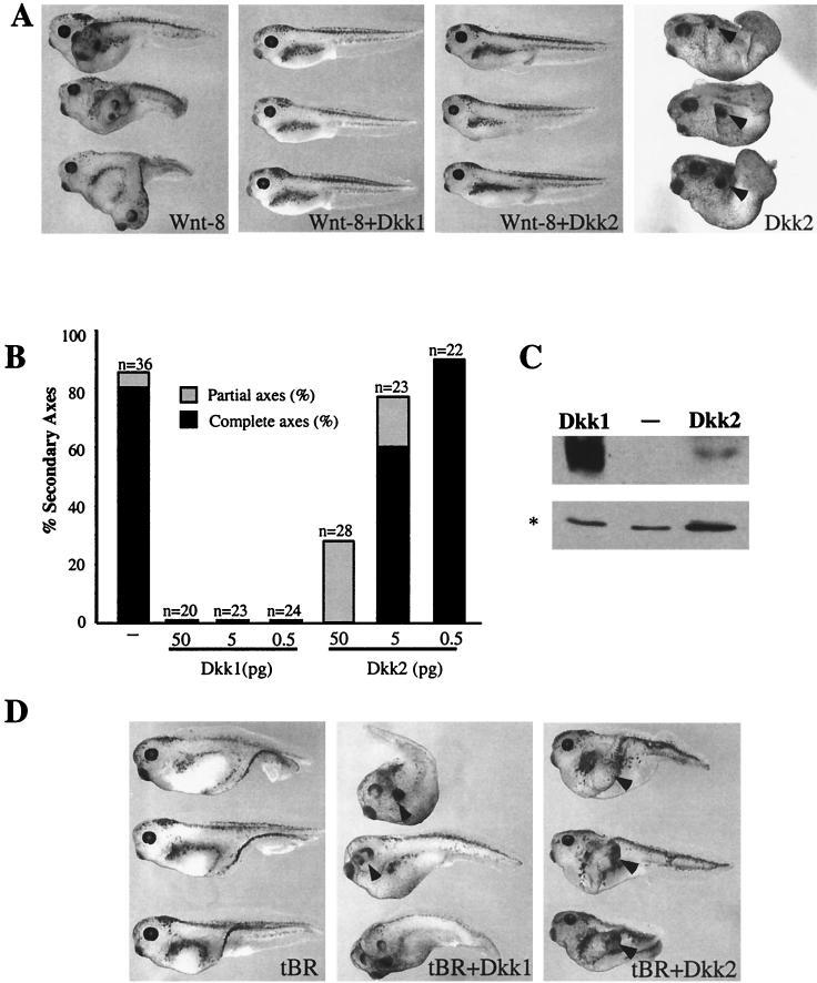
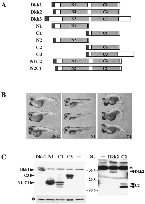
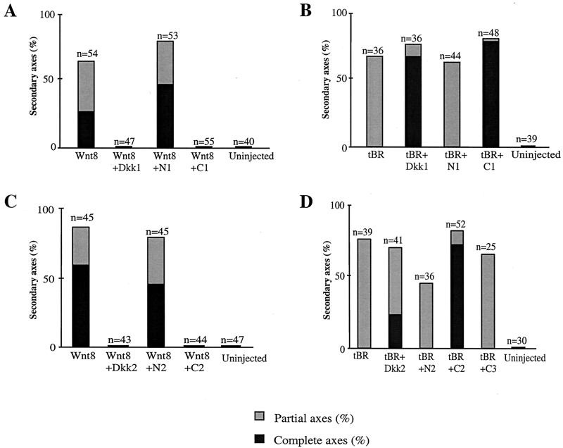
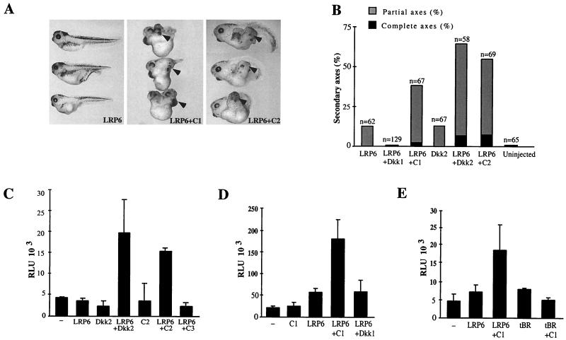
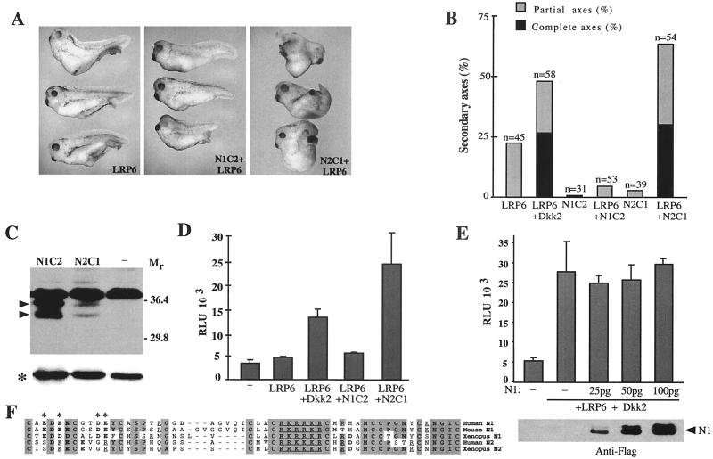
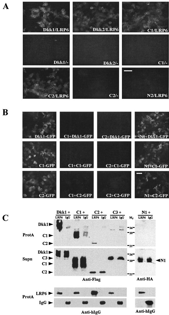
Dickkopfs (Dkks) are secreted developmental regulators composed of two cysteine-rich domains. We report that the effects of Dkks depend on molecular context. Although Wnt8 signaling is inhibited by both Dkk1 and Dkk2 in Xenopus embryos, the same pathway is activated upon interaction of Dkk2 with the Wnt coreceptor LRP6. Analysis of individual Dkk domains and chimeric Dkks shows that the carboxy-terminal domains of both Dkks associate with LRP6 and are necessary and sufficient for Wnt8 inhibition, whereas the amino-terminal domain of Dkk1 plays an inhibitory role in Dkk-LRP interactions. Our study illustrates how an inhibitor of a pathway may be converted into an activator and is the first study to suggest a molecular mechanism for how a ligand other than Wnt can positively regulate beta-catenin signaling.


Figures

FIG. 1.
FIG. 1.
Dkk1 and Dkk2 inhibit Wnt8-dependent secondary axes and synergize with tBR in head induction. (A) Both Dkk1 and Dkk2 inhibit Wnt8-induced secondary axes. Embryos at the four- to eight-cell stage were injected into one ventro-vegetal blastomere with Wnt8 mRNA (1 pg) and Dkk1 (2.5 pg) or Dkk2 (50 pg) mRNAs. The dose of Dkk2 mRNA is higher than that of Dkk1 RNA to compensate for lower protein expression. (B) Dose-dependent inhibition of Wnt8 by Dkk1 and Dkk2. Embryos were injected as described above with Wnt8 mRNA (1 pg) and Dkk1 or Dkk2 mRNAs at the indicated doses. Results are representative of three separate experiments. Complete axes are defined by the presence of a second head with eyes; partial axes are composed of a secondary trunk without anterior head structures. (C) Expression levels of Flag-tagged Dkk1 and Dkk2 in Xenopus embryos. Each blastomere of four-cell embryos was injected with 5 ng of the indicated Dkk RNAs. Embryo lysates prepared at stage 9 were separated in SDS-12% polyacrylamide gels, transferred to Immobilon P membrane, and probed with anti-Flag M2 antibodies. A nonspecific protein band marked by an asterisk reflects loading. (D) Both Dkk1 and Dkk2 synergize with tBR to induce head structures. Embryos were injected with tBR mRNA (20 pg) and Dkk1 (2.5 pg) or Dkk2 (20 pg) mRNA, as indicated. Arrowheads indicate secondary head structures.
FIG. 2.
FIG. 2.
Dkk2, but not Dkk1, synergizes with LRP6 to activate β-catenin signaling pathways. (A and B) Embryos were injected ventro-vegetally with LRP6 mRNA (3 ng) and Dkk1 (5 pg) or Dkk2 (50 pg) mRNAs, as shown. (A) Morphology of injected embryos. (B) Combined results of three separate experiments. (C) Activation of the Siamois promoter by coexpression of LRP6 and Dkk1 or Dkk2. Embryos were injected into two ventral-animal blastomeres with pSia-Luc plasmid (20 pg) and with the following RNAs: LRP6 (5 ng), Dkk1 (20 pg), or Dkk2 (50 pg), as indicated. −, no RNA was coinjected with the pSia-Luc plasmid. (D) Dkk2 inhibits Wnt8, but synergizes with LRP6. Embryos were injected with pSia-Luc plasmid (20 pg), Wnt8 (1 pg), LRP6 (2 ng), and Dkk2 (50 pg), as indicated. (C and D) Luciferase activity was measured in embryonic lysates at stages 10 to 10+. Bars depict the means ± standard errors of triplicate samples, containing five embryos each. Each experiment was performed at least three times; a representative experiment is shown.
FIG. 3.
FIG. 3.
Structure, protein levels, and overexpression phenotypes of Dickkopf constructs. (A) Dkk constructs used in this study. (B) Overexpression phenotypes of Dkk1 constructs. Two dorsal-marginal blastomeres were injected with 50 pg of mRNA encoding Dkk1, N1, or C1, as indicated. Embryos injected with C1 developed enlarged heads and cement glands, impaired eyes, and shortened trunks. Embryos injected with N1, N2, and C2 mRNAs are not significantly different from uninjected controls. (C) Expression levels of Flag-tagged Dkk constructs in Xenopus embryos. Each blastomere of four-cell embryos was injected with 5 ng of the indicated Dkk RNAs. Embryo lysates prepared at stage 9 were separated in SDS-12% polyacrylamide gels, transferred to Immobilon P membrane, and probed with anti-Flag antibodies. The membrane on the right was exposed for 10 times longer than the one on the left to detect lower levels of Dkk2 and C2. A nonspecific protein band marked by an asterisk reflects equal loading.
FIG. 4.
FIG. 4.
The C-terminal domains of Dkks inhibit Wnt8-dependent secondary axes and synergize with tBR in head induction. (A and C) C1 and C2, but not N1 or N2, inhibit secondary axis formation by Wnt8. Embryos were injected into one ventral-vegetal blastomere with Wnt8 mRNA (1 pg) and 2.5 pg of Dkk1, N1, or C1 mRNA (A) or 20 pg of Dkk2, N2, or C2 mRNA (C). Combined results of three separate experiments are shown. (B and D) C1 and C2, but not N1 or N2, induce head structures when expressed with tBR. Embryos were injected with tBR mRNA (20 pg) and 2.5 pg of Dkk1, N1, or C1 mRNA (B) or 20 pg of Dkk2, N2, or C2 mRNA. (D) Combined results of three separate experiments are shown for each graph.
FIG. 5.
FIG. 5.
C1 and C2, but not Dkk1, synergize with LRP6 in axis induction. (A and B) C1 and C2 synergize with LRP6 in axis induction. Embryos were injected with LRP6 mRNA (3 ng) and either 10 pg of Dkk1 or C1 or 1 ng of Dkk2 or C2 mRNAs, as shown. (A) Representative embryos. (B) Combined results of three separate experiments. (C to E) Activation of the Siamois promoter by coexpression of LRP6 and the C-terminal Dkk constructs. Embryos were injected into two ventral-animal blastomeres with pSia-Luc DNA (20 pg) and the following RNAs: LRP6 at 2 ng; Dkk2, C2, or C3 at 50 pg; tBR at 20 pg; and Dkk1 or C1 at 10 pg, as shown. Luciferase activity was measured as described in Fig. 2. Each experiment was performed at least three times; representative experiments are shown.
FIG. 6.
FIG. 6.
Role of N-terminal domain in regulation of Dkk signaling. (A, B, and D) Synergistic effects of LRP6 and chimeric Dkk proteins. Fifty picograms of N1C2 and N2C1 RNAs was injected as described in the legend to Fig. 5. (A) Representative embryos. (B) Combined results of three separate experiments. (C) Expression levels of the N1C2 and N2C1 proteins in injected embryos. Arrowheads indicate N1C2 and N2C1 doublets; an asterisk indicates a loading control. (D) Activation of the Siamois promoter by coinjection of LRP6 and N1C2 or N2C1 chimeric RNAs. (E) N1 does not interfere with signaling by LRP6 and Dkk2. RNAs were injected at the following doses: LRP6 at 2 ng, Dkk2 at 50 pg, and N1 as indicated. N1 expression levels in injected embryos are shown below. (F) Comparison of the N-terminal domains of Dkk1 and Dkk2. Shaded areas indicate identical or highly conserved residues. Acidic residues are in boldface; basic residues are underlined. Asterisks mark charged residues that differ between N1 and N2.
FIG. 7.
FIG. 7.
Association of C1 and C2 with LRP6. (A) Cell culture media with Dkk-GFP proteins were incubated for 1 h with HEK293T cells transfected either with LRP6 (LRP6) or a control vector (−) as indicated. GFP fluorescence reflects Dkk-GFP binding to cells. Bar, 65 μm. (B) LRP6-transfected HEK293T cells were preincubated for 30 min with the culture media containing C1, C2, or N1, followed by additional 30-min incubation with Dkk-GFP-containing media as indicated. Bar, 65 μm. (C) Biochemical association of C1 and C2 with LRP6. Media from HEK293T cells, expressing the extracellular domain of LRP6 fused to the IgG heavy chain (LRP6) or control secreted IgG heavy chain (IgG) were mixed in equal volumes with Dkk-containing media as indicated and incubated with protein A-Sepharose beads (ProtA) to bind IgG or LRP6. The presence of Dkks in ProtA pellets and original supernatants (Supn) was assessed with anti-Flag or anti-HA antibodies, and LRP6 and IgG were detected with anti-human IgG (Fc) (Anti-hIgG) antibody.

References

    1. Aravind, L., and E. V. Koonin. 1998. A colipase fold in the carboxy-terminal domain of the Wnt antagonists—the Dickkopfs. Curr. Biol. 8:477-478. - PubMed
    1. Bafico, A., G. Liu, A. Yaniv, A. Gazit, and S. A. Aaronson. 2001. Novel mechanism of Wnt signalling inhibition mediated by Dickkopf-1 interaction with LRP6/Arrow. Nat. Cell Biol. 3:683-686. - PubMed
    1. Bhanot, P., M. Brink, C. H. Samos, J. C. Hsieh, Y. Wang, J. P. Macke, D. Andrew, J. Nathans, and R. Nusse. 1996. A new member of the frizzled family from Drosophila functions as a Wingless receptor. Nature 382:225-230. - PubMed
    1. Bouwmeester, T., S. Kim, Y. Sasai, B. Lu, and E. M. De Robertis. 1996. Cerberus is a head-inducing secreted factor expressed in the anterior endoderm of Spemann's organizer. Nature 382:595-601. - PubMed
    1. Brannon, M., M. Gomperts, L. Sumoy, R. T. Moon, and D. Kimelman. 1997. A beta-catenin/XTcf-3 complex binds to the siamois promoter to regulate dorsal axis specification in Xenopus. Genes Dev. 11:2359-2370. - PMC - PubMed

Publication types

MeSH terms

Substances

LinkOut - more resources