Comparative Study
. 2002 Sep;22(17):6170-82.
doi: 10.1128/MCB.22.17.6170-6182.2002.

Hypophosphorylation of Mdm2 augments p53 stability

Affiliations
Comparative Study

Hypophosphorylation of Mdm2 augments p53 stability

Christine Blattner et al. Mol Cell Biol. 2002 Sep.

Abstract

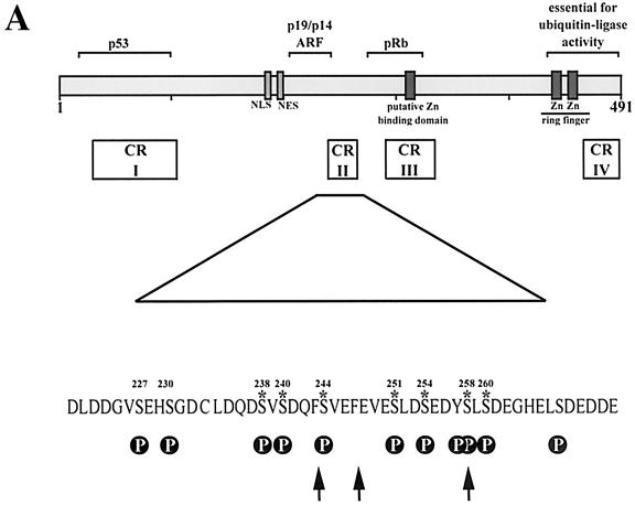
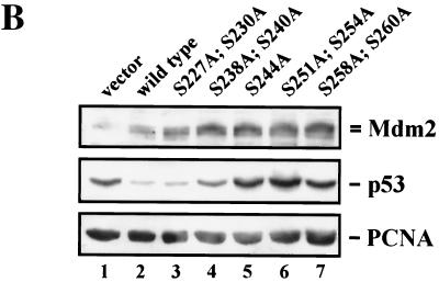
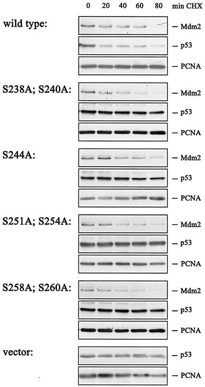
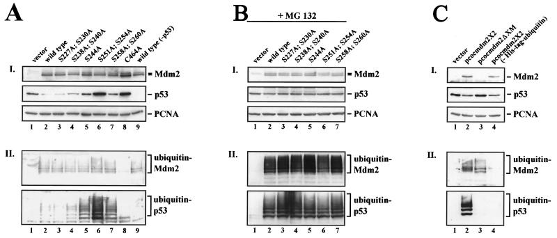
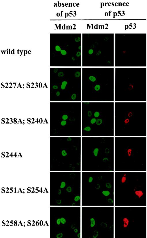
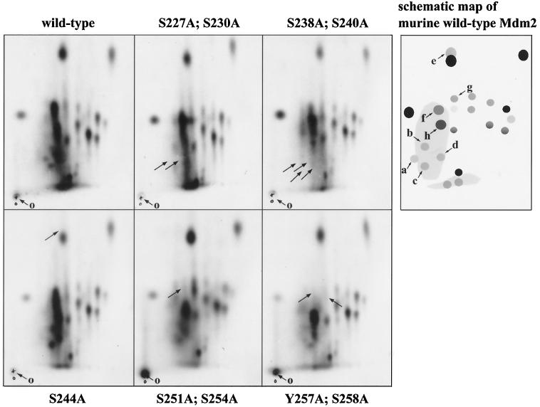
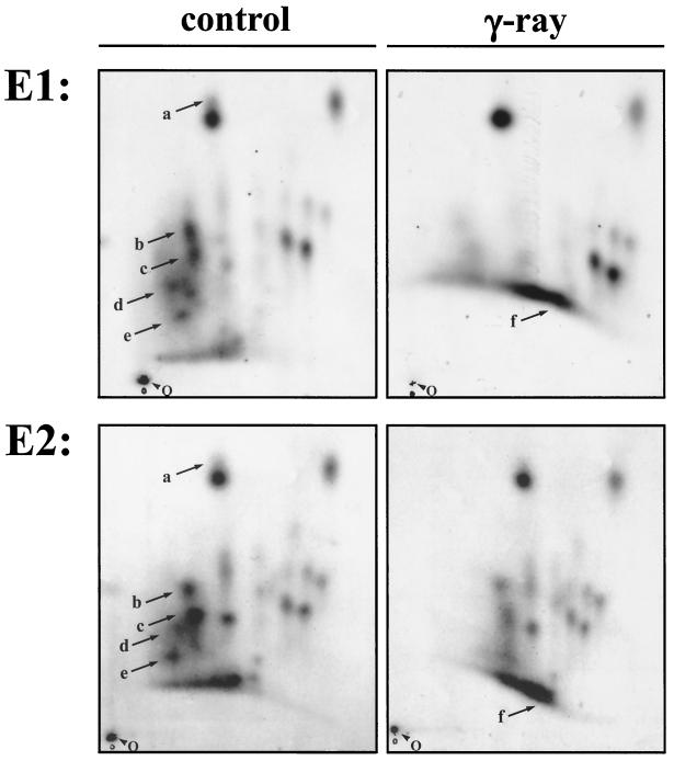
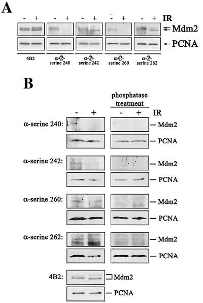
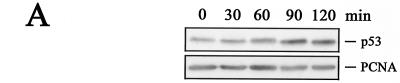
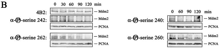
The Mdm2 protein mediates ubiquitylation and degradation of p53 and is a key regulator of this tumor suppressor. More recently, it has been shown that Mdm2 is highly phosphorylated within its central acidic domain. In order to address the issue of how these modifications might regulate Mdm2 function, putative phosphorylation sites within this domain were substituted, individually or in pairs, with alanine residues. Mutants with serine-to-alanine substitutions between residues 244 and 260 abolished or at least reduced the capacity of Mdm2 to promote p53 degradation. In each case, loss of degradation function was independent of the ability to bind to p53 or p14ARF. Moreover, each of the Mdm2 mutants completely retained the capacity to act as a ubiquitin ligase in vivo. Thus, ubiquitylation and degradation can be uncoupled. Two-dimensional phosphopeptide mapping coupled with the use of phospho-specific antibodies revealed that Mdm2 is phosphorylated physiologically at several sites within this region, consistent with the idea that phosphorylation is important for Mdm2 activity. Strikingly, treatment of cells with ionizing radiation resulted in a significant decrease in the phosphorylation of residues that are important for p53 turnover. This hypophosphorylation preceded p53 accumulation. These findings indicate that Mdm2 contributes an additional function toward the degradation of p53 that is distinct from its ubiquitin ligase activity and is regulated by phosphorylation. Our model suggests that hypophosphorylation of Mdm2 in response to ionizing irradiation inactivates this novel function, thereby contributing to p53 stabilization.


Figures

FIG. 1.
FIG. 1.
Degradation of p53 by Mdm2 serine→alanine mutants. (A) Diagram of Mdm2 wild-type protein showing the functional domains, conserved regions (CR), and interacting proteins. Arrows point to chymotrypsin cleavage sites. Putative phosphorylation sites (P) are indicated. Asterisks denote serines important for p53 degradation. (B) H1299 cells were transiently transfected with 1 μg of pcDNA3-p53 DNA (lanes 1 through 7) and 5 μg of wild-type pcDNA3-mdm2 DNA (lane 2), the pcDNA3-mdm2 DNA possessing the indicated single- and double-point mutations (lanes 3 through 7) or 5 μg of pcDNA3 vector DNA (lane 1). Thirty-six hours after transfection, cells were harvested, lysed in NP-40 lysis buffer, and analyzed for the presence of p53 and Mdm2. Forty micrograms of crude cellular extract was separated on an SDS-10% PAGE minigel and transferred to a nitrocellulose membrane. The Western blot was serially probed with the anti-p53 antibody DO-1 (ascites, diluted 1:750), the anti-Mdm2 antibody 4B2 (ascites, diluted 1:500), and the anti-PCNA antibody PC-10 (ascites, diluted 1:3,000), developed by enhanced chemiluminescence, and exposed.
FIG. 1.
FIG. 1.
Degradation of p53 by Mdm2 serine→alanine mutants. (A) Diagram of Mdm2 wild-type protein showing the functional domains, conserved regions (CR), and interacting proteins. Arrows point to chymotrypsin cleavage sites. Putative phosphorylation sites (P) are indicated. Asterisks denote serines important for p53 degradation. (B) H1299 cells were transiently transfected with 1 μg of pcDNA3-p53 DNA (lanes 1 through 7) and 5 μg of wild-type pcDNA3-mdm2 DNA (lane 2), the pcDNA3-mdm2 DNA possessing the indicated single- and double-point mutations (lanes 3 through 7) or 5 μg of pcDNA3 vector DNA (lane 1). Thirty-six hours after transfection, cells were harvested, lysed in NP-40 lysis buffer, and analyzed for the presence of p53 and Mdm2. Forty micrograms of crude cellular extract was separated on an SDS-10% PAGE minigel and transferred to a nitrocellulose membrane. The Western blot was serially probed with the anti-p53 antibody DO-1 (ascites, diluted 1:750), the anti-Mdm2 antibody 4B2 (ascites, diluted 1:500), and the anti-PCNA antibody PC-10 (ascites, diluted 1:3,000), developed by enhanced chemiluminescence, and exposed.
FIG. 2.
FIG. 2.
Determination of p53 and Mdm2 half-lives in the presence of wild-type or mutant Mdm2. H1299 cells were transfected with 0.5 μg of pcDNA3-p53 DNA and 3.75 μg of wild-type pcDNA3-mdm2 DNA, mutant mdm2 DNA, or vector DNA. Thirty-six hours after transfection, cycloheximide (CHX) was added (20 μg/ml) and cells were harvested at the indicated time points after CHX addition. The cells were lysed in NP-40 lysis buffer. Forty micrograms of cellular extract were separated on an SDS-10% PAGE gel and transferred to a nitrocellulose membrane. Western blot membranes were cut into strips which were probed with the monoclonal antibodies DO-1 (anti-p53), 4B2 (anti-Mdm2), and PC-10 (anti-PCNA) and developed by enhanced chemiluminescence.
FIG. 3.
FIG. 3.
Interaction of mutant Mdm2 with p53 and p14ARF. H1299 cells were transfected with 5 μg of pcDNA3-p53 DNA (A) or 5 μg of pcDNA3-ARF-FLAG DNA (B) in the absence (lane 1) or presence (lane 2) of 5 μg of wild-type pcDNA3-mdm2 DNA or the indicated Mdm2 mutants (lanes 3 through 6). Twenty-four hours after transfection, cells were treated with the proteasome inhibitor MG132 for 6 h, prior to lysis. (A, panel I) p53/Mdm2 complexes were immunoprecipitated (IP) out of 350 μg of cellular lysate with either the CM-1 anti-p53 antibody coupled to protein A+G-Sepharose, an anti-Flag antibody, or preimmune serum coupled to protein A+G-Sepharose. (B, panel I) ARF/Mdm2 complexes were precipitated out of 350 μg of cellular lysate with an anti-p53 antibody, an anti-Flag antibody, or preimmune serum coupled to protein A+G-Sepharose The protein/antibody complexes were washed three times in NP-40 lysis buffer and resolved in 1× sample buffer. The complexes were separated on an SDS-10% PAGE gel, transferred to a nitrocellulose membrane, and probed for the presence of Mdm2 with the 4B2 (anti-Mdm2) antibody. Western blots were developed by enhanced chemiluminescence. (A and B, panels II) Forty micrograms of the remaining cell lysate was separated on an SDS-10% (A, panel II) or -13% (B, panel II) PAGE gel, transferred to a nitrocellulose membrane, and probed for the presence of p53 and Mdm2 (A, panel II) by using the DO-1 (anti-p53) and 4B2 (anti-Mdm2) antibodies or for the presence of ARF and Mdm2 (B, panel II) by using 4B2 (anti-Mdm2) and the M1 anti-Flag antibody.
FIG. 4.
FIG. 4.
Ubiquitylation is not restrained by Mdm2 serine→alanine mutants. H1299 cells were transiently transfected with 2 μg of His-tagged ubiquitin DNA, 1 μg of pcDNA3-p53 DNA (lanes 1 through 8), and 10 μg of wild-type pcDNA3-mdm2 DNA (lanes 2 and 9), the pcDNA3-mdm2 DNA possessing the indicated single- and double-point mutations (lanes 3 through 8) or 10 μg of pcDNA3 vector DNA (lane 1). Thirty-six hours after transfection, cells were harvested. (A, panel I) Aliquots of the cells were lysed in NP-40 lysis buffer and analyzed for the presence of p53 and Mdm2. Forty micrograms of crude cellular extract were separated on an SDS-10% PAGE minigel and transferred to a nitrocellulose membrane. The Western blot membrane was cut into strips and probed with the anti-p53 antibody DO-1 (ascites, diluted 1:750), the anti-Mdm2 antibody 4B2 (ascites, diluted 1:500), and the anti-PCNA antibody PC-10 (ascites, diluted 1:3,000), developed by enhanced chemiluminescence, and exposed. (A, panel II) The remaining cells were lysed in guanidinium lysis buffer and probed for the presence of ubiquitylated p53 and Mdm2. Ubiquitylated proteins were purified by adsorption to Ni2+-NTA-agarose beads and resolved on an SDS-8% PAGE minigel. After transfer of the proteins to a nitrocellulose membrane, the Western blot was probed with the anti p53-antibody DO-1 (ascites, diluted 1:750) or anti-Mdm2 antibody 4B2 (ascites, diluted 1:500) and developed by enhanced chemiluminescence. (B) H1299 cells were transiently transfected with 2 μg of His-tagged ubiquitin DNA, 1 μg of pcDNA3-p53 DNA (lanes 1 through 8), and 10 μg of wild-type pcDNA3-mdm2 DNA (lane 2), the pcDNA3-mdm2 DNA possessing the indicated single- and double-point mutations (lanes 3 through 7) or 10 μg of pcDNA3 vector DNA (lane 1). Thirty-six hours after transfection, cells were treated with 10 μM MG132 for 6 h, harvested, and processed as described above. (C) H1299 cells were transiently transfected with 2 μg of His-tagged ubiquitin DNA (lanes 1 through 3), 1 μg of pcDNA3-p53 DNA, and 10 μg of wild-type pcocmdm2X2 DNA (lanes 2 and 4), pcocmdm2ΔXM possessing an amino-terminal deletion encompassing the p53-binding site as well as the 4B2 antibody epitope (lane 3) or 10 μg of vector DNA (lane 1). Thirty-six hours after transfection, cells were harvested and treated as described above except that ubiquitylated Mdm2 (panel II) was detected with the anti-Mdm2 antibody 2A10.
FIG. 5.
FIG. 5.
Localization of wild-type and mutant Mdm2. H1299 cells were transfected with 5 μg of wild-type or mutant pcDNA3-mdm2 DNA/2.5 × 105 cells in the absence or presence of 1 μg of pcDNA3-p53 DNA. Twenty-four hours after transfection, cells were fixed with acetone-methanol (1:1) and stained for expression of Mdm2 with the mouse monoclonal anti-Mdm2 antibody 4B2 and for p53 with the rabbit polyclonal anti-p53 antibody CM-1. Fluorescein isothiocyanate-coupled anti-mouse IgG and tetramethyl rhodamine isothiocyanate-coupled anti-rabbit IgG were used as secondary antibodies. The two left-hand columns (in green) show expression of Mdm2, and the right-hand column (in red) shows expression of p53.
FIG. 6.
FIG. 6.
Mdm2 is phosphorylated in vivo in conserved domain II. Myc-tagged mouse wild-type mdm2 DNA (pDWM659) or mdm2 DNA possessing the indicated mutations was transiently transfected into COS-7 cells. Twelve hours prior to irradiation, cells were incubated with the proteasome inhibitor MG132, and 48 h after transfection, cells were radioactively labeled with [32P]orthophosphate for 3 h. Cells were harvested and lysed in NP-40 lysis buffer, and Myc-tagged Mdm2 was precipitated with the anti-Myc monoclonal antibody 9E10. After washing, the Mdm2-antibody complexes were resolved on an SDS-8% PAGE minigel. The Mdm2 protein was eluted from the gel and digested with chymotrypsin, and the resulting peptides were separated by electrophoresis in buffer (pH 1.9; first dimension) and chromatography buffer (second dimension). The origin (o) where the phosphopeptides were loaded is indicated. Arrows point to changes which resulted from the serine substitutions. The schematic map shows the following phosphopeptides, identified by serine→alanine substitutions: a through d, aa 189 through 243; e, aa 244 through 247; f, aa 248 through 257; g and h, aa 248 through 274.
FIG. 7.
FIG. 7.
Mdm2 is posttranslationally modified in response to irradiation. Cells were irradiated with 10 Gy 3 h prior to harvest and processed as described in the legend to Fig. 6. Arrows point to irradiation-mediated changes in phosphorylation. E1, experiment 1; E2, experiment 2, O, origin.
FIG. 8.
FIG. 8.
Mdm2 is hypophosphorylated in the central conserved domain upon irradiation (IR). GM1604 cells were treated with MG132 for 10 h. Prior to lysis (1.5 h), cells were irradiated with 10 Gy. (A) Forty micrograms of lysate was separated on an SDS-10% PAGE gel, transferred to a nitrocellulose membrane, and probed with 4B2 or phosphorylation-specific anti-Mdm2 antibodies. Hybridization with an anti-PCNA antibody was used for loading control. Western blots were developed by enhanced chemiluminescence. (B) Forty micrograms of lysate was separated on an SDS-10% PAGE gel and transferred to a nitrocellulose membrane. After transfer, membranes were incubated with calf intestinal phosphatase at 37°C for 2 h prior to antibody incubation. Membranes were then probed with 4B2 or phosphorylation-specific anti-Mdm2 antibodies and with an anti-PCNA antibody for loading control.
FIG. 9.
FIG. 9.
Hypophosphorylation precedes p53 accumulation. (A) GM1604 cells were irradiated with 10 Gy and harvested at the indicated time points. Forty micrograms of lysate was separated on an SDS-10% PAGE gel, transferred to a nitrocellulose membrane, and probed with Pab1801. Hybridization with an anti-PCNA antibody was used for loading control. Western blots were developed by enhanced chemiluminescence. (B) GM1604 cells were treated with MG132 for 10 h. At the indicated times prior to lysis, cells were irradiated with 10 Gy. Forty micrograms of lysate was separated on an SDS-10% PAGE gel, transferred to a nitrocellulose membrane, and probed with phosphorylation-specific antibodies. Hybridization with the anti-Mdm2 antibody 4B2 shows that Mdm2 levels remained unchanged, and the anti-PCNA antibody was used for loading control. Western blots were developed by enhanced chemiluminescence.
FIG. 9.
FIG. 9.
Hypophosphorylation precedes p53 accumulation. (A) GM1604 cells were irradiated with 10 Gy and harvested at the indicated time points. Forty micrograms of lysate was separated on an SDS-10% PAGE gel, transferred to a nitrocellulose membrane, and probed with Pab1801. Hybridization with an anti-PCNA antibody was used for loading control. Western blots were developed by enhanced chemiluminescence. (B) GM1604 cells were treated with MG132 for 10 h. At the indicated times prior to lysis, cells were irradiated with 10 Gy. Forty micrograms of lysate was separated on an SDS-10% PAGE gel, transferred to a nitrocellulose membrane, and probed with phosphorylation-specific antibodies. Hybridization with the anti-Mdm2 antibody 4B2 shows that Mdm2 levels remained unchanged, and the anti-PCNA antibody was used for loading control. Western blots were developed by enhanced chemiluminescence.

References

    1. Argentini, M., N. Barboule, and B. Wasylyk. 2001. The contribution of the acidic domain of MDM2 to p53 and MDM2 stability. Oncogene 20:1267-1275. - PubMed
    1. Attardi, L. D., and T. Jacks. 1999. The role of p53 in tumor suppression: lessons from mouse models. Cell. Mol. Life Sci. 55:48-63. - PMC - PubMed
    1. Barak, Y., E. Gottlieb, G. T. Juven, and M. Oren. 1994. Regulation of mdm2 expression by p53: alternative promoters produce transcripts with non-identical translation potential. Genes Dev. 8:1739-1749. - PubMed
    1. Blattner, C., A. Sparks, and D. P. Lane. 1999. Transcription factor E2F-1 is upregulated in response to DNA damage in a manner analogous to that of p53. Mol. Cell. Biol. 19:3704-3713. - PMC - PubMed
    1. Bottger, A., V. Bottger, A. Sparks, W.-L. Liu, S. F. Howard, and D. P. Lane. 1997. Design of a synthetic Mdm2-binding mini protein that activates the p53 response in vivo. Curr. Biol. 7:860-869. - PubMed

Publication types

MeSH terms