

Neuropathologic and MR imaging correlation in a neonatal case of cerebellar cortical dysplasia
- PMID: 12169464
- PMCID: PMC8185714
Neuropathologic and MR imaging correlation in a neonatal case of cerebellar cortical dysplasia
Abstract
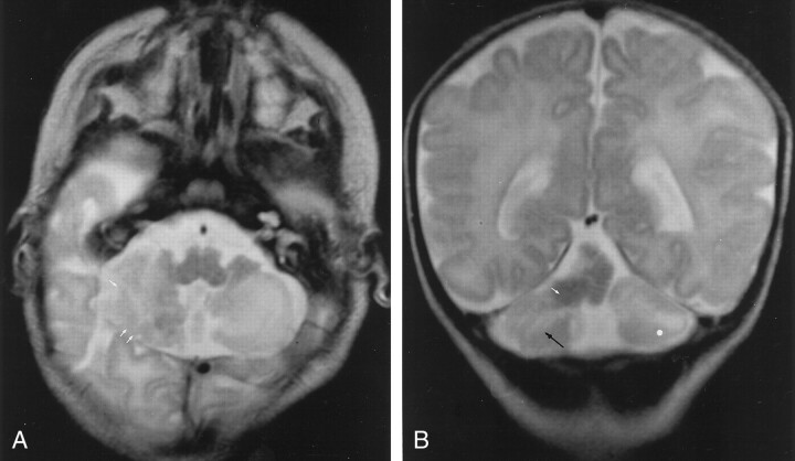
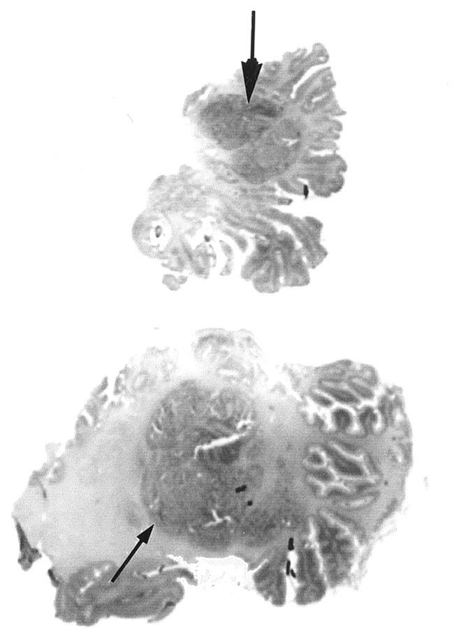
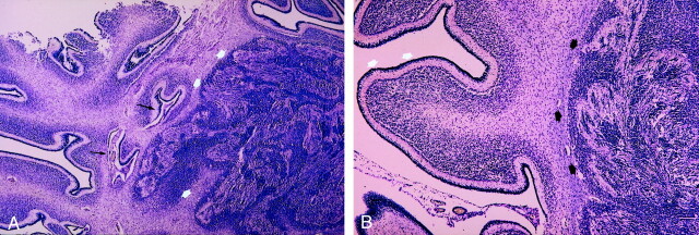
Little documentation of the correlation between MR imaging findings in isolated cerebellar cortical dysplasia (CCD) and its neuropathologic characteristics exists in the recent literature. We documented a postmortem neuropathologic study of a clinically and radiologically well-documented case of CCD in a neonate with severe hypotonia and status epilepticus. MR imaging revealed a global vermian hypoplasia with marked cortical dysplasia. CCD was associated with a voluminous heterotopic mass. The postmortem neuropathologic study confirmed vermian hypoplasia and CCD, which consisted of right cerebellar cortical polymicrogyria with subcortical heterotopia. CCD is a pathologic entity that could be well diagnosed with MR imaging even in the neonatal period.
Figures
Similar articles
-
Cerebellar cortical dysplasia: MR findings in a complex entity.AJNR Am J Neuroradiol. 2000 Sep;21(8):1511-9. AJNR Am J Neuroradiol. 2000. PMID: 11003288 Free PMC article.
-
[Cerebellar cortical dysplasia: MRI aspects and significance].J Radiol. 2004 Jun;85(6 Pt 1):729-40. doi: 10.1016/s0221-0363(04)97675-5. J Radiol. 2004. PMID: 15243373 Review. French.
-
Supratentorial functional disturbances in two children with cerebellar cortical dysplasia.J Neuroradiol. 2004 Dec;31(5):399-405. doi: 10.1016/s0150-9861(04)97024-1. J Neuroradiol. 2004. PMID: 15687960
-
Prenatal diagnosis of cerebellar cortical dysplasia associated with abnormalities of foliation.Ultrasound Obstet Gynecol. 2012 Aug;40(2):243-4. doi: 10.1002/uog.11210. Epub 2012 Jul 13. Ultrasound Obstet Gynecol. 2012. PMID: 22689128 No abstract available.
-
Cleidocranial dysplasia with neonatal death due to central nervous system injury in utero: case report and literature review.Pediatr Dev Pathol. 1998 Jul-Aug;1(4):314-8. doi: 10.1007/s100249900045. Pediatr Dev Pathol. 1998. PMID: 10463294 Review.
Cited by
-
A developmental and genetic classification for midbrain-hindbrain malformations.Brain. 2009 Dec;132(Pt 12):3199-230. doi: 10.1093/brain/awp247. Brain. 2009. PMID: 19933510 Free PMC article. Review.
-
Prenatal Diagnosis of Cerebellar Cortical Dysplasia: Case Report.Cerebellum. 2024 Oct;23(5):1942-1949. doi: 10.1007/s12311-024-01688-9. Epub 2024 Apr 12. Cerebellum. 2024. PMID: 38607531
-
Analysis and classification of cerebellar malformations.AJNR Am J Neuroradiol. 2003 Jan;24(1):153; author reply 153. AJNR Am J Neuroradiol. 2003. PMID: 12533349 Free PMC article. No abstract available.
-
Brain regional glucose uptake changes in isolated cerebellar cortical dysplasia: qualitative assessment using coregistrated FDG-PET/MRI.Cerebellum. 2012 Mar;11(1):280-8. doi: 10.1007/s12311-011-0309-7. Cerebellum. 2012. PMID: 21833659
-
MRI of the fetal posterior fossa.Pediatr Radiol. 2005 Feb;35(2):124-40. doi: 10.1007/s00247-004-1316-3. Epub 2004 Nov 23. Pediatr Radiol. 2005. PMID: 15565345 Review.
References
-
- Rorke LB, Fogelson MH, Riggs HE. Cerebellar heterotopia in infancy Dev Med Child Neurol 1968;10:644–650 - PubMed
-
- Friede RL. Developmental neuropathology. 2nd ed. Berlin Heidelberg New York: Springer-Verlag;1989. :361–371
-
- Sugita K, Ando M, Makino M, Takanashi J, Fujimoto N, Niimi H. Magnetic resonance imaging of the brain in congenital rubella virus and cytomegalovirus infections Neuroradiology 1991;33:239–242 - PubMed
-
- Marques Dias MJ, Harmant-van Rijckeevorsel G, Landrieu P, et al. Prenatal cytomegalovirus disease and cerebral microgyria: evidence for perfusion failure, not disturbance of histogenesis, as the major cause of fetal cytomegalovirus encephalopathy Neuropediatrics 1984;15:18–24 - PubMed
Publication types
MeSH terms
LinkOut - more resources
Full Text Sources
Medical