

Measuring the axial rotation of lumbar vertebrae in vivo with MR imaging
- PMID: 12169466
- PMCID: PMC8185728
Measuring the axial rotation of lumbar vertebrae in vivo with MR imaging
Abstract
Background and purpose: Flexion-extension radiography is neither sensitive nor specific in the diagnosis of degenerative spinal instability, a presumed cause of back pain and an indication for spinal fusion. We tested the hypothesis that with MR imaging and a device to rotate the torso, axial rotations of lumbar vertebrae can be measured with sufficient accuracy and that significantly different rotations can be detected between lumbar segments with degenerated disks and those with normal disks.
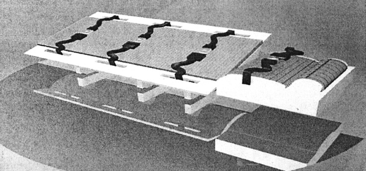
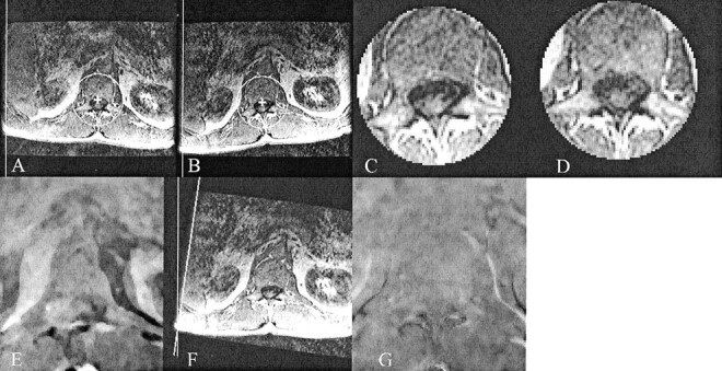
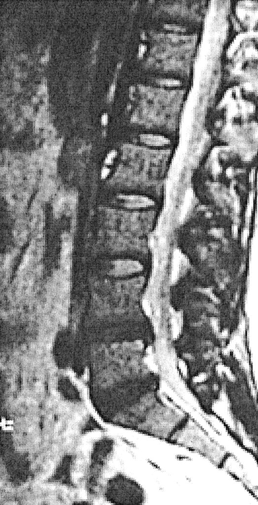
Methods: We studied five volunteers without back pain (group 1), five patients who underwent MR imaging because of back pain but were not considered candidates for fusion (group 2), and five patients in whom diskography identified one or more disks with concordant pain (group 3). Each participant was placed on a specially built table that provided separate supports for the torso and for the hips and legs. Series of sagittal images were acquired with a T2-weighted fast spin-echo sequence, with the torso rotated clockwise and then counterclockwise. The amount of rotation was calculated from axial images with use of an automated program.
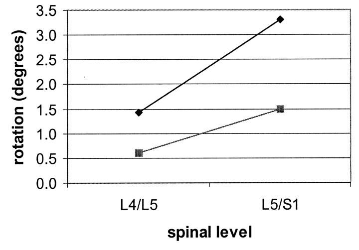
Results: In the five volunteers, rotations of the lumbar motion segments varied between -1.8 degrees and 5.7 degrees, with an average of 0.8 degrees. The abnormal disks in five patients in group 2 rotated from -0.9 degrees to 5.6 degrees, with an average of 3.2 degrees. In group 3, the disks in which concordant pain was elicited rotated from 0.8 degrees to 4.4 degrees, with an average of 2.2 degrees. Difference in rotation between abnormal and normal disks was statistically significant.
Conclusion: Measurements of rotations of lumbar vertebrae with MR imaging may have value for determining levels that move abnormally in axial rotation.
Figures

References
-
- Quinnell R, Stockdale HR. Flexion and extension radiography of the lumbar spine: comparison with lumbar discography. Clin Radiol 1983;34:405–411 - PubMed
-
- McCormick PC. Selection criteria for degenerative lumbar spine instability. Clin Neurosurg 1997;44:29–39 - PubMed
-
- Posner I, White AA, Edwards WT, Hayes WC. A biomechanical analysis of the clinical stability of the lumbar and lumbosacral spine. Spine 1982;7:374–389 - PubMed
-
- Stokes IA, Frymoyer JW. Segmental motion and instability. Spine 1987;12:688–691 - PubMed
Publication types
MeSH terms
LinkOut - more resources
Full Text Sources
Other Literature Sources
Medical