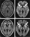
Conspicuity and evolution of lesions in Creutzfeldt-Jakob disease at diffusion-weighted imaging
- PMID: 12169476
- PMCID: PMC8185711
Conspicuity and evolution of lesions in Creutzfeldt-Jakob disease at diffusion-weighted imaging
Abstract
Background and purpose: Diffusion-weighted imaging can disclose distinct hyperintense lesions in Creutzfeldt-Jakob disease (CJD). However, these findings and chronologic changes of CJD at diffusion-weighted imaging have not been fully investigated. Our purpose was to assess the diagnostic value of diffusion-weighted imaging in depicting CJD-related lesions and in tracking the evolution of these lesions. We also compared the sensitivity of diffusion-weighted imaging in depicting CJD-related lesions to that of fluid-attenuated inversion recovery (FLAIR) imaging.
Methods: We reviewed findings in 13 patients with a diagnosis of CJD who underwent MR imaging, including diffusion-weighted imaging. Nine patients were initially examined within 4 months of onset of symptoms (early stage), and eight were examined 4 months or later (late stage). We evaluated four items: 1) distribution of lesions at diffusion-weighted imaging, 2) conspicuity of lesions at diffusion-weighted imaging and FLAIR imaging, 3) chronologic changes in lesions at diffusion-weighted imaging, and 4) chronologic changes in lesions revealed by apparent diffusion coefficient (ADC) maps.
Results: Patients had striatal lesions or cerebral cortical lesions or both. The thalamus was involved in only one patient, and the globus pallidus was spared in all patients. The sensitivity of diffusion-weighted imaging in depicting lesions was superior or at least equal to that of FLAIR imaging. Hyperintense lesions at diffusion-weighted imaging changed in extent and intensity over time. Unlike infarction, lesional ADC decreased for 2 weeks or longer.
Conclusion: The progressively hyperintense changes in the striata and cerebral cortices at diffusion-weighted imaging are considered characteristic of CJD. Diffusion-weighted imaging may be useful for the early diagnosis of CJD.
Figures


Comment in
-
Progress in understanding Creutzfeldt-Jakob disease.AJNR Am J Neuroradiol. 2002 Aug;23(7):1070-2. AJNR Am J Neuroradiol. 2002. PMID: 12169459 Free PMC article. No abstract available.
Similar articles
-
Diagnostic value of diffusion-weighted brain magnetic resonance imaging in patients with sporadic Creutzfeldt-Jakob disease: a systematic review and meta-analysis.Eur Radiol. 2021 Dec;31(12):9073-9085. doi: 10.1007/s00330-021-08031-4. Epub 2021 May 12. Eur Radiol. 2021. PMID: 33982159
-
Creutzfeldt-Jakob disease: comparative analysis of MR imaging sequences.AJNR Am J Neuroradiol. 2006 Aug;27(7):1459-62. AJNR Am J Neuroradiol. 2006. PMID: 16908558 Free PMC article.
-
Diffusion-weighted and fluid-attenuated inversion recovery imaging in Creutzfeldt-Jakob disease: high sensitivity and specificity for diagnosis.AJNR Am J Neuroradiol. 2005 Jun-Jul;26(6):1551-62. AJNR Am J Neuroradiol. 2005. PMID: 15956529 Free PMC article.
-
Isolated cortical signal increase on MR imaging as a frequent lesion pattern in sporadic Creutzfeldt-Jakob disease.AJNR Am J Neuroradiol. 2008 Sep;29(8):1519-24. doi: 10.3174/ajnr.A1122. Epub 2008 Jul 3. AJNR Am J Neuroradiol. 2008. PMID: 18599580 Free PMC article.
-
Diffusion-weighted MRI in Creutzfeldt-Jakob disease: a better diagnostic marker than CSF protein 14-3-3?J Neuroimaging. 2003 Apr;13(2):147-51. J Neuroimaging. 2003. PMID: 12722497 Review.
Cited by
-
Diagnostic value of diffusion-weighted brain magnetic resonance imaging in patients with sporadic Creutzfeldt-Jakob disease: a systematic review and meta-analysis.Eur Radiol. 2021 Dec;31(12):9073-9085. doi: 10.1007/s00330-021-08031-4. Epub 2021 May 12. Eur Radiol. 2021. PMID: 33982159
-
Neuroanatomical correlates of prion disease progression - a 3T longitudinal voxel-based morphometry study.Neuroimage Clin. 2016 Nov 2;13:89-96. doi: 10.1016/j.nicl.2016.10.021. eCollection 2017. Neuroimage Clin. 2016. PMID: 27942451 Free PMC article.
-
Prion disease diagnosis using subject-specific imaging biomarkers within a multi-kernel Gaussian process.Neuroimage Clin. 2019;24:102051. doi: 10.1016/j.nicl.2019.102051. Epub 2019 Oct 25. Neuroimage Clin. 2019. PMID: 31734530 Free PMC article.
-
Diffusion-weighted MRI findings and clinical correlations in sporadic Creutzfeldt-Jakob disease.J Neurol. 2015 Jun;262(6):1440-6. doi: 10.1007/s00415-015-7723-6. Epub 2015 Apr 11. J Neurol. 2015. PMID: 25860342
-
Creutzfeldt-jakob disease involvement of rolandic cortex: a quantitative apparent diffusion coefficient evaluation.AJNR Am J Neuroradiol. 2006 Sep;27(8):1755-9. AJNR Am J Neuroradiol. 2006. PMID: 16971630 Free PMC article.
References
-
- Barboriak DP, Provenzale JM, Boyko OB. MR diagnosis of Creutzfeldt-Jakob disease: significance of high signal intensity of the basal ganglia. AJR Am J Roentgenol 1994;162:137–140 - PubMed
-
- Finkenstaedt M, Szudra A, Zerr I, et al. MR imaging of Creutzfeldt-Jakob disease. Radiology 1996. Jun;199:793–798 - PubMed
-
- Demaerel P, Baert AL, Vanopdenbosch L, Robberecht W, Dom R. Diffusion-weighted magnetic resonance imaging in Creutzfeldt-Jakob disease. Lancet 1997;349:847–848 - PubMed
-
- Bahn MM, Kido DK, Lin W, Pearlman AL. Brain magnetic resonance diffusion abnormalities in Creutzfeldt-Jakob disease. Arch Neurol1997;54:1411–1415 - PubMed
Publication types
MeSH terms
LinkOut - more resources
Full Text Sources
Medical
