

The ascending pharyngeal artery: branches, anastomoses, and clinical significance
- PMID: 12169487
- PMCID: PMC8185735
The ascending pharyngeal artery: branches, anastomoses, and clinical significance
Abstract
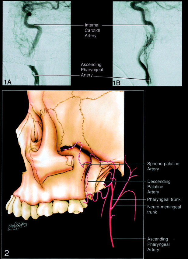
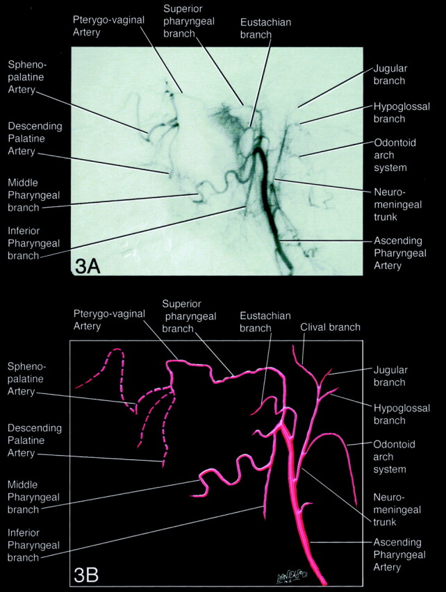
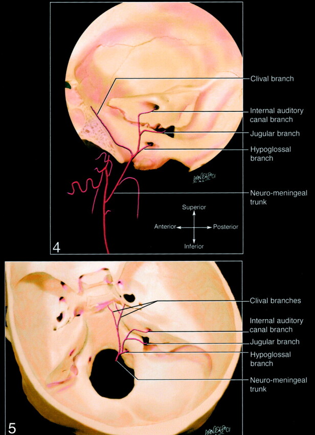
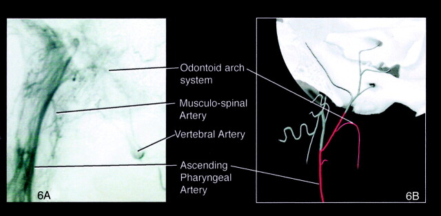
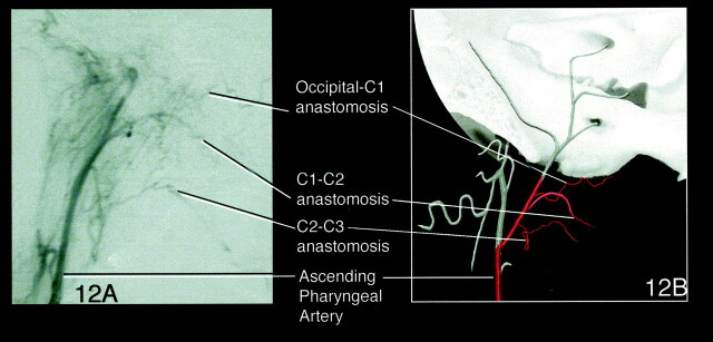
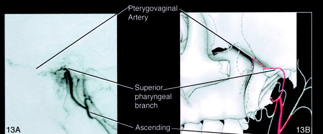
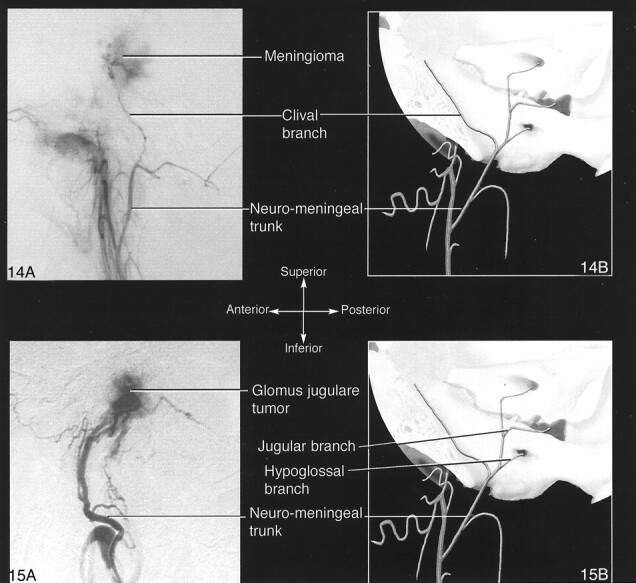
Neuroradiologists generally do not fully appreciate the importance of the territory of the ascending pharyngeal artery. The ascending pharyngeal artery is a small but important artery that supplies multiple cranial nerves and anastomotic channels to the anterior and posterior cerebral circulations. Several disease processes in the head and neck involve the ascending pharyngeal artery. To evaluate and treat such diseases, it is necessary for neuroradiologists not only to know selective angiography and embolization techniques, but also the territory of the ascending pharyngeal artery, anastomoses, and vascular supply to the vasa nervorum of lower cranial nerves. Herein, the normal angiographic anatomy of the ascending pharyngeal artery, its relationship with neighboring territories, its importance in clinical situations, and research models are reviewed.
Figures
Similar articles
-
[The Ascending Pharyngeal Artery, Accessory Meningeal Artery, Lingual Artery, Vasa Nervorum, and Potential Anastomotic Channels].No Shinkei Geka. 2024 May;52(3):539-548. doi: 10.11477/mf.1436204947. No Shinkei Geka. 2024. PMID: 38783497 Review. Japanese.
-
The ascending pharyngeal artery and the blood supply of the lower cranial nerves.J Neuroradiol. 1978 Dec;5(4):287-301. J Neuroradiol. 1978. PMID: 755101 No abstract available.
-
Balloon-augmented Onyx embolization of a dural arteriovenous fistula arising from the neuromeningeal trunk of the ascending pharyngeal artery: technical report.J Neurointerv Surg. 2011 Sep;3(3):300-3. doi: 10.1136/jnis.2010.003095. Epub 2010 Dec 8. J Neurointerv Surg. 2011. PMID: 21990848
-
Dangerous extracranial-intracranial anastomoses and supply to the cranial nerves: vessels the neurointerventionalist needs to know.AJNR Am J Neuroradiol. 2009 Sep;30(8):1459-68. doi: 10.3174/ajnr.A1500. Epub 2009 Mar 11. AJNR Am J Neuroradiol. 2009. PMID: 19279274 Free PMC article. Review.
-
Posterior circulation stroke following embolization of glomus tympanicum?relevance of anatomy and anastomoses of ascending pharyngeal artery. A case report.Interv Neuroradiol. 2009 Jul 29;15(2):229-36. doi: 10.1177/159101990901500216. Epub 2009 Sep 1. Interv Neuroradiol. 2009. PMID: 20465905 Free PMC article.
Cited by
-
Operative Technique: Angiomatoid Fibrous Histiocytoma-Unique Case and Management.J Neurol Surg Rep. 2022 Sep 20;83(3):e110-e118. doi: 10.1055/s-0042-1754320. eCollection 2022 Jul. J Neurol Surg Rep. 2022. PMID: 36148089 Free PMC article.
-
Severe epistaxis after nasogastric tube insertion requiring arterial embolisation.BMJ Case Rep. 2013 Jan 18;2013:bcr2012007278. doi: 10.1136/bcr-2012-007278. BMJ Case Rep. 2013. PMID: 23334489 Free PMC article.
-
Patulous eustachian tubes and an unusual case of fused retropharangeal internal carotid arteries with an aberrant course through the clivus and dorsum sellae.BJR Case Rep. 2020 Feb 12;6(1):20190017. doi: 10.1259/bjrcr.20190017. eCollection 2020 Mar. BJR Case Rep. 2020. PMID: 32201598 Free PMC article.
-
Onyx embolization of dural arteriovenous fistulas of the cavernous sinus through the superior pharyngeal branch of the ascending pharyngeal artery.BMJ Case Rep. 2014 Apr 23;2014:bcr2013011067. doi: 10.1136/bcr-2013-011067. BMJ Case Rep. 2014. PMID: 24759156 Free PMC article.
-
Solid hemangioblastoma in the cerebellopontine angle: Importance of external carotid blood supply with regard to the probable site of origin and preoperative embolization.Surg Neurol Int. 2016 Jan 7;7(Suppl 1):S1-4. doi: 10.4103/2152-7806.173553. eCollection 2016. Surg Neurol Int. 2016. Retraction in: Surg Neurol Int. 2016 Apr 14;7:42. doi: 10.4103/2152-7806.180467. PMID: 26862451 Free PMC article. Retracted.
References
-
- Lasjaunias P, Moret J The ascending pharyngeal artery: normal and pathological radioanatomy. Neuroradiology 1976;11:77–82 - PubMed
-
- Lasjaunias P, Berenstein A. Surgical Neuroangiography, Vol 1: Functional Anatomy of Craniofacial Arteries. Berlin-Heidelberg: Springer-Verlag;1986. :123–154
-
- Kaneko K, Akita M, Murata E, Imai M, Sowa K. Unilateral anomalous left common carotid artery; a case report. Anat Anz 1996;178:477–480 - PubMed
-
- Haffajee MR. A contribution by the ascending pharyngeal artery to the arterial supply of the odontoid process of the axis vertebra. Clin Anat 1997;10:14–18 - PubMed
-
- Lasjaunias P, Doyon D. The ascending pharyngeal artery and the blood supply of the lower cranial nerves. J Neuroradiol 1978;5:287–301 - PubMed
Publication types
MeSH terms
LinkOut - more resources
Full Text Sources