

Structure and function of Hib pili from Haemophilus influenzae type b
- PMID: 12169612
- PMCID: PMC135281
- DOI: 10.1128/JB.184.17.4868-4874.2002
Structure and function of Hib pili from Haemophilus influenzae type b
Abstract
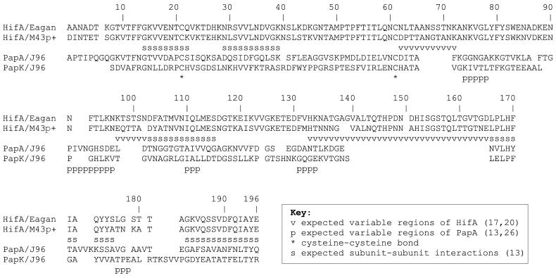
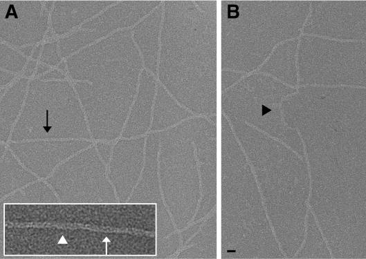
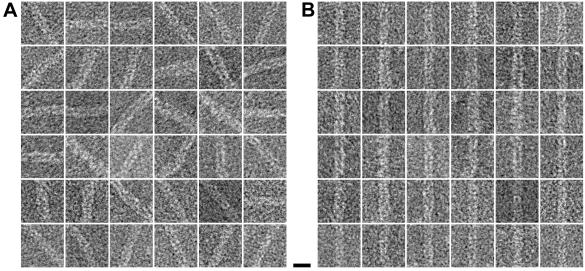
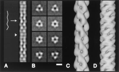
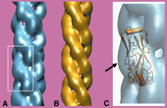
Pathogenic bacteria are specifically adapted to bind to their customary host. Disease is then caused by subsequent colonization and/or invasion of the local environmental niche. Initial binding of Haemophilus influenzae type b to the human nasopharynx is facilitated by Hib pili, filaments expressed on the bacterial surface. With three-dimensional reconstruction of electron micrograph images, we show that Hib pili comprise a helix 70 A in diameter with threefold symmetry. The Hib pilus filament has 3.0 subunits per turn, with each set of three subunits translated 26.9 A along and rotated 53 degrees about the helical axis. Amino acid sequence analysis of pilins from Hib pili and from P-pili expressed on uropathogenic Escherichia coli were used to predict the physical location of the highly variable and immunogenic region of the HifA pilin in the Hib pilus structure. Structural differences between Hib pili and P-pili suggest a difference in the strategies by which bacteria remain bound to their host cells: P-pili were shown to be capable of unwinding to five times their original length (E. Bullitt and L. Makowski, Nature 373:164-167, 1995), while damage to Hib pili occurs by slight shearing of subunits with respect to those further along the helical axis. This capacity to resist unwinding may be important for continued adherence of H. influenzae type b to the nasopharynx, where the three-stranded Hib pilus filaments provide a robust tether to withstand coughs and sneezes.
Figures
Similar articles
-
Evidence for donor strand complementation in the biogenesis of Haemophilus influenzae haemagglutinating pili.Mol Microbiol. 2000 Mar;35(6):1335-47. doi: 10.1046/j.1365-2958.2000.01816.x. Mol Microbiol. 2000. PMID: 10760135
-
Identification of a gene essential for piliation in Haemophilus influenzae type b with homology to the pilus assembly platform genes of gram-negative bacteria.Infect Immun. 1994 Feb;62(2):468-75. doi: 10.1128/iai.62.2.468-475.1994. Infect Immun. 1994. PMID: 7905461 Free PMC article.
-
Identification of two minor subunits in the pilus of Haemophilus influenzae.J Bacteriol. 1997 Jul;179(13):4227-31. doi: 10.1128/jb.179.13.4227-4231.1997. J Bacteriol. 1997. PMID: 9209037 Free PMC article.
-
Chaperone-assisted assembly and molecular architecture of adhesive pili.Annu Rev Microbiol. 1991;45:383-415. doi: 10.1146/annurev.mi.45.100191.002123. Annu Rev Microbiol. 1991. PMID: 1683764 Review.
-
Longus pilus of enterotoxigenic Escherichia coli and its relatedness to other type-4 pili--a minireview.Gene. 1997 Jun 11;192(1):39-43. doi: 10.1016/s0378-1119(97)00039-5. Gene. 1997. PMID: 9224872 Review.
Cited by
-
Fourier-Bessel reconstruction of helical assemblies.Methods Enzymol. 2010;482:131-65. doi: 10.1016/S0076-6879(10)82005-1. Methods Enzymol. 2010. PMID: 20888960 Free PMC article.
-
Nanoscale structural and mechanical properties of nontypeable Haemophilus influenzae biofilms.J Bacteriol. 2009 Apr;191(8):2512-20. doi: 10.1128/JB.01596-08. Epub 2009 Feb 13. J Bacteriol. 2009. PMID: 19218382 Free PMC article.
-
An open or closed case for the conformation of calponin homology domains on F-actin?J Muscle Res Cell Motil. 2004;25(4-5):351-8. doi: 10.1007/s10974-004-0690-7. J Muscle Res Cell Motil. 2004. PMID: 15548864 Review.
-
Nanoscale characterization and determination of adhesion forces of Pseudomonas aeruginosa pili by using atomic force microscopy.J Bacteriol. 2006 Jan;188(2):370-7. doi: 10.1128/JB.188.2.370-377.2006. J Bacteriol. 2006. PMID: 16385026 Free PMC article.
-
A structural basis for sustained bacterial adhesion: biomechanical properties of CFA/I pili.J Mol Biol. 2012 Feb 3;415(5):918-28. doi: 10.1016/j.jmb.2011.12.006. Epub 2011 Dec 9. J Mol Biol. 2012. PMID: 22178477 Free PMC article.
References
-
- Bullitt, E., and L. Makowski. 1995. Structural polymorphism of bacterial adhesion pili. Nature 373:164-167. - PubMed
-
- Choudhury, D., A. Thompson, V. Stojanoff, S. Langermann, J. Pinkner, S. J. Hultgren, and S. D. Knight. 1999. X-ray structure of the FimC-FimH chaperone-adhesin complex from uropathogenic Escherichia coli. Science 285:1061-1066. - PubMed
-
- DeRosier, D. J., and P. B. Moore. 1970. Reconstruction of three-dimensional images from electron micrographs of structures with helical symmetry. J. Mol. Biol. 52:355-369. - PubMed
Publication types
MeSH terms
Substances
LinkOut - more resources
Full Text Sources
