. 2002 Sep;184(17):4906-11.
doi: 10.1128/JB.184.17.4906-4911.2002.

Surface loop motion in FepA

Affiliations

Surface loop motion in FepA

Daniel C Scott et al. J Bacteriol. 2002 Sep.

Abstract

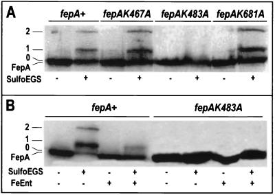
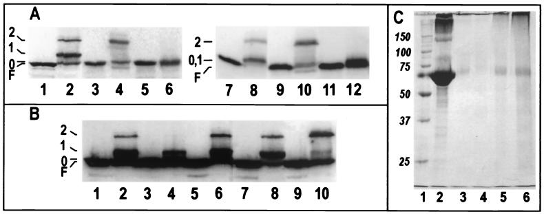
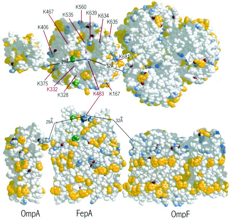
Using a lysine-specific cleavable cross-linking reagent ethylene glycolbis(sulfosuccimidylsuccinate) (Sulfo-EGS), we studied conformational motion in the surface loops of Escherichia coli FepA during its transport of the siderophore ferric enterobactin. Site-directed mutagenesis determined that Sulfo-EGS reacted with two lysines, K332 and K483, and at least two other unidentified Lys residues in the surface loops of the outer membrane protein. The reagent cross-linked K483 in FepA L7 to either K332 in L5, forming a product that we designated band 1, or to the major outer membrane proteins OmpF, OmpC, and OmpA, forming band 2. Ferric enterobactin binding to FepA did not prevent modification of K483 by Sulfo-EGS but blocked its cross-linking to OmpF/C and OmpA and reduced its coupling to K332. These data show that the loops of FepA undergo conformational changes in vivo, with an approximate magnitude of 15 A, from a ligand-free open state to a ligand-bound closed state. The coupling of FepA L7 to OmpF, OmpC, or OmpA was TonB independent and was unaffected by the uncouplers CCCP (carbonyl cyanide m-chlorophenylhydrazone) and DNP (2,4-dinitrophenol) but completely inhibited by cyanide.


Figures

FIG. 1.
FIG. 1.
FepAK483 participates in the formation of complex 2. (A) KDF541containing pITS23 or its derivatives that expressed wild-type or mutant FepA proteins were incubated in the the absence (−) or presence (+) of Sulfo-EGS. Cell lysates were resolved by SDS-PAGE, transferred to nitrocellulose, and visualized with α-FepA MAb 45 and 125I-labeled protein A. (B). KDF541/pITS23 and KDF541/pfepAK483A were incubated in the absence (−) or presence (+) of Sulfo-EGS, either with or without prior incubation with FeEnt (5 μM).
FIG. 2.
FIG. 2.
(A) Reactivity of K332 and the effects of energy poisons. KDF541 containing pITS23 (lanes 1, 2, 7, and 8), pfepAK332A (lanes 3, 4, 9, and 10), or pfepAK483A (lanes 5, 6, 11, and 12) was incubated in the the absence (odd-numbered lanes) or presence (even-numbered lanes) of Sulfo-EGS. Cell lysates were resolved on 10% acrylamide-0.26% bisacrylamide (lanes 1 to 6) or 10% acrylamide-0.067% bisacrylamide (lanes 7 to 12)slab gels, transferred to nitrocellulose, and visualized with a mixture of α-FepA MAbs 41 and 45 and 125I-labeled protein A. (B). Sulfo-EGS treatment of bacteria prepared in the presence of energy inhibitors. KDF541/pITS23 was either untreated (odd lanes) or exposed to Sulfo-EGS (even lanes) in the presence energy inhibitors at the following concentrations: no energy inhibitors (lanes 1 and 2), 20 mM cyanide (lanes 3 and 4), 0.1 mM CCCP (lanes 5 and 6), 2 mM DNP (lanes 7 and 8), and 10 mM azide (lanes 9 and 10). (C) Sulfo-EGS cross-linking of purified BSA in the presence or absence of energy inhibitors. A total of 25 μg of BSA was left untreated (lane 2) or was cross-linked in the absence (lane 3) or presence of 20 mM cyanide (lane 4), 10 mM azide (lane 5), or 100 mM azide (lane 6). The samples were solubiized in sample buffer, subjected to SDS-PAGE, and stained with Coomassie blue. Cross-linked BSA was unable to enter the gel slab (lanes 3 to 6); image analysis (not shown) revealed that only sodium azide had a small inhibitory effect on the cross-linking reaction (<1%). Molecular mass markers appear in lane 1.
FIG. 3.
FIG. 3.
Space-filling representations of OmpA, FepA, and OmpF showing a view of their surface Lys residues, including those in FepA that we mutagenized (enumerated) (top), and a side view in which the outer membrane proteins are aligned by their girdles of aromatic amino acids (yellow) at the internal and external interfaces of the outer membrane bilayer (bottom). Lys residues are shown in CPK colors. FepA residues 323 and 335, which define the terminal crystallographically solved portions of L4, are green. K332 exists somewhere between these two amino acids, close to residue 335. K483 is highlighted in the center of FepA.

Similar articles

Cited by

References

    1. Abdella, P. M., P. K. Smith, and G. P. Royer. 1979. A new cleavable reagent for cross-linking and reversible immobilization of proteins. Biochem. Biophys. Res. Commun. 87:734-742. - PubMed
    1. Ames, G. F. 1974. Resolution of bacterial proteins by polyacrylamide gel electrophoresis on slabs: membrane, soluble, and periplasmic fractions. J. Biol. Chem. 249:634-644. - PubMed
    1. Bos, C., D. Lorenzen, and V. Braun. 1998. Specific in vivo labeling of cell surface-exposed protein loops: reactive cysteines in the predicted gating loop mark a ferrichrome binding site and a ligand-induced conformational change of the Escherichia coli FhuA protein. J. Bacteriol. 180:605-613. - PMC - PubMed
    1. Bradbeer, C. 1993. The proton motive force drives the outer membrane transport of cobalamin in Escherichia coli. J. Bacteriol. 175:3146-3150. - PMC - PubMed
    1. Buchanan, S. K., B. S. Smith, L. Venkatramani, D. Xia, L. Esser, M. Palnitkar, R. Chakraborty, D. van der Helm, and J. Deisenhofer. 1999. Crystal structure of the outer membrane active transporter FepA from Escherichia coli. Nat. Struct. Biol. 6:56-63. - PubMed

Publication types

LinkOut - more resources