. 2002 Aug 15;21(16):4207-18.
doi: 10.1093/emboj/cdf417.

Structure of the periplasmic domain of Pseudomonas aeruginosa TolA: evidence for an evolutionary relationship with the TonB transporter protein

Affiliations

Structure of the periplasmic domain of Pseudomonas aeruginosa TolA: evidence for an evolutionary relationship with the TonB transporter protein

Michael Witty et al. EMBO J. .

Abstract

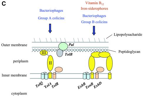
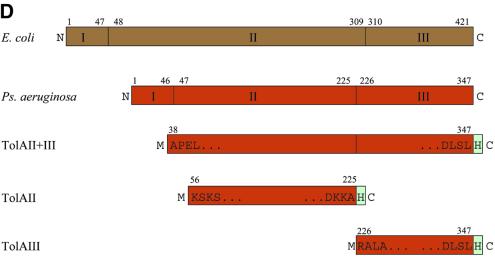
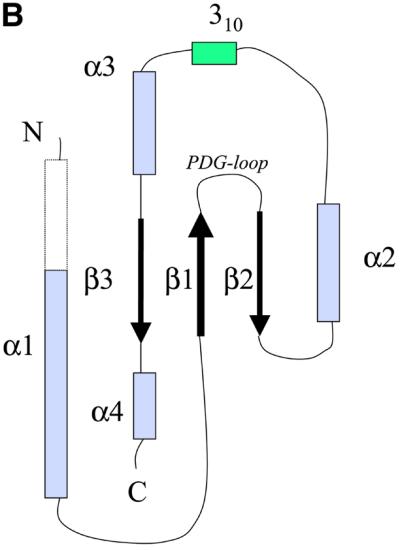
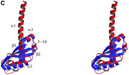
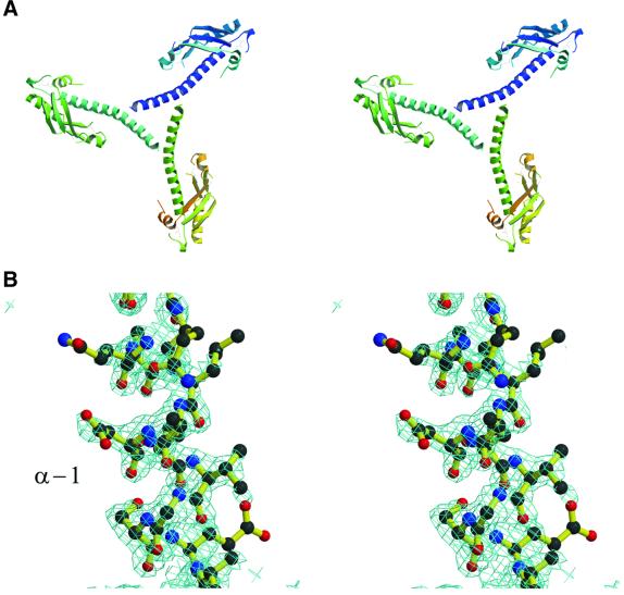
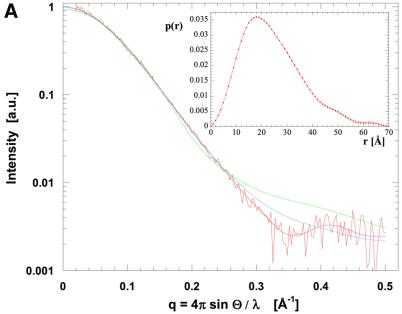
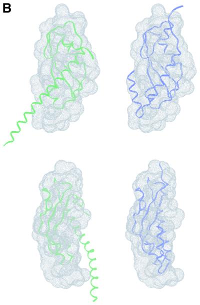
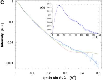
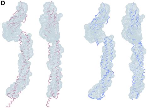
The crystal structure of the C-terminal domain III of Pseudomonas aeruginosa TolA has been determined at 1.9 A resolution. The fold is similar to that of the corresponding domain of Escherichia coli TolA, despite the limited amino acid sequence identity of the two proteins (20%). A pattern was discerned that conserves the fold of domain III within the wider TolA family and, moreover, reveals a relationship between TolA domain III and the C-terminal domain of the TonB transporter proteins. We propose that the TolA and TonB C-terminal domains have a common evolutionary origin and are related by means of domain swapping, with interesting mechanistic implications. We have also determined the overall shape of the didomain, domains II + III, of P.aeruginosa TolA by solution X-ray scattering. The molecule is monomeric-its elongated, stalk shape can accommodate the crystal structure of domain III at one end, and an elongated helical bundle within the portion corresponding to domain II. Based on these data, a model for the periplasmic domains of P.aeruginosa TolA is presented that may explain the inferred allosteric properties of members of the TolA family. The mechanisms of TolA-mediated entry of bateriophages in P.aeruginosa and E.coli are likely to be similar.


Figures

None
Fig. 1. (A) Schematic representation of the periplasmic localization of the E.coli TolA protein and its putative and known partners. TolA forms an assembly with the TolQ and TolR inner membrane proteins, and interacts with the Pal protein in the outer membrane. TolA domain III has been shown to interact with the phage g3p protein that infects E.coli (Lubkowski et al., 1999), as suggested in (B). As represented in (A), the g3p protein binds to the tip of the F pilus and triggers depolymerization with the result that virus and bacteria move into contact. (C) Parallels between the TolA/Q/R and TonB/ExbB/ExbD systems, which are both conserved in Gram-negative bacteria. The Tol system is the conduit for bacteriophages and group A colicins (left). The Exb/Ton system is required for the energy-dependent uptake of vitamin B12 and iron chelates, and is the conduit for group B colicins and certain classes of bacteriophage. (D) Domain organization of E.coli and P.aeruginosa TolA proteins. Also shown are schematic representations of the three constructs studied here: domain II, domain III and domain II + III. H indicated in the boxes represents the His6 tag; M originates from the initiation codon. E.coli residue numbers relate to the Swiss-Prot file P19934. P.aeruginosa residue numbers relate to the Swiss-Prot file P50600. For example R in RALA is residue 226 and the second L in DLSL is residue 347.
None
Fig. 2. (A) Two stereoscopic views of the ribbon trace of P.aeruginosa TolA domain III. (B) The convention used here to identify the secondary structural elements of P.aeruginosa TolA domain III. The N-terminal helix is longer in the crystal of the Se-methionine derivative compared with the native as demarcated by the broken lines. The helix begins at residue 2 in the Se-methionine form, but at residue 19 in the native form. The conserved PDG loop, which was used to align the TolA and TonB proteins (Figure 4), is indicated. (C) An overlay of the traces of the E.coli (blue) and P.aeruginosa (red) proteins. The E.coli protein (PDB code 1TOL) was solved as a fusion with the phage g3p protein, and the phage protein has been removed here. The perspective is the same as in the upper portion of (A). The figure was prepared using MOLSCRIPT (Kraulis, 1991) and Raster3D (Meritt and Murphy, 1994).
None
Fig. 3. The N-terminal helical extension of P.aeruginosa TolAIII. (A) Intermolecular contacts of the N-terminal helices of three neighbouring molecules in the crystal lattice of the Se-methionine derivative. The helices form a distinctive bundle. (B) Electron density in the vicinity of residue 20 in the N-terminal extension of the Se-methinone crystal. In the native crystal, the helical region is not as well defined and only begins at residue 19. The electron density map was calculated with minimal bias coefficients using REFMAC (Murshudov et al., 1997). The figure was generated using BOBSCRIPT (Esnouf, 1999) and Raster3D (Meritt and Murphy, 1994).
None
Fig. 4. Structural relationship of the TolA and TonB proteins. (A) A structure-based sequence alignment of TolA domain III from E.coli and P.aeruginosa, and the TonB protein. Residues in the loop between β1 and β2 are highly conserved amongst the available TolA and TonB homologues (not shown). The consensus secondary structure is shown below the alignment: α for α-helical positions; 3 for 310-helical positions; β for β-sheet positions. The formatting convention of JOY is as follows: red, α helices; blue, β strands; maroon, 310 helices; upper-case letters, solvent inaccessible; lower-case letters, solvent accessible; bold type, hydrogen bonds to main chain amides; underlining, hydrogen bonds to main chain carbonyls; tilde (∼), hydrogen bonds to other side chain/heteroatoms; italic, positive main chain torsion angles φ; cedilla (ç), disulfide-bonded cystine residues. (B) The TonB homodimer structure (PDB entry 1IHR). The individual protomers have been coloured purple and blue. The secondary structural elements have been labelled to correspond to the convention used for P.aeruginosa TolAIII (see Figure 22). (C) An overlay of the ribbon traces of a TonB pseudo- protomer (blue) with the P.aeruginosa TolA domain III protein (red). The TonB pseudo-promoter was generated by consolidating the elements of the dimer that are common with one TolA molecule and trimming back strands corresponding to β1 and β3. The perspective is similar to that used in the top portion of Figure 2A. The PDG loop is in the upper right.
None
Fig. 5. Molecular shape of the TolA periplasmic domains. (A) Solution X-ray scattering profile for P.aeruginosa TolAII domain. The smooth red curve through the experimental scattering data represents the profile from the restored shape shown in (B). Scattering profiles have been calculated based on the crystallographic structure of TolAIII (green curve, see Figure 2A) and a TolAIII model with changed N-terminal helical conformation (blue curve). (B) Shape of TolAIII as deduced from the experimental scattering profile alone with superimposed ribbons for the models introduced in (A). The top and bottom sets are different views of the two models. (C) Scattering profile and (D) shape reconstruction for the protein comprising both domains II and III. Two ribbon models have been superimposed: spectrin repeat of actinin (red) and two repeats of the alanine-rich helical bundles (blue) from the enzyme IIAlactose have been built into the molecular envelope. Two views are shown for each model. Their corresponding simulated scattering profiles have been given in (C).

Similar articles

Cited by

References

    1. Bennett M.J., Schlunegger,M.P. and Eisenberg,D. (1995) 3D domain swapping: a mechanism for oligomer assembly. Protein Sci., 4, 2455–2468. - PMC - PubMed
    1. Bernadac A., Gavioli,M., Lazzaroni,J.C., Raina,S. and Lloubes,R. (1998) Escherichia coli tol–pal mutants form outer membrane vesicles. J. Bacteriol., 180, 4872–4878. - PMC - PubMed
    1. Boulin C., Kempf,R., Koch,M.H.J. and McLaughlin,S.M. (1986) Data appraisal, evaluation and display for synchrotron radiation experiments: hardware and software. Nucl. Instrum. Methods Phys. Res. A, 249, 399–407.
    1. Braun V. and Herrmann,C. (1993) Evolutionary relationship of uptake systems for biopolymers in Escherichia coli: cross-complementation between TonB-ExbB-ExbD and the TolA-TolQ-TolR proteins. Mol. Microbiol., 8, 261–268. - PubMed
    1. Brown J.H., Kim,K.H., Jun,G., Greenfield,N.J., Dominguez,R., Volkmann,N., Hitchcock-DeGregori,S.E. and Cohen,C. (2001) Deciphering the design of the tropomysin molecule. Proc. Natl Acad. Sci. USA, 98, 8496–8501. - PMC - PubMed

Publication types

MeSH terms

Associated data