. 2002 Aug 15;21(16):4229-39.
doi: 10.1093/emboj/cdf426.

Maternally transmitted severe glucose 6-phosphate dehydrogenase deficiency is an embryonic lethal

Affiliations

Maternally transmitted severe glucose 6-phosphate dehydrogenase deficiency is an embryonic lethal

Letizia Longo et al. EMBO J. .

Abstract

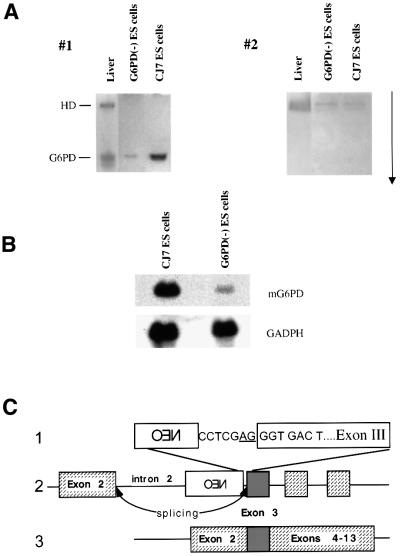
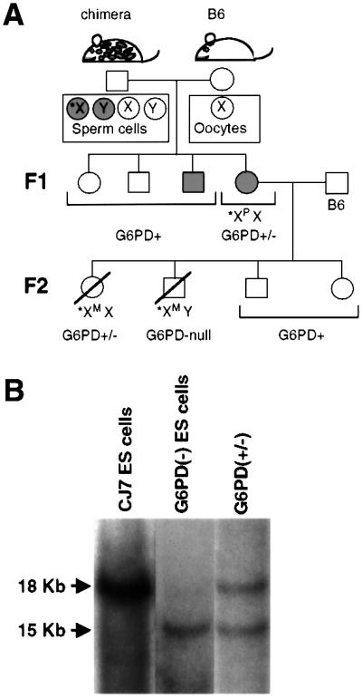
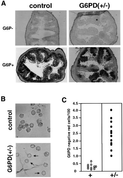
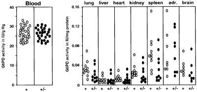
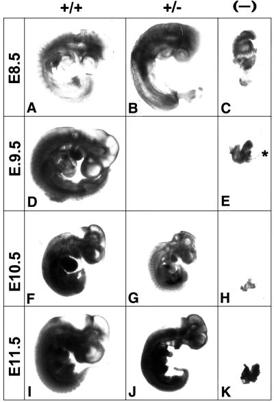
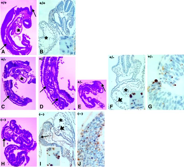
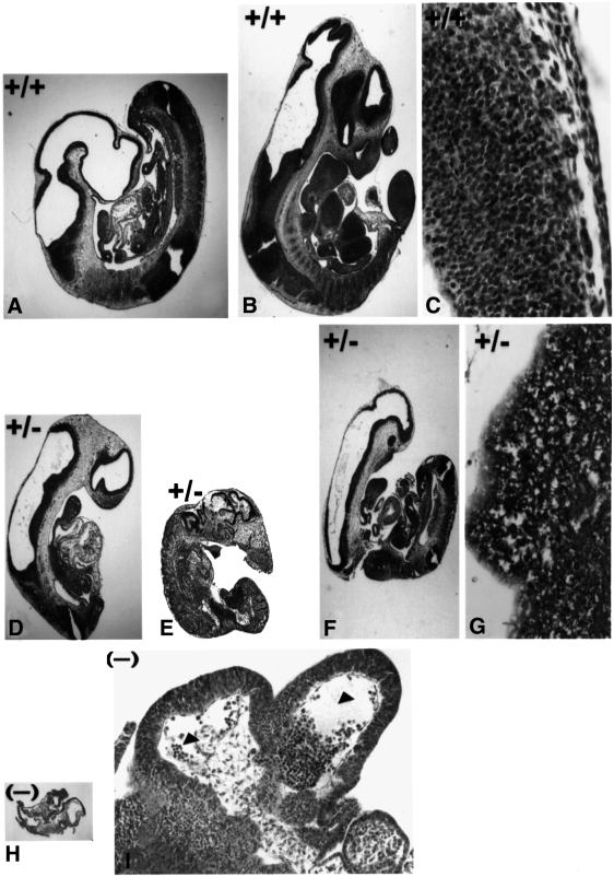
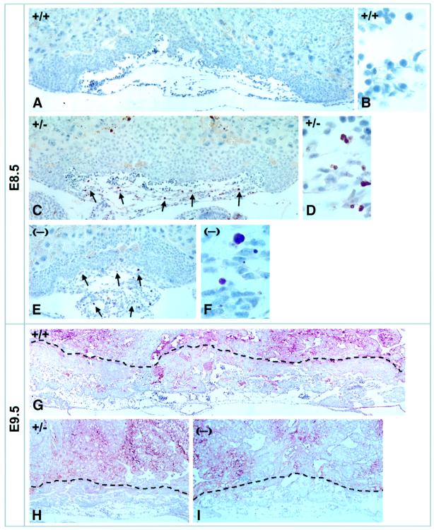
Mouse chimeras from embryonic stem cells in which the X-linked glucose 6-phosphate dehydrogenase (G6PD) gene had been targeted were crossed with normal females. First-generation (F(1)) G6PD(+/-) heterozygotes born from this cross were essentially normal; analysis of their tissues demonstrated strong selection for cells with the targeted G6PD allele on the inactive X chromosome. When these F(1) G6PD(+/-) females were bred to normal males, only normal G6PD mice were born, because: (i) hemizygous G6PD(-) male embryos died by E10.5 and their development was arrested from E7.5, the time of onset of blood circulation; (ii) heterozygous G6PD(+/-) females showed abnormalities from E8.5, and died by E11.5; and (iii) severe pathological changes were present in the placenta of both G6PD(-) and G6PD(+/-) embryos. Thus, G6PD is not indispensable for early embryo development; however, severe G6PD deficiency in the extraembryonic tissues (consequent on selective inactivation of the normal paternal G6PD allele) impairs the development of the placenta and causes death of the embryo. Most importantly, G6PD is indispensable for survival when the embryo is exposed to oxygen through its blood supply.


Figures

None
Fig. 1. Analysis of residual dehydrogenase activity in G6PD(–) ES cells. (A) Lysates from mouse liver, CJ7 and G6PD(–) ES cells were applied to a polyacrylamide gel and electrophoresis was carried out at 4°C and 40 W for 24 h. The arrow indicates the direction of migration. Gel 1 was stained with glucose 6-phosphate as substrate; gel 2 was stained with galactose 6-phosphate as substrate. The HD band is visible in gel 2 in both the wild-type and G6PD(–) ES cells. (B) Northern blot analysis of G6PD mRNA. By densitometry, the band in the G6PD(–) ES cells is estimated to be ∼15% of normal. (C) Diagram of the genomic murine G6PD locus after integration of the targeting vector (not drawn to scale). This structure of the targeting vector is revised from that shown in Figure 1 of the original description (Pandolfi et al., 1995). Exon 3 is highlighted in gray. (1) Junction between the NEO cassette and G6PD exon 3: an artificial splicing acceptor site is underlined upstream of exon 3. (2) Diagram of the primary transcript from the targeted G6PD. (3) Normal G6PD mRNA is produced at a low level because a splicing acceptor site has been recreated artificially by the insertion of an XhoI linker between the NEO cassette and exon 3.
None
Fig. 2. Generation of G6PD-deficient mice from G6PD(–) ES cells. (A) Design of the genetic crosses. *X indicates the X chromosome bearing the knock-out allele. Note that in the F1 heterozygotes, this is paternal (*XP); whereas in the F2 heterozygotes it is maternal (*XM). (B) Southern blot analysis (after DNA digestion with HpaI; Pandolfi et al., 1995) is diagnostic of the heterozygotes genotype [G6PD(+/–)] in the right lane by comparison in the other two lanes with normal CJ7 ES cells and with G6PD(–) ES cells, respectively.
None
Fig. 3. Histochemical staining for G6PD activity in cells from F1 heterozygotes. (A) Large intestine from first-generation heterozygotes and wild-type female littermates. The two top sections of this panel are negative controls stained without the substrate G6P. Mosaicism in G6PD expression is evident in heterozygotes. Similar results have been obtained in the small intestine. (B) Cytochemical analysis of peripheral blood red cells in a heterozygous mouse shows a small but significant proportion of G6PD-deficient red cells. (C) Scattergram of quantitative cytochemical analysis carried out on peripheral blood red cells: there is a statistically significant difference between heterozygotes and wild-type mice (Fisher’s exact test, P < 0.00001).
None
Fig. 4. Quantitative analysis of G6PD expression in F1 heterozygotes in various tissues. Adr, adrenal glands.
None
Fig. 5. Different deadly impacts of the G6PD(–) allele on the development of G6PD(–) and G6PD(+/–) embryos in the F2 generation. Embryos from mating of F1 G6PD(+/–) females with wild-type males (see Figure 2) were dissected, and examples of all genotypes (indicated at the top) are shown. The d.p.c. are shown on the left. It is seen that G6PD(–) embryos (C, E, H and K) arrest at around E8, fail to turn and then die. The asterisk in (E) indicates the heart. On the other hand, G6PD(+/–) embryos exhibit abnormalities as early as E8.5, but they continue to grow until E11.5 (compare B, G and J with A, F and I, respectively). Magnification: (A–C) 5×; (D and E) 2.5×; (F–K) 1.25×.
None
Fig. 6. Histological analysis of E8.5 mutants demonstrates major damage in the central nervous sytem and heart. All sections are from F2 embryos at E8.5; asterisks in (A), (B), (C), (F) and (I) indicate the heart; long arrows in (A), (C), (D), (E), (H) and (I) point to the neuroepithelium. Short arrows in (F) and (I) indicate the areas magnified in the corresponding insets. Sections from G6PD(–) hemizygous embryos confirm a major defect in the neuroepithelium (compare H with A), which is associated with extensive apoptosis (J). Apoptosis is also seen in the heart (compare I with B). Similar but less severe changes are seen in heterozygous G6PD(+/–) embryos (CG). (A), (C), (D), (E) and (H) are stained with H&E. (B), (F), (G), (I) and (J) show TUNEL staining. Magnification: (A), (C), (D) and (E) 10×; (B), (F), (H) and (I) 20×; (G) and (J) and insets in (B), (F) and (I) 100×.
None
Fig. 7. Progression of pathological alterations eventually leads to necrosis in all mutant embryos. H&E-stained sections of E10.5 (A, D, E, H and I) and E11.5 (B, C, F and G) embryos. The G6PD(–) hemizygous embryo in (H) is nearly reabsorbed; at higher magnification (I), one sees markedly dilated blood vessels and hemorrhages in the mesenchyme of the head. (D) and (E) (compared with A) demonstrate the variable extent of tissue damage in heterozygous embryos (more severe in E than in D). At E11.5, the damage is already substantial (compare F with B), with extensive lysis of tissue (compare G with C). Magnification: (A), (B), (D), (E), (F) and (H) 2.5×; (I) 10×; (C) and (G) 40×.
None
Fig. 8. Severe abnormalities of the placenta are similar in F2 G6PD(–) and G6PD(+/–) embryos. At day 8.5, TUNEL staining demonstrates widespread apoptosis in allantoic cells (arrows) in (C) and (E) (compare with A). Magnification: (A), (C) and (E) 20×; (B), (D) and (F) 100×. At day 9.5, the development of the placenta is obviously markedly impaired (see H and I compared with G; magnification 10×). The sections were stained with an anti-CD31 antibody.

Similar articles

Cited by

References

    1. Beutler E. (1978) Glucose 6-phosphate dehydrogenase deficiency. In Beutler,E. (ed.), Hemolytic Anemia in Disorders of Red Cell Metabolism. Plenum Medical Book Co., New York, NY, pp. 23–167.
    1. Beutler E. (1991) Glucose 6-phosphate dehydrogenase deficiency. N. Engl. J. Med., 324, 169–174. - PubMed
    1. Beutler E. (1995) Glucose 6-Phosphate Dehydrogenase Deficiency. McGraw-Hill, New York, NY.
    1. Beutler E. and Vulliamy,T. (2002) Hematologically important mutations: glucose 6-phosphate dehydrogenase. Blood Cells Mol. Dis., 28, 93–103. - PubMed
    1. Campbell F., Fuller,C.E., Williams,G.T. and Williams,E.D. (1994) Human colonic stem cell mutation frequency with and without irradiation. J. Pathol., 174, 175–182. - PubMed

Publication types

MeSH terms

Substances