. 2002 Aug 15;21(16):4327-37.
doi: 10.1093/emboj/cdf432.

Antagonistic regulation of flowering-time gene SOC1 by CONSTANS and FLC via separate promoter motifs

Affiliations

Antagonistic regulation of flowering-time gene SOC1 by CONSTANS and FLC via separate promoter motifs

Shelley R Hepworth et al. EMBO J. .

Abstract

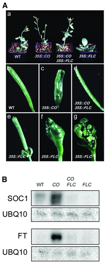
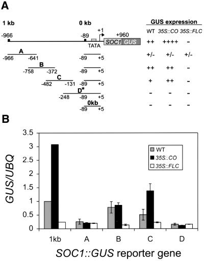
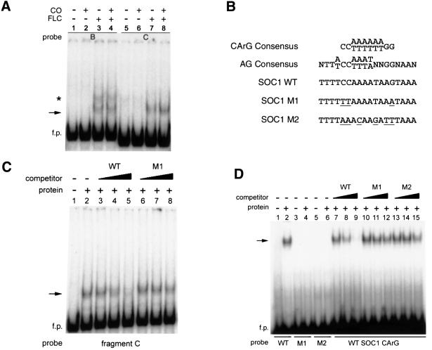
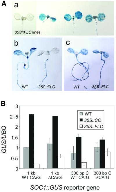
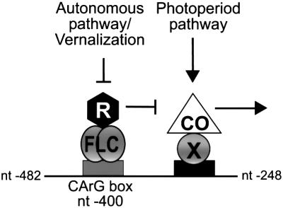
Flowering in Arabidopsis is controlled by endogenous and environmental signals relayed by distinct genetic pathways. The MADS-box flowering-time gene SOC1 is regulated by several pathways and is proposed to co-ordinate responses to environmental signals. SOC1 is directly activated by CONSTANS (CO) in long photoperiods and is repressed by FLC, a component of the vernalization (low-temperature) pathway. We show that in transgenic plants overexpressing CO and FLC, these proteins regulate flowering time antagonistically and FLC blocks transcriptional activation of SOC1 by CO. A series of SOC1::GUS reporter genes identified a 351 bp promoter sequence that mediates activation by CO and repression by FLC. A CArG box (MADS-domain protein binding element) within this sequence was recognized specifically by FLC in vitro and mediated repression by FLC in vivo, suggesting that FLC binds directly to the SOC1 promoter. We propose that CO is recruited to a separate promoter element by a DNA-binding factor and that activation by CO is impaired when FLC is bound to an adjacent CArG motif.


Figures

None
Fig. 1. Antagonistic effect of 35S::CO and 35S::FLC on flowering time, floral morphology, and expression of SOC1 and FT. (A) Phenotype of wild-type (WT), 35S::CO, 35S::CO 35S::FLC, and 35S::FLC plants. (a) Thirty-two-day-old plants grown in LDs; (b– f) morphology of siliques; (g) dissection of silique in (f) to show an ectopic inflorescence. (B) Northern analysis of SOC1 and FT mRNA in WT, 35S::CO, 35S::CO 35S::FLC, and 35S::FLC plants. One filter made with RNA harvested 8 h after dawn was hybridized with probes for SOC1 and UBQ10 (upper rows). A second filter made with RNA harvested 16 h after dawn was sequentially hybridized with probes for FT and UBQ10 (lower rows).
None
Fig. 2. Analysis of expression of SOC1::GUS reporter genes in wild-type plants. (A) Diagram of SOC1::GUS reporter genes with full-length (4 kb) or truncated promoters (2, 1 and 0 kb). The 5′ endpoints of constructs are numbered relative to the transcription start site (+1). (B) Expression of SOC1::GUS reporter genes in seedlings, cauline leaves, flowers and siliques as monitored by GUS staining. Days of growth are in the lower right of panels. Four kilobase SOC1::GUS reporter gene expression in (a–g) seedlings, (h) cauline leaves, (i) flowers and (j) siliques; 2 kb SOC1::GUS reporter gene expression in (k and l) seedlings, (m) cauline leaves, (n) flowers and (o) siliques; 1 kb SOC1::GUS expression in (p and q) seedlings, (r) cauline leaves, (s) flowers and (t) siliques; 0 kb SOC1::GUS expression in (u and v) seedlings, (w) cauline leaves, (x) flowers and (y) siliques. (C) Time course of 4, 2 and 1 kb SOC1::GUS reporter gene expression in wild-type seedlings as determined by GUS activity assays (see Materials and methods). RFU, relative fluorescence units.
None
Fig. 3. One kilobase SOC1::GUS reporter gene expression is activated in 35S::CO lines and repressed by FLC. (A) Analysis of 1 kb SOC1::GUS expression in wild-type (WT), 35S::CO and 35S::FLC plants by GUS staining: (a) 10-day-old seedlings, 1 kb SOC1::GUS WT (left) and 1 kb SOC1::GUS 35S::CO (right); (b) 14-day-old seedlings, 1 kb SOC1::GUS WT (left) and 1 kb SOC1::GUS 35S::FLC (right); 1 kb SOC1::GUS 35S::CO in (c) cauline leaves and (d) flower. (B) Northern analysis of 1 and 0 kb SOC1::GUS expression. RNA was purified from 1 kb SOC1::GUS or 0 kb SOC1::GUS seedlings: wild type (WT), 35S::CO (CO), 35S::CO 35S::FLC (CO FLC), 35S::FLC (FLC) and 35S::CO:GR. Dexamethasone (Dex) treatment, (+) or (–). The filter was sequentially hybridized with probes for GUS, SOC1 and β-TUB (loading control). (C) Transient expression assays of 1 kb SOC1::LUC. Leaves of Columbia (Col), flc-1 (in Col background), Landsberg erecta (Ler) and fca-1 (in Ler) plants were bombarded with beads coated with DNA of plasmids carrying 1 kb SOC1::LUC and 35S::GFP (Materials and methods). The ratio of luciferase to GFP expression is shown for each genotype (Materials and methods). In each case, the column represents the mean value, with the standard error.
None
Fig. 4. Identification of a 234 bp region of the SOC1 promoter that mediates activation by CO and repression by FLC. (A) Summary of expression of SOC1::GUS reporter genes in wild-type (WT), 35S::CO and 35S::FLC lines. Relative activities were determined by GUS staining. Top line, 1 kb SOC1::GUS reporter gene; second to fifth lines, 300 bp SOC1::GUS reporter genes containing overlapping fragments A, B, C or D from the SOC1 promoter. Fragments of 300 bp were each inserted upstream of the minimal 0 kb reporter gene at a unique BamHI site (see Materials and methods). Asterisk denotes that fragment D contains two copies of the minimal promoter region between –89 and +5. Bottom line, minimal 0 kb SOC1::GUS reporter gene that contains the TATA box and transcription start site (+1) for SOC1. (B) Promoter fragment C mediates activation in 35S::CO plants and repression in 35S::FLC plants. Expression of 1 kb SOC1::GUS (1 kb) and 300 bp SOC1::GUS (A, B, C, D) reporter genes was monitored in WT, 35S::CO and 35S::FLC seedlings by northern blotting using probes for GUS and UBQ10 (loading control). These data were quantified and are presented in histogram format. The amount of GUS/UBQ transcript in WT plants containing the 1 kb SOC1::GUS reporter gene was given an arbitrary value of 1. The relative level of transcript for each reporter gene construct in each background is presented as an average.
None
Fig. 5. Specific binding of FLC protein to a CArG box at nt –400 of the SOC1 promoter. (A) FLC protein forms a gel retardation complex (arrow) with 300 bp fragments B and C. FLC and/or CO protein were incubated with promoter fragments B or C as probes. The protein contained in each reaction is indicated above the panel. f.p., free probe. The asterisk indicates an apparent non-specific protein–DNA complex that was not consistently observed and was competed with non-specific competitor. (B) Comparison of wild-type (WT) and mutant MADS-box protein binding sites (CArG boxes): includes CArG box at nt –400 of the SOC1 promoter (CCAAAATAAG), as well as mutant versions of SOC1 CArG box. (C) Specific binding of FLC protein to fragment C probe. The presence of FLC protein is indicated above the panels and competitor DNAs are described in the text. Lane 1, no protein and no competitor DNA; lanes 3–5, SOC1 WT fragment as competitor DNA; lanes 6–8, SOC1 M1 fragment as competitor DNA. Non-labelled DNA in molar excess was used as competitor in lanes 3 and 6 (10-fold), lanes 4 and 7 (100-fold), and lanes 5 and 8 (1000-fold). (D) Specific binding of FLC protein to a 30 bp fragment containing the CArG sequence at nt –400. FLC protein was incubated with 30 bp fragment probes as indicated below the panel. The presence of FLC protein is indicated above the panels and competitor DNAs are described in the text. Lanes 1, 3, 5, no protein and no competitor DNA; lanes 2, 4, 6, FLC and no competitor DNA; lanes 7–9, SOC1 WT fragment as competitor DNA; lanes 10–12, SOC1 M1 fragment as competitor DNA; lanes 13–15, SOC1 M2 fragment as competitor DNA. Non-labelled DNA in molar excess was used as competitor in lanes 7, 10 and 13 (10-fold), lanes 8, 11 and 14 (100-fold), and lanes 9, 12 and 15 (1000-fold).
None
Fig. 6. The CArG box at nt –400 of the SOC1 promoter mediates repression of SOC1::GUS expression in 35S::FLC lines. (A) Mutation of the CArG box at nt –400 leads to derepression of SOC1::GUS expression in 35S::FLC lines. Ten-day-old seedlings were stained for GUS activity. (a) One kilobase SOC1::GUS expression in a 35S::FLC control seedling (left); three independent lines showing 1 kb ΔCArG SOC1::GUS expression in 35S::FLC seedlings (right); (b) 300 bp C SOC1::GUS expression in a wild-type (WT) seedling and in a 35S::FLC seedling; (c) mutated 300 bp C ΔCArG SOC1::GUS expression in a WT seedling and in a 35S::FLC seedling. (B) Analysis of the effect of the CArG box mutation on SOC1::GUS activation in 35S::CO lines and repression in 35S::FLC lines. Expression of WT and mutated ΔCArG reporter genes was monitored in WT, 35S::CO and 35S::FLC seedlings by northern blotting using probes for GUS and UBQ10 (loading control). The data were quantified and are presented in histogram format.
None
Fig. 7. Model for the antagonistic regulation of SOC1 expression by CO and FLC. The horizontal line represents a 234 bp region of the SOC1 promoter with the indicated nucleotide endpoints. Arrows indicate promotion and T-bars indicate repression of SOC1 expression. We propose that FLC binds directly to a CArG box motif at nt –400 of the SOC1 promoter and may recruit a global repression complex (R) to DNA to repress transcription. CO is proposed to be recruited by a sequence-specific DNA-binding factor (X) to an adjacent motif located between nt –372 and –248 in the SOC1 promoter to mediate activation. Deletion of the CArG box does not abolish FLC-mediated repression, suggesting that FLC also represses SOC1 expression by other more indirect mechanisms.

Similar articles

Cited by

References

    1. Alvarez-Buylla E.R., Liljegren,S.J., Pelaz,S., Gold,S.E., Burgeff,C., Ditta,G.S., Vergara-Silva,F. and Yanofsky,M.F. (2000) MADS-box gene evolution beyond flowers: expression in pollen, endosperm, guard cells, roots and trichomes. Plant J., 24, 457–466. - PubMed
    1. Araki T. (2001) Transition from vegetative to reproductive phase. Curr. Opin. Plant Biol., 4, 63–68. - PubMed
    1. Blázquez M.A. and Weigel,D. (2000) Integration of floral inductive signals in Arabidopsis. Nature, 404, 889–892. - PubMed
    1. Blázquez M.A., Soowal,L.N., Lee,I. and Weigel,D. (1997) LEAFY expression and flower initiation in Arabidopsis. Development, 124, 3835–3844. - PubMed
    1. Borden K.L.B. (1998) RING fingers and B-boxes: zinc-binding protein–protein interaction domains. Biochem. Cell Biol., 76, 351–358. - PubMed

Publication types

MeSH terms