. 2002 Aug 20;99(17):11079-84.
doi: 10.1073/pnas.162232399. Epub 2002 Aug 8.

Crystal structures of native and thrombin-complexed heparin cofactor II reveal a multistep allosteric mechanism

Affiliations

Crystal structures of native and thrombin-complexed heparin cofactor II reveal a multistep allosteric mechanism

Trevor P Baglin et al. Proc Natl Acad Sci U S A. .

Abstract

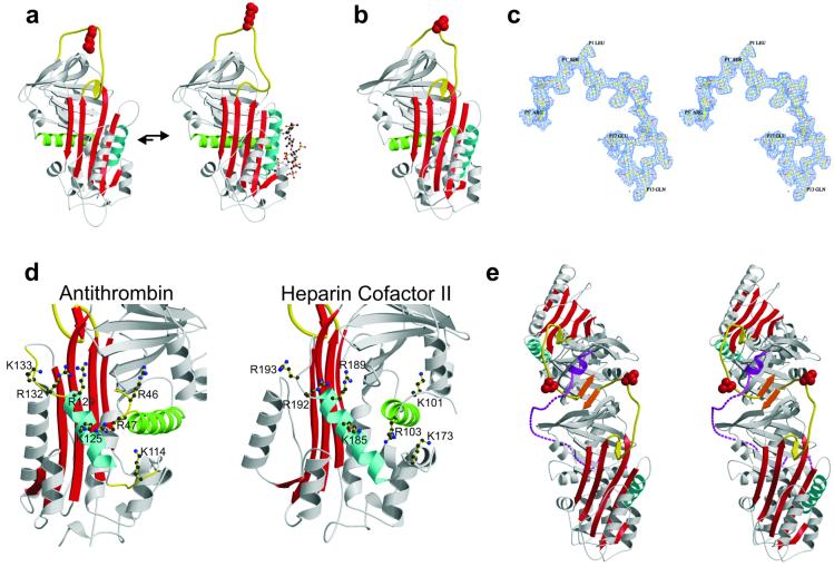
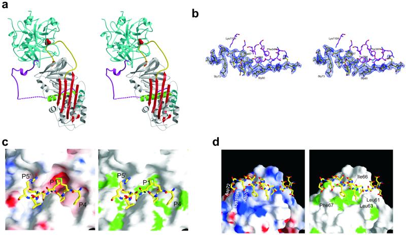
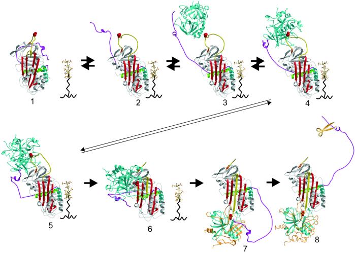
The serine proteases sequentially activated to form a fibrin clot are inhibited primarily by members of the serpin family, which use a unique beta-sheet expansion mechanism to trap and destroy their targets. Since the discovery that serpins were a family of serine protease inhibitors there has been controversy as to the role of conformational change in their mechanism. It now is clear that protease inhibition depends entirely on rapid serpin beta-sheet expansion after proteolytic attack. The regulatory advantage afforded by the conformational mobility of serpins is demonstrated here by the structures of native and S195A thrombin-complexed heparin cofactor II (HCII). HCII inhibits thrombin, the final protease of the coagulation cascade, in a glycosaminoglycan-dependent manner that involves the release of a sequestered hirudin-like N-terminal tail for interaction with thrombin. The native structure of HCII resembles that of native antithrombin and suggests an alternative mechanism of allosteric activation, whereas the structure of the S195A thrombin-HCII complex defines the molecular basis of allostery. Together, these structures reveal a multistep allosteric mechanism that relies on sequential contraction and expansion of the central beta-sheet of HCII.


Figures

Fig 1.
Fig 1.
The AT-like structure of native HCII. (a) The native structure of AT is different from all other know serpins with the partial incorporation of the reactive center loop (yellow) into β-sheet A (red), resulting in an unfavorable orientation of the reactive center arginine side chain (red, space-filling). Binding of heparin (ball-and-stick) to helix D (cyan) results in the expulsion of the reactive center loop from β-sheet A and its freeing for the formation of a productive Michaelis complex. (b) The structure of native HCII resembles that of native AT, with its reactive center loop partially inserted into β-sheet A. (c) Stereo depiction of the electron density for the entire reactive center loop contoured at 1σ. (d) The high degree of conservation of heparin binding residues on helix D (cyan) and helix A (green) with AT indicates a similar mode of binding and suggests similar conformational changes in response to binding. (e) A stereo representation of the crystallographic dimer of HCII formed by the antiparallel β-sheet interaction between strands 1C (orange) of each monomer. This contact is the probable cause of the displacement of the acidic N-terminal tail (magenta). The bottom monomer is molecule A.
Fig 2.
Fig 2.
Crystallographic structure of the HCII–thrombin Michaelis complex. (a) A stereo representation of the Michaelis complex between S195A thrombin (cyan) and HCII (colored as described for Fig. 1), with the γ-loop in front and the 60-insertion loop behind the reactive center loop. (b) Stereo representation of the electron density covering the portion of the acidic tail (yellow) that interacts with thrombin (magenta), contoured at 1σ. (c) The subsite interactions between the reactive center residues of HCII (rods) and the active site cleft of thrombin (surface representation) are extensive and complementary in both electrostatic (Left, negative potential is red, and positive is blue) and hydrophobic (Right, green for hydrophobic side chains) properties. (d) The interaction between exosite I of thrombin (surface representation as described for c) and the hirudin-like N-terminal tail of HCII (rods, the side chains of residues not interacting with thrombin were removed for clarity) is primarily hydrophobic. The only ionic interactions are between Asp-70 and Asp-72 of HCII and L110H of thrombin. Sulfated tyrosines, 60 and 73, make no contacts with thrombin and are not shown.
Fig 3.
Fig 3.
The sequential mechanism of GAG-mediated thrombin inhibition by HCII (as described in Results and Discussion). For a video of this mechanism, see Movie 2.

References

    1. Gettins P. G. W., Patston, P. A. & Olson, S. T., (1996) Serpins: Structure, Function, and Biology (Landes, Austin, TX).
    1. Huntington J. A. & Carrell, R. W. (1901) Sci. Prog. 84, 125-136. - PMC - PubMed
    1. Huntington J. A., Read, R. J. & Carrell, R. W. (2000) Nature (London) 407, 923-926. - PubMed
    1. Atchley W. R., Lokot, T., Wollenberg, K., Dress, A. & Ragg, H. (2001) Mol. Biol. Evol. 18, 1502-1511. - PubMed
    1. Ragg H., Lokot, T., Kamp, P. B., Atchley, W. R. & Dress, A. (2001) Mol. Biol. Evol. 18, 577-584. - PubMed

Publication types

LinkOut - more resources