

Rapid detection of IgH/BCL2 rearrangement in follicular lymphoma by interphase fluorescence in situ hybridization with bacterial artificial chromosome probes
- PMID: 12169675
- PMCID: PMC1906983
- DOI: 10.1016/S1525-1578(10)60695-2
Rapid detection of IgH/BCL2 rearrangement in follicular lymphoma by interphase fluorescence in situ hybridization with bacterial artificial chromosome probes
Abstract
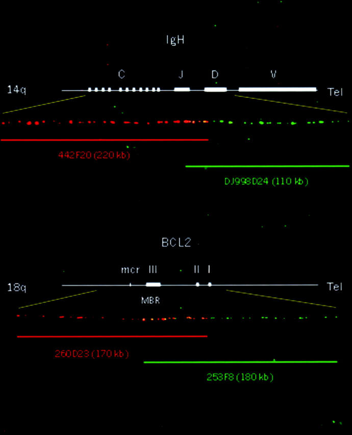
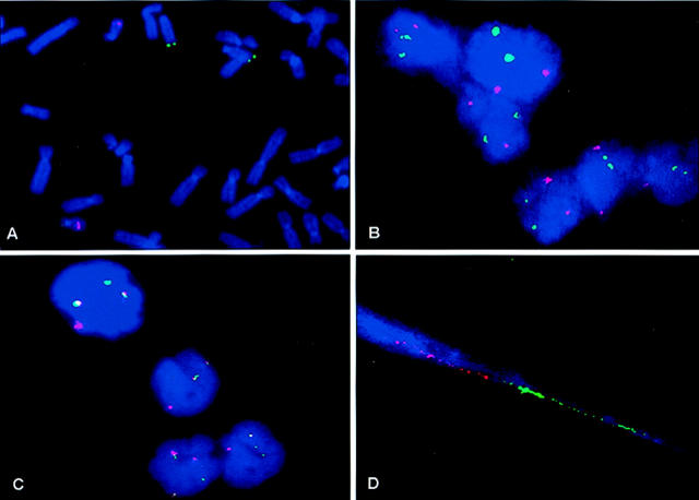
Follicular lymphomas (FLs) can be difficult to diagnose on aspirated specimens since the architectural pattern is not present. FLs characteristically have rearrangements in the IgH and BCL2 genes resulting from the reciprocal t(14;18) (q32; q21) translocation. Because of the dispersed distribution of breakpoints, fluorescence in situ hybridization (FISH) using genomic probes that span or flank the breakpoints is ideal for detecting this rearrangement in fine-needle aspiration (FNA) biopsies. To develop a set of probes, a bacterial artificial chromosome library was screened and the clones were mapped by fiber FISH. The probes were produced by the direct incorporation of fluorochrome-labeled nucleotides. The colocalization base FISH assay was applied to Cytospin preparations from FNA biopsies of lymph nodes from 26 patients with FL and 10 patients without FL. In those with FL, the percentage of cells with at least one IgH/BCL2 fusion signal ranged from 22% to 100% (mean, 63%), which was statistically significantly higher than that in FL-negative samples (mean, 2.7%). The probes demonstrated a significantly lower cutoff value (7%) in normal controls and effectively reduced the false-positive rate in FL-negative cases. These results were confirmed with fiber FISH assays on the same specimens. This interphase FISH assay is rapid and reliable for detecting rearrangements in the IGH/BCL2 gene, thereby aiding in the diagnosis of FL on FNA biopsy specimens.
Figures
References
-
- Hockenbery D, Nunez G, Milliman C, Schreiber RD, Korsmeyer SJ: Bcl-2 is an inner mitochondrial membrane protein that blocks programmed cell death. Nature 1990, 348:334-336 - PubMed
-
- Buchonnet G, Lenain P, Ruminy P, Lepretre S, Stamatoullas A, Parmentier F, Jardin F, Duval C, Tilly H, Bastard C: Characterization of BCL2-JH rearrangements in follicular lymphoma: PCR detection of 3′ BCL2 breakpoints and evidence of a new cluster. Leukemia 2000, 14:1563-1569 - PubMed
-
- Yunis JJ, Frizzera G, Oken MM, McKenna J, Theologides A, Arnesen M: Multiple recurrent genomic defects in follicular lymphoma: a possible model for cancer. N Engl J Med 1987, 316:79-84 - PubMed
-
- Vaandrager JW, Schuuring E, Raap T, Philippo K, Kleiverda K, Kluin P: Interphase FISH detection of BCL2 rearrangement in follicular lymphoma using breakpoint-flanking probes. Genes Chromosomes Cancer 2000, 27:85-94 - PubMed
-
- Poetsch M, Weber-Matthiesen K, Plendl HJ, Grote W, Schlegelberger B: Detection of the t(14;18) chromosomal translocation by interphase cytogenetics with yeast-artificial-chromosome probes in follicular lymphoma and non-neoplastic lymphoproliferation. J Clin Oncol 1996, 14:963-969 - PubMed
Publication types
MeSH terms
Substances
Grants and funding
LinkOut - more resources
Full Text Sources
Research Materials
