. 2002 Aug 20;99(17):11305-10.
doi: 10.1073/pnas.172382899. Epub 2002 Aug 9.

Chk2 regulates irradiation-induced, p53-mediated apoptosis in Drosophila

Affiliations

Chk2 regulates irradiation-induced, p53-mediated apoptosis in Drosophila

Malte Peters et al. Proc Natl Acad Sci U S A. .

Abstract

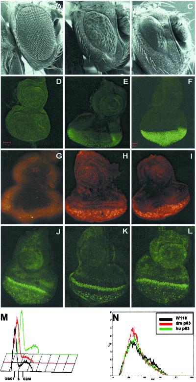
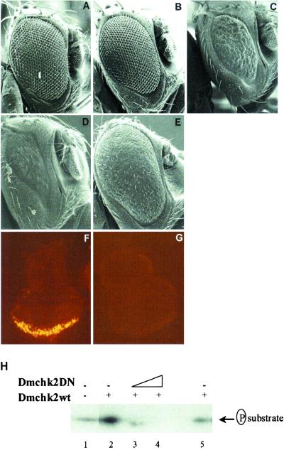
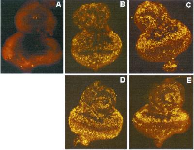
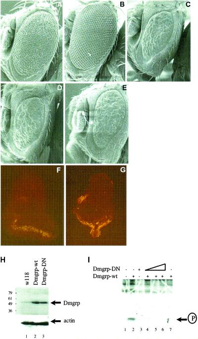
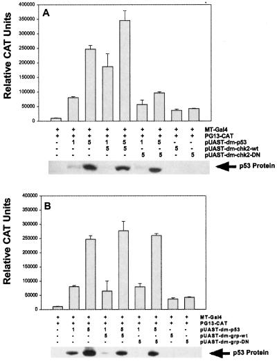
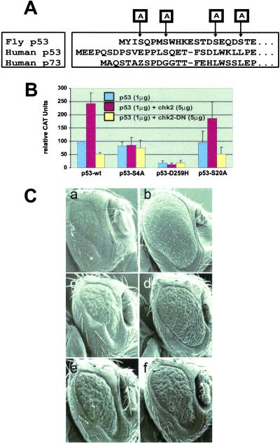
The tumor suppressor function of p53 has been attributed to its ability to regulate apoptosis and the cell cycle. In mammals, DNA damage, aberrant growth signals, chemotherapeutic agents, and UV irradiation activate p53, a process that is regulated by several posttranslational modifications. In Drosophila melanogaster, however, the regulation modes of p53 are still unknown. Overexpression of D. melanogaster p53 (Dmp53) in the eye induced apoptosis, resulting in a small eye phenotype. This phenotype was markedly enhanced by coexpression with D. melanogaster Chk2 (DmChk2) and was almost fully rescued by coexpression with a dominant-negative (DN), kinase-dead form of DmChk2. DN DmChk2 also inhibited Dmp53-mediated apoptosis in response to DNA damage, whereas overexpression of Grapes (Grp), the Drosophila Chk1-homolog, and its DN mutant had no effect on Dmp53-induced phenotypes. DmChk2 also activated the Dmp53 transactivation activity in cultured cells. Mutagenesis of Dmp53 amino terminal Ser residues revealed that Ser-4 is critical for its responsiveness toward DmChk2. DmChk2 activates the apoptotic activity of Dmp53 and Ser-4 is required for this effect. Contrary to results in mammals, Grapes, the Drosophila Chk1-homolog, is not involved in regulating Dmp53. Chk2 may be the ancestral regulator of p53 function.


Figures

Fig 1.
Fig 1.
Effects of hp53 or Dmp53 overexpression in the fly eye. (AC) Scanning electron micrographs of eyes from GMR-Gal4 flies crossed to a wild-type fly (A), UAS-Dmp53 fly (B), or UAS-hp53 fly (C). (Magnification: ×220.) (DF) Expression of p53 in eye discs. Immunohistochemical detection of Dmp53 in wild-type (D), and UAS-Dmp53 (E) or hp53 in UAS-hp53 (F) flies crossed to GMR-Gal4. (GI) Distribution of dying cells in eye discs by using acridine orange. Wild-type larva (G), UAS-Dmp53 larva (H), or hp53 larva (I) crossed to GMR-Gal4 flies. (JL) Cellular proliferation shown by BrdUrd labeling. Wild-type flies (J), Dmp53 overexpressing flies (K), and hp53 overexpressing flies (L). (M and N) Cell cycle analysis in single cell suspensions dissected from eye discs. (Magnification: ×400.) (M) Histogram displaying DNA content and cell numbers from single cell suspensions from eye discs dissected from wild-type (black), Dmp53 (red), and human p53 (green) overexpressing flies. (N) Forward side scatter displaying cell size and cell number.
Fig 2.
Fig 2.
Chk2 regulates p53 in Drosophila. (AE) Scanning electron micrographs of eyes from flies overexpressing DmChk2 (A), DN-DmChk2 (B), Dmp53 (C), DmChk2 and Dmp53 (D), or DN-DmChk2 and Dmp53 (E), driven by the GMR-Gal4 promoter. (Magnification: ×220.) (F and G) Chk2-regulation of p53 induced apoptosis. Eye discs from a fly overexpressing DmChk2 and Dmp53 (F) or DN-DmChk2 and Dmp53 (G), driven by the GMR-Gal4 promoter, were stained with the vital dye acridine orange. (Magnification: ×400.) (H) DN-DmChk2 inhibits DmChk2 kinase activity. Myc/His-tagged DmChk2, alone (lane 2) or together with increasing amounts of DN-DmChk2 (lanes 3 and 4), immunoprecipitated from 293T cells, was incubated with synthetic Chk1/Chk2 substrate peptide and the resulting phosphorylation was assessed by autoradiography. As a control (lane 5), the anti-Myc/His Ab was omitted in the immunoprecipitation step.
Fig 3.
Fig 3.
DN forms of p53 and Chk2 prevent irradiation-induced apoptosis. Acridine orange-stained eye discs from transgenic flies treated without (A) or with (BE) 40 Gy of γ-irradiation. Wild type, unirradiated (A), wild type (B), Dmp53–259H (C), DmChk2 (D), and DN-DmChk2 (E), driven by GMR-Gal4. (Magnifications: ×400.)
Fig 4.
Fig 4.
Grp does not affect Dmp53 activity. (AE) Scanning electron micrographs of eyes from flies overexpressing Grp (A), DN-Grp (B), Dmp53 (C), Grp and Dmp53 (D), or DN-Grp and Dmp53 (E) directed by GMR-Gal4. (Magnification: ×220.) (FG) Acridine orange staining of eye discs overexpressing Grp and Dmp53 (F) or DN-Grp and Dmp53 (G). (Magnification: ×400.) (H) Expression of Grp and DN-Grp. Protein extracts from w118 (lane 1), Grp (lane 2), and DN-Grp (lane 3) flies crossed to armGal4 flies were analyzed by Western blot using anti-Chk1 Ab. Equal loading was confirmed by actin (Lower). (I) DN-Grp inhibits Grp kinase activity. Myc/His-tagged Grp, alone (lane 2) or together with increasing amounts of DN-Grp (lanes 4–6), immunoprecipitated from 293T cells, was incubated with synthetic Chk1/Chk2 substrate peptide, and the resulting phosphorylation was assessed by autoradiography. As a control (lane 7), the anti-Myc/His Ab was omitted in the immunoprecipitation step.
Fig 5.
Fig 5.
Chk2 regulates p53 transactivation in cultured cells. Drosophila S2 cells were transfected with 1 or 5 μg of wild-type Dmp53 to test for transcriptional activation from PG13, a promoter containing wild-type binding sites for human p53. (A) Cells were cotransfected with 5 μg of wild-type DmChk2 or a DN, kinase-dead form of DmChk2. (B) Cells were cotransfected with 5 μg of either wild-type Grp or a DN, kinase-dead form of Grp. Error bars indicate the SEM from three independent experiments. (A Lower and B Lower) Expression of Dmp53 protein in the respective lysates.
Fig 6.
Fig 6.
Regulation of Dmp53 by DmChk2 via Ser-4. (A) Amino acid sequence alignment of Drosophila and hp53 and hp73. Arrows indicate mutated Ser residues. (B) Drosophila S2 cells were transfected with 1 μg of wild-type Dmp53, Dmp53-S4A, Dmp53-D259H, and Dmp53-S20A, alone (blue bars), and with 5 μg of wild-type or DN-DmChk2 (yellow bars) and p53 responsive PG13-CAT. (C) Scanning electron micrographs of eyes of a fly expressing Dmp53 (a and b), Dmp53-S20A (c and d), Dmp53-S4A (e and f) together with one copy of the DmChk2 (a, c, and e), or DN DmChk2 (b, d, and f) directed by GMR-Gal4. (Magnifications: ×220.)

Similar articles

Cited by

References

    1. Levine A. J. (1997) Cell 88, 323-331. - PubMed
    1. Vousden K. H. (2000) Cell 103, 691-694. - PubMed
    1. Walworth N. C. (2000) Curr. Opin. Cell. Biol. 12, 697-704. - PubMed
    1. Zhou B. S. & Elledge, S. J. (2000) Nature (London) 408, 433-439. - PubMed
    1. Chehab N. H., Malikzay, A., Appel, M. & Halazonetis, T. D. (2000) Genes Dev. 14, 278-288. - PMC - PubMed

Publication types

MeSH terms