. 2002 Aug;14(8):1817-32.
doi: 10.1105/tpc.003681.

A geminivirus replication protein interacts with a protein kinase and a motor protein that display different expression patterns during plant development and infection

Affiliations

A geminivirus replication protein interacts with a protein kinase and a motor protein that display different expression patterns during plant development and infection

Ling-Jie Kong et al. Plant Cell. 2002 Aug.

Abstract

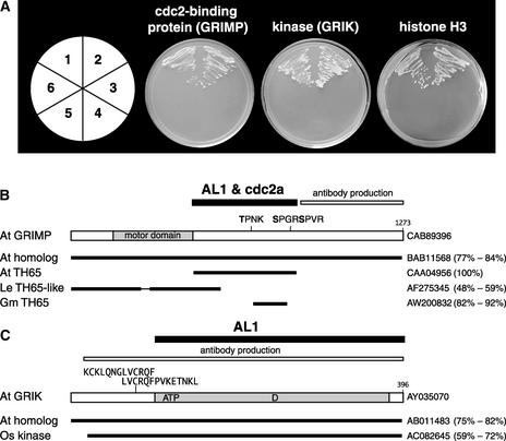
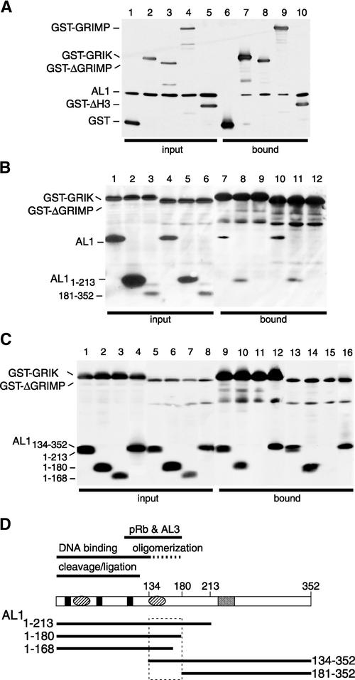
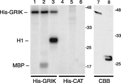
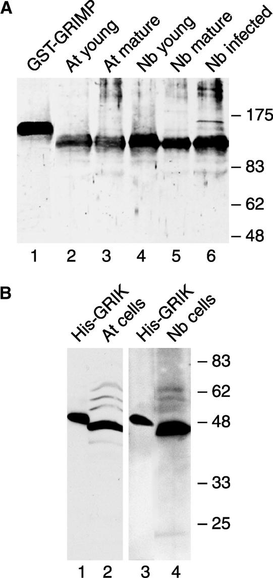
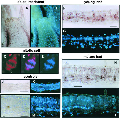
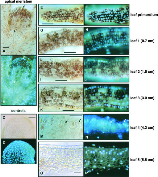
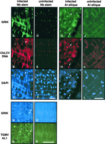
The geminivirus protein AL1 initiates viral DNA replication, regulates its own expression, and induces plant gene transcription. To better understand how AL1 interacts with host proteins during these processes, we used yeast two-hybrid library screening and a baculovirus protein interaction system to identify plant proteins that interact with AL1. These studies identified a Ser/Thr kinase, a kinesin, and histone H3 as AL1 partners. The kinase is autophosphorylated and can phosphorylate common kinase substrates in vitro. The kinesin is phosphorylated in insect cells by a cyclin-dependent kinase. Immunostaining of Nicotiana benthamiana and Arabidopsis showed that kinase protein levels and subcellular location are regulated during plant development and geminivirus infection. By contrast, the kinesin is ubiquitous even though it is associated with the spindle apparatus in mitotic cells. Together, our results establish that AL1 interacts with host proteins involved in plant cell division and development. Possible functions of these host factors in healthy and geminivirus-infected plants are discussed.


Figures

Figure 1.
Figure 1.
Identification of Three Plant Proteins That Interact with AL1. (A) Protein interactions in the yeast two-hybrid system. Arabidopsis cDNA sequences fused to the Gal4 activation domain (AD) vector isolated by library screening are shown. Center left, cdc2 binding protein (GRIMP473-866); center right, kinase (GRIK107-396); far right, histone (H358-136). The AD fusions were cotransformed with the various Gal4 DNA binding domain (BD) vectors into yeast strain AH109 and grown in the absence of Leu, Trp, His, and adenine. Section 1, AD-cDNA + BD-CbLCV AL1; section 2, AD-cDNA + BD-TGMV AL1; section 3, pGADT7 + pGBKT7; section 4, pGADT7 + BD-CbLCV AL1; section 5, pGADT7 + BD-TGMV AL1; section 6, AD-cDNA + pGBKT7. (B) Diagram of GRIMP showing the motor domain (amino acids 108 to 369) and three consensus CDK recognition sites at positions 698, 841, and 845. The regions that interact with AL1 and Arabidopsis cdc2a and that were used for antibody production are indicated. Related sequences from Arabidopsis (At), tomato (Le), and soybean (Gm) are shown with their GenBank accession numbers at right. The percentage identity and homology are shown in parentheses. (C) Diagram of GRIK showing the AL1-interacting region and the fragment used for antibody production. The ATP binding site (amino acids 114 to 137), the kinase active site D residue (amino acid 240), and the two overlapping calmodulin binding motifs (amino acids 73 to 85 and 80 to 93) are marked. Related sequences from Arabidopsis and rice (Os) are shown with their GenBank accession numbers at right. The percentage identity and homology are shown in parentheses.
Figure 2.
Figure 2.
AL1 Interacts with the Three Host Factors in Insect Cells. Protein extracts from Sf9 cells coexpressing TGMV AL1 and GST fusion proteins were purified on glutathione-Sepharose resin. Total and bound proteins were resolved by SDS-PAGE and analyzed by immunoblotting using anti-AL1 and anti-GST antibodies. GST-GRIK includes amino acids 12 to 396 of GRIK. GST-GRIMP corresponds to full-length GRIMP. GST-ΔGRIMP contains amino acids 473 to 866 of GRIMP. GST-ΔH3 includes amino acids 58 to 136 of unprocessed histone H3. (A) Verification of protein interactions in insect cells. Total (lanes 1 to 5) and bound (lanes 6 to 10) proteins are shown for assays containing full-length TGMV AL1 and GST (lanes 1 and 6), GST-GRIK (lanes 2 and 7), GST-ΔGRIMP (lanes 3 and 8), GST-GRIMP (lanes 4 and 9), or GST-ΔH3 (lanes 5 and 10). (B) AL1 domains that interact with GRIK and GRIMP. Total (lanes 1 to 6) and bound (lanes 7 to 12) fractions from insect cells coexpressing TGMV AL1 (lanes 1, 4, 7, and 10) or the AL1 truncations AL11-213 (lanes 2, 5, 8, and 11) and AL1181-352 (lanes 3, 6, 9, and 12), with GST-GRIK (lanes 1 to 3 and 7 to 9) or GST-ΔGRIMP (lanes 4 to 6 and 10 to 12), are shown. (C) Fine mapping of the AL1 domains that interact with GRIK and GRIMP. Total (lanes 1 to 8) and bound (lanes 9 to 16) fractions from insect cells coexpressing the TGMV truncations AL11-213 (lanes 1, 5, 9, and 13), AL11-180 (lanes 2, 6, 10, and 14), AL11-168 (lanes 3, 7, 11, and 15), and AL1134-352 (lanes 4, 8, 12, and 16), with GST-GRIK (lanes 1 to 4 and 9 to 12) or GST-GRIMP473-866 (lanes 5 to 8 and 13 to 16), are shown. (D) The domain of AL1 that interacts with GRIK and GRIMP. Diagram of the AL1 protein showing the previously mapped domains for binding to pRb and AL3, oligomerization (dashed line), DNA binding (solid lines), and DNA cleavage/ligation activities. Three conserved DNA cleavage motifs (solid boxes), two predicted pairs of α-helices (ovals), and the ATP binding site (open box) are marked. Solid lines below the diagram show the positions of the AL1 truncations designated by their N- and C-terminal amino acids. The boxed region indicates the domain that interacts with GRIK and GRIMP.
Figure 3.
Figure 3.
Phosphorylation of Recombinant GRIK and GRIMP in Insect Cells. Sf9 cells infected with recombinant baculoviruses were labeled with 32P-orthophosphate. Radiolabeled proteins were resolved by SDS-PAGE. The positions of recombinant proteins are indicated at left. The migration and masses of protein markers are shown at right. The recombinant protein nomenclature is described for Figure 1. The various ΔGRIMP and GRIK fusions are nearly identical in size and migrate differently relative to each other depending on the tag. Their identities were verified using anti-GRIK and anti-GRIMP antibodies (data not shown). (A) Phosphoproteins from cells infected with recombinant baculoviruses expressing His-CAT (lane1), His-GRIK (lane 2), His-ΔGRIMP (lane 3), GST (lane 4), and GST-GRIMP (lane 5) as indicated. The positions of GST and His-CAT are marked by dots at right of their respective lanes. (B) A CDK inhibitor reduces GST-ΔGRIMP phosphorylation. Sf9 cells were infected with recombinant baculoviruses expressing GST-GRIK (lanes 1 to 3) or GST-ΔGRIMP (lanes 4 to 6) and labeled with 32P-orthophosphate in the presence of the CDK inhibitor olomoucine. The final concentrations of the inhibitor were 0 μM (lanes 1 and 4), 10 μM (lanes 2 and 5), and 20 μM (lanes 3 and 6), as indicated. The bottom panel shows an immunoblot using anti-GST antibody and 10% of the total protein analyzed by autoradiography.
Figure 4.
Figure 4.
GRIK Acts as a Protein Kinase in Vitro. His-GRIK (lanes 1 to 3) or His-CAT (lanes 4 to 6) was incubated in kinase reactions with no substrate (lanes 1 and 4), myelin basic protein (lanes 2 and 5 [MBP]), or histone H1 (lanes 3 and 6). The reactions were resolved by SDS-PAGE, and the 32P-labeled products marked at left were visualized by autoradiography. The right panel shows Coomassie blue staining (CBB) of the purified His-GRIK (lane 7; 0.6 μg) and His-CAT (lane 8; 1.8 μg) used in the kinase assays. The migration and masses of protein markers are shown at right. The recombinant protein nomenclature is as described for Figure 1.
Figure 5.
Figure 5.
Immunodetection of GRIK and GRIMP in Plant Tissues and Cultured Cells. (A) Total protein extracts from young leaves (lanes 2 and 4), mature leaves (lanes 3 and 5), or infected mature leaves (lane 6) of Arabidopsis (lanes 2 and 3) or N. benthamiana (lanes 4 to 6) were separated by SDS-PAGE and detected using anti-GRIMP antibody. Recombinant GST-GRIMP (lane 1) was used as a control. (B) Total protein extracts from Arabidopsis (lane 2) or N. benthamiana (lane 4) cultured cells were resolved by SDS-PAGE and detected using anti-GRIK antibody. Recombinant His-GRIK (lanes 1 and 3) was used as a control.
Figure 6.
Figure 6.
Immunolocalization of GRIMP. N. benthamiana tissue sections were analyzed using anti-GRIMP IgG ([A] to [I]) or preimmune IgG ([J] to [M]) followed by anti-rabbit secondary antibody and peroxidase staining ([A], [B], and [F] to [M]) or Texas red–conjugated secondary antibody ([C] to [E]). All sections also were stained with DAPI. Bars = 50 μm. (A) and (B) Meristem showing GRIMP (A) and DAPI (B) staining. (C) to (E) Confocal image of a mitotic cell treated with anti-GRIMP IgG and a Texas red–labeled secondary antibody (C), DAPI staining (E), and the merged image (D). Open arrowheads show GRIMP staining associated with the spindle poles. (F) and (G) Young leaf showing GRIMP (F) and DAPI (G) staining. (H) and (I) Mature leaf showing GRIMP (H) and DAPI (I) staining. (J) and (K) Young leaf treated with preimmune IgG showing no peroxidase staining (J) but stained with DAPI (K). (L) and (M) Mature leaf treated with preimmune IgG showing no peroxidase staining (L) but stained with DAPI (M).
Figure 7.
Figure 7.
Developmental Expression of GRIK. N. benthamiana sections were analyzed using anti-GRIK IgG ([A], [B], and [E] to [P]) or preimmune IgG ([C] and [D]). Peroxidase-conjugated secondary antibody was used, followed by peroxidase and DAPI staining. The leaves are numbered from top to bottom of the plant, and their lengths are indicated at right. Bars = 50 μm. (A) and (B) Meristem showing GRIK (A) and DAPI (B) staining. The arrow indicates a cell with GRIK in the cytosol, and the open arrowhead marks a cell with GRIK in the nucleus. (C) and (D) Meristem treated with preimmune IgG showing no peroxidase staining (C) but stained with DAPI (D). (E) and (F) Leaf primordium showing GRIK (E) and DAPI (F) staining. (G) and (H) First leaf showing GRIK (G) and DAPI (H) staining. (I) and (J) Second leaf showing GRIK (I) and DAPI (J) staining. The open arrowheads designate a cell negative for GRIK surrounded by cells containing high levels of the kinase. (K) and (L) Third leaf showing GRIK (K) and DAPI (L) staining. (M) and (N) Fourth leaf showing GRIK (M) and DAPI (N) staining. The arrows identify cells with nuclear GRIK staining. (O) and (P) Fifth leaf showing GRIK (O) and DAPI (P) staining.
Figure 8.
Figure 8.
GRIK Induction in Geminivirus-Infected Plants. Sections of N. benthamiana stem (left two columns) or Arabidopsis silique tissues (right two columns) were fixed and analyzed using anti-GRIK IgG. In the same experiments, Texas red–labeled CbLCV DNA probe ([A] to [L]) or mouse anti-TGMV AL1 antibody ([M] to [P]) was used to detect the viruses. In (A) to (L), Alexa 488–conjugated goat anti-rabbit secondary antibody and DAPI were used to show specific proteins and nuclei, respectively. In (M) to (P), Cascade blue–conjugated goat anti-rabbit secondary antibody and Alexa 488–conjugated goat anti-mouse secondary antibody were used to show GRIK and TGMV AL1, respectively. The mock-inoculated controls ([D], [E], [F], [J], [K], [L], [O], and [P]) were exposed 15 times longer than infected tissues to show cell structure. Bars = 50 μm. (A) to (C) CbLCV-infected N. benthamiana stem showing GRIK staining (A), CbLCV hybridization (B), and DAPI staining (C). The arrows mark an infected cell positive for GRIK, and the open arrowheads identify a nucleus with no detectable GRIK. (D) to (F) Uninfected N. benthamiana stem tissue showing no GRIK staining (D) or CbLCV hybridization (E) but stained with DAPI (F). (G) to (I) CbLCV-infected Arabidopsis silique showing GRIK staining (G), CbLCV hybridization (H), and DAPI staining (I). The arrows mark infected cells positive for GRIK. (J) to (L) Uninfected Arabidopsis silique tissue showing no GRIK staining (J) or CbLCV hybridization (K) but stained with DAPI (L). (M) and (N) TGMV-infected N. benthamiana stem stained for GRIK (M) and AL1 (N). The arrows show a nucleus that contains GRIK, and the open arrowheads mark a nucleus without detectable GRIK. Both nuclei are positive for AL1. (O) and (P) Uninfected N. benthamiana stem showing no GRIK (O) or AL1 (P) staining.

Similar articles

Cited by

References

    1. Abouzid, A.M., Hiebert, E., and Strandberg, J.O. (1992). Cloning, identification, and partial sequencing of the genomic components of a geminivirus infecting the Brassicaceae. Phytopathology 82, 1070.
    1. Ach, R.A., Durfee, T., Miller, A.B., Taranto, P., Hanley-Bowdoin, L., Zambriski, P.C., and Gruissem, W. (1997). RRB1 and RRB2 encode maize retinoblastoma-related proteins that interact with a plant D-type cyclin and geminivirus replication protein. Mol. Cell. Biol. 17, 5077–5086. - PMC - PubMed
    1. Alexiadis, V., Halmer, L., and Gruss, C. (1997). Influence of core histone acetylation on SV40 minichromosome replication in vitro. Chromosoma 105, 324–331. - PubMed
    1. Amundson, S.A., Myers, T.G., and Fornace, A.J. (1998). Roles for p53 in growth arrest and apoptosis: Putting on the brakes after genotoxic stress. Oncogene 17, 3287–3299. - PubMed
    1. Bass, H.W., Nagar, S., Hanley-Bowdoin, L., and Robertson, D. (2000). Chromosome condensation induced by geminivirus infection of mature plant cells. J. Cell Sci. 113, 1149–1160. - PubMed

MeSH terms

Associated data

LinkOut - more resources