

B-ZIP proteins encoded by the Drosophila genome: evaluation of potential dimerization partners
- PMID: 12176927
- PMCID: PMC186634
- DOI: 10.1101/gr.67902
B-ZIP proteins encoded by the Drosophila genome: evaluation of potential dimerization partners
Abstract
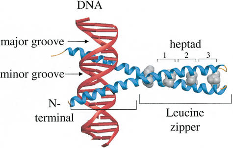
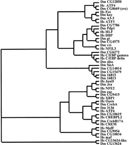
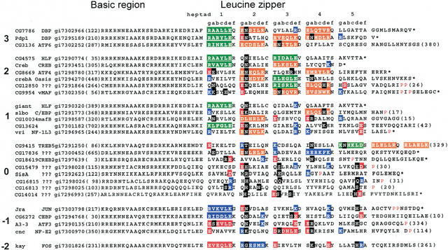
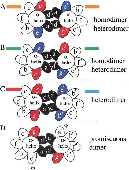
The basic region-leucine zipper (B-ZIP) (bZIP) protein motif dimerizes to bind specific DNA sequences. We have identified 27 B-ZIP proteins in the recently sequenced Drosophila melanogaster genome. The dimerization specificity of these 27 B-ZIP proteins was evaluated using two structural criteria: (1) the presence of attractive or repulsive interhelical g<-->e' electrostatic interactions and (2) the presence of polar or charged amino acids in the 'a' and 'd' positions of the hydrophobic interface. None of the B-ZIP proteins contain only aliphatic amino acids in the'a' and 'd' position. Only six of the Drosophila B-ZIP proteins contain a "canonical" hydrophobic interface like the yeast GCN4, and the mammalian JUN, ATF2, CREB, C/EBP, and PAR leucine zippers, characterized by asparagine in the second 'a' position. Twelve leucine zippers contain polar amino acids in the first, third, and fourth 'a' positions. Circular dichroism spectroscopy, used to monitor thermal denaturations of a heterodimerizing leucine zipper system containing either valine (V) or asparagine (N) in the 'a' position, indicates that the V-N interaction is 2.3 kcal/mole less stable than an N-N interaction and 5.3 kcal/mole less stable than a V-V interaction. Thus, we propose that the presence of polar amino acids in novel positions of the 'a' position of Drosophila B-ZIP proteins has led to leucine zippers that homodimerize rather than heterodimerize.
Figures
Comment in
-
Intraproteomic networks: new forays into predicting interaction partners.Genome Res. 2002 Aug;12(8):1156-8. doi: 10.1101/gr.353302. Genome Res. 2002. PMID: 12176922 Review. No abstract available.
Similar articles
-
Dimerization specificity of all 67 B-ZIP motifs in Arabidopsis thaliana: a comparison to Homo sapiens B-ZIP motifs.Nucleic Acids Res. 2004 Jun 29;32(11):3435-45. doi: 10.1093/nar/gkh653. Print 2004. Nucleic Acids Res. 2004. PMID: 15226410 Free PMC article.
-
A heterodimerizing leucine zipper coiled coil system for examining the specificity of a position interactions: amino acids I, V, L, N, A, and K.Biochemistry. 2002 Dec 3;41(48):14122-31. doi: 10.1021/bi020486r. Biochemistry. 2002. PMID: 12450375
-
Experimental identification of homodimerizing B-ZIP families in Homo sapiens.J Struct Biol. 2006 Aug;155(2):130-9. doi: 10.1016/j.jsb.2006.02.018. Epub 2006 May 6. J Struct Biol. 2006. PMID: 16725346
-
Deciphering B-ZIP transcription factor interactions in vitro and in vivo.Biochim Biophys Acta. 2006 Jan-Feb;1759(1-2):4-12. doi: 10.1016/j.bbaexp.2005.12.005. Epub 2006 Jan 31. Biochim Biophys Acta. 2006. PMID: 16580748 Review.
-
Interactions of coiled coils in transcription factors: where is the specificity?Curr Opin Genet Dev. 1993 Apr;3(2):278-85. doi: 10.1016/0959-437x(93)90035-n. Curr Opin Genet Dev. 1993. PMID: 8504253 Review.
Cited by
-
Lessons from the genome sequence of Neurospora crassa: tracing the path from genomic blueprint to multicellular organism.Microbiol Mol Biol Rev. 2004 Mar;68(1):1-108. doi: 10.1128/MMBR.68.1.1-108.2004. Microbiol Mol Biol Rev. 2004. PMID: 15007097 Free PMC article. Review.
-
Roles of C/EBP class bZip proteins in the growth and cell competition of Rp ('Minute') mutants in Drosophila.Elife. 2020 Jan 7;9:e50535. doi: 10.7554/eLife.50535. Elife. 2020. PMID: 31909714 Free PMC article.
-
Physiologic and anatomic characterization of the brain surface glia barrier of Drosophila.Glia. 2011 Sep;59(9):1322-40. doi: 10.1002/glia.21147. Epub 2011 Feb 23. Glia. 2011. PMID: 21351158 Free PMC article. Review.
-
Predicting specificity in bZIP coiled-coil protein interactions.Genome Biol. 2004;5(2):R11. doi: 10.1186/gb-2004-5-2-r11. Epub 2004 Jan 16. Genome Biol. 2004. PMID: 14759261 Free PMC article.
-
The role of bZIP transcription factors in green plant evolution: adaptive features emerging from four founder genes.PLoS One. 2008 Aug 13;3(8):e2944. doi: 10.1371/journal.pone.0002944. PLoS One. 2008. PMID: 18698409 Free PMC article.
References
-
- Adams MD, Celniker SE, Holt RA, Evans CA, Gocayne JD, Amanatides PG, Scherer SE, Li PW, Hoskins RA, Galle RF, et al. The genome sequence of Drosophila melanogaster. Science. 2000;287:2185–2195. - PubMed
-
- Alber T. Structure of the leucine zipper. Curr Opin Genet Dev. 1992;2:205–210. - PubMed
-
- Altschul SF, Gish W, Miller W, Myers EW, Lipman DJ. Basic local alignment search tool. J Mol Biol. 1990;215:403–410. - PubMed
-
- Apweiler R, Biswas M, Fleischmann W, Kanapin A, Karavidopoulou Y, Kersey P, Kriventseva EV, Mittard V, Mulder N, Phan I, et al. Proteome Analysis Database: Online application of InterPro and CluSTr for the functional classification of proteins in whole genomes. Nucleic Acids Res. 2001;29:44–48. - PMC - PubMed
Publication types
MeSH terms
Substances
LinkOut - more resources
Full Text Sources
Molecular Biology Databases
Miscellaneous