. 2002 Aug 19;158(4):787-99.
doi: 10.1083/jcb.200204041. Epub 2002 Aug 12.

Distinct molecular and cellular contributions to stabilizing selectin-mediated rolling under flow

Affiliations

Distinct molecular and cellular contributions to stabilizing selectin-mediated rolling under flow

Tadayuki Yago et al. J Cell Biol. .

Abstract

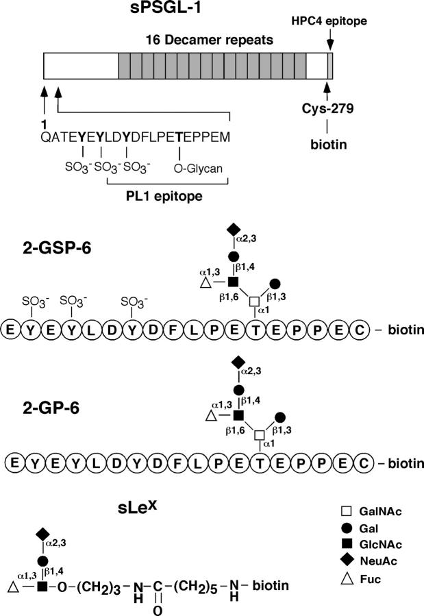
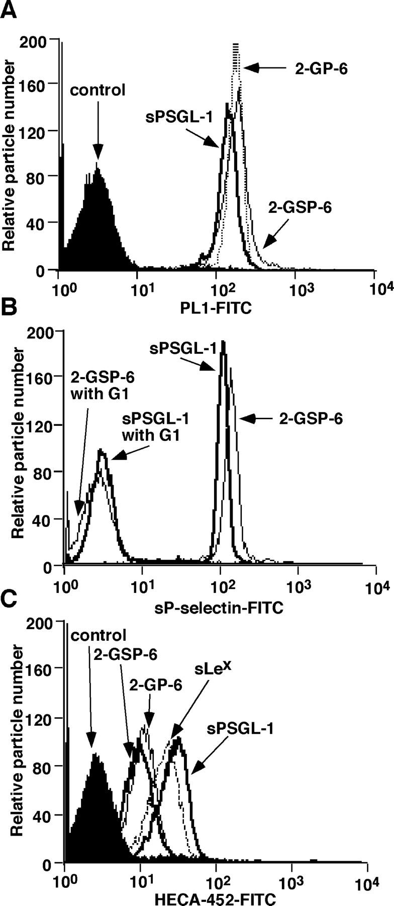
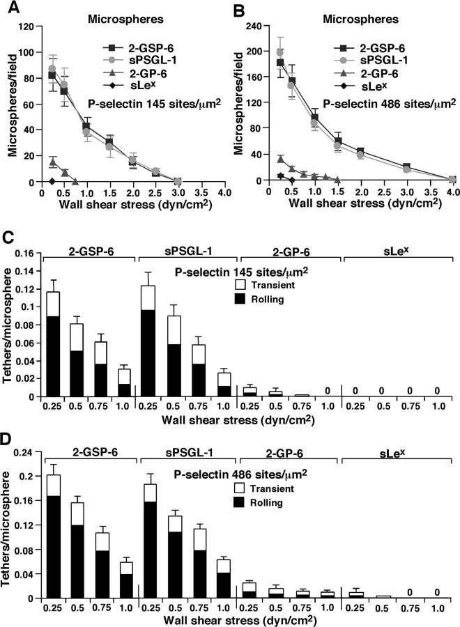
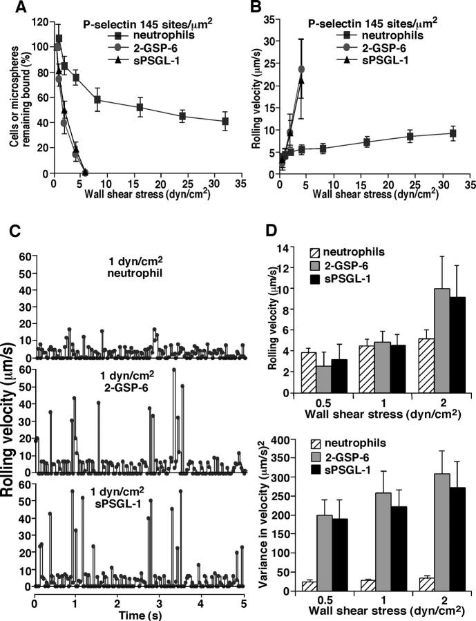
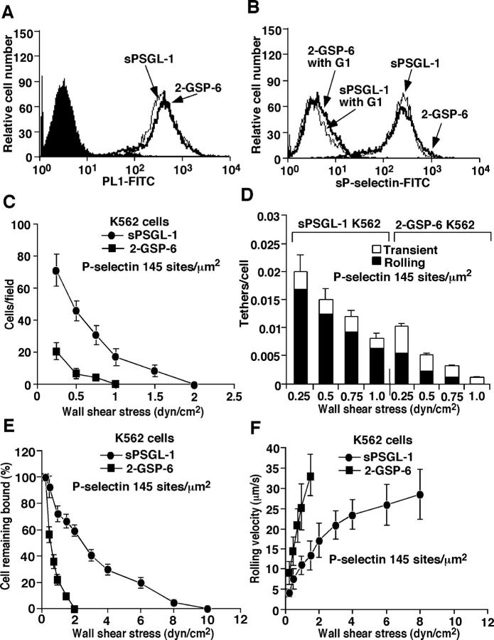
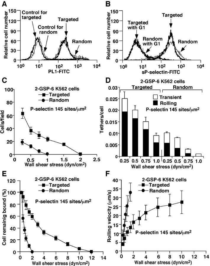
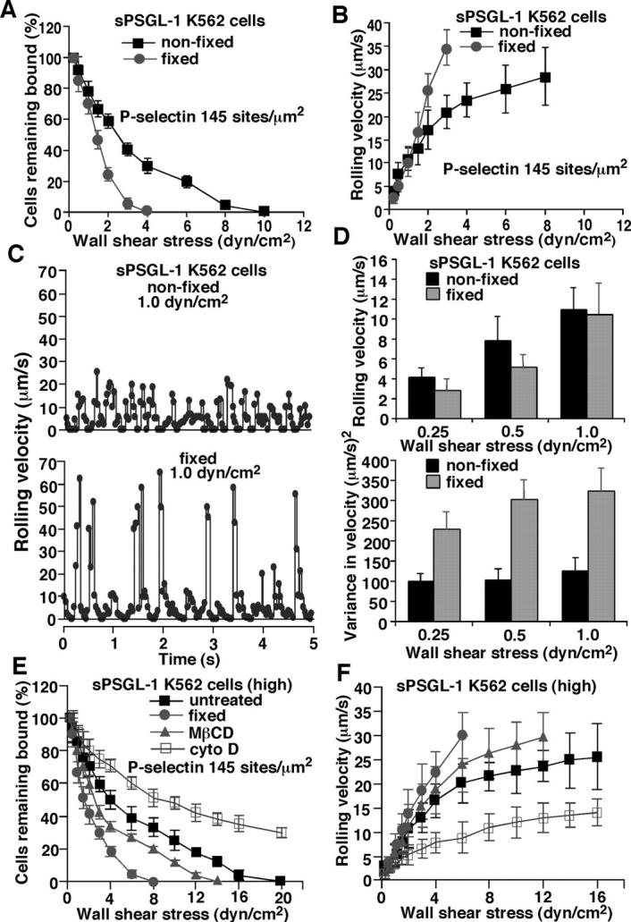
Leukocytes roll on selectins at nearly constant velocities over a wide range of wall shear stresses. Ligand-coupled microspheres roll faster on selectins and detach quickly as wall shear stress is increased. To examine whether the superior performance of leukocytes reflects molecular features of native ligands or cellular properties that favor selectin-mediated rolling, we coupled structurally defined selectin ligands to microspheres or K562 cells and compared their rolling on P-selectin. Microspheres bearing soluble P-selectin glycoprotein ligand (sPSGL)-1 or 2-glycosulfopeptide (GSP)-6, a GSP modeled after the NH2-terminal P-selectin-binding region of PSGL-1, rolled equivalently but unstably on P-selectin. K562 cells displaying randomly coupled 2-GSP-6 also rolled unstably. In contrast, K562 cells bearing randomly coupled sPSGL-1 or 2-GSP-6 targeted to a membrane-distal region of the presumed glycocalyx rolled more like leukocytes: rolling steps were more uniform and shear resistant, and rolling velocities tended to plateau as wall shear stress was increased. K562 cells treated with paraformaldehyde or methyl-beta-cyclodextrin before ligand coupling were less deformable and rolled unstably like microspheres. Cells treated with cytochalasin D were more deformable, further resisted detachment, and rolled slowly despite increases in wall shear stress. Thus, stable, shear-resistant rolling requires cellular properties that optimize selectin-ligand interactions.


Figures

Figure 1.
Figure 1.
Selectin ligands coupled to microspheres or K562 cells. Biotin was covalently attached to the COOH-terminal cysteine of sPSGL-1, 2-GSP-6, or 2-GP-6, or to a spacer group on sLex. The biotinylated ligands were bound to streptavidin-coated microspheres or K562 cells.
Figure 2.
Figure 2.
Surface densities of selectin ligands on microspheres measured by flow cytometry. (A) Microspheres were incubated with anti–PSGL-1 mAb PL1 or with an isotype-matched control mAb. Bound antibody was detected with FITC-conjugated goat anti–mouse IgG. (B) Microspheres were incubated with sP-selectin complexed with the nonblocking anti–P-selectin mAb S12 in the presence or absence of the blocking anti–P-selectin mAb G1. Bound sP-selectin was detected with FITC-conjugated goat anti–mouse IgG. (C) Microspheres were incubated with the anti-sLex mAb HECA-452 or with an isotype-matched control mAb. Bound mAb was detected with FITC-conjugated goat anti–rat IgM.
Figure 3.
Figure 3.
Rolling and tethering of ligand-coupled microspheres on P-selectin. (A and B) Microspheres coupled with the indicated ligand were perfused over P-selectin at the indicated density. After 5 min, the number of rolling microspheres was quantified. (C and D) The measured number of microspheres that tethered during the first 60 s was normalized by dividing by the number of cells delivered across the field of view in the focal plane of the substrate. The percentage of tethers that were transient or that were converted to rolling adhesion is also indicated. The data represent the mean ± SD of five experiments.
Figure 4.
Figure 4.
Detachment resistance and rolling properties of neutrophils or ligand-coupled microspheres on P-selectin. Neutrophils or microspheres were allowed to accumulate on P-selectin at 0.5 dyn/cm2. Wall shear stress was then increased every 30 s, and the percentage of remaining adherent cells (A) and their rolling velocities (B) was determined. (C) Frame-by-frame velocities of representative neutrophils or microspheres rolling on P-selectin at 145 sites/μm2. (D) Mean velocities and variances of velocities for neutrophil or microsphere populations rolling on P-selectin at 145 sites/μm2. The data represent the mean ± SD of five experiments.
Figure 5.
Figure 5.
Tethering and rolling of ligand-coupled K562 cells on P-selectin. Binding of anti-PSGL-1 mAb PL1 (A) or sP-selectin (B) was measured as in Fig. 2. The accumulated number of rolling cells (C), the tethering rates (D), the detachment resistance (E), and the mean rolling velocities (F) were measured as in Fig. 3. The data represent the mean ± SD of five experiments.
Figure 6.
Figure 6.
Rolling and tethering of K562 cells with 2-GSP-6 coupled to randomly distributed streptavidin or with 2-GSP-6 targeted to a membrane-distal region of a glycoform of PSGL-1 that does not bind selectins. (A) Surface densities of random or targeted 2-GSP-6 were measured by binding of anti–PSGL-1 mAb PL1 as described in Materials and methods. The binding of sP-selectin (B), the accumulated number of rolling cells (C), the tethering rates (D), the detachment resistance (E), and the mean rolling velocities (F) were measured as in Figs. 2 and 3. The data represent the mean ± SD of five experiments.
Figure 7.
Figure 7.
Effects of cellular perturbations on detachment resistance and rolling properties of sPSGL-1–coupled K562 cells on P-selectin. Cells were fixed with paraformaldehyde or treated with MβCD or cytochalasin D before coupling of sPSGL-1. Untreated and treated cells displayed equivalent surface densities of sPSGL-1 (unpublished data). (A and B) Detachment resistance and mean rolling velocities of nonfixed or fixed cells were measured as in Fig. 3. (C) Frame-by-frame velocities of representative nonfixed and fixed cells rolling on P-selectin at 145 sites/μm2. (D) Mean velocities and variances of velocities for populations of nonfixed or fixed cells rolling on P-selectin at 145 sites/μm2. (E and F) sPSGL-1 was coupled to approximately sevenfold higher density on untreated cells, fixed cells, or cells treated with MβCD or cytochalasin D. Detachment resistance and mean rolling velocities were measured as in Fig. 3. Cells treated with αCD, an inactive analogue of MβCD, or with DMSO, the diluent for cytochalasin D, rolled identically to untreated cells (unpublished data). The data represent the mean ± SD of five experiments.

References

    1. Alon, R., D.A. Hammer, and T.A. Springer. 1995. Lifetime of the P-selectin: carbohydrate bond and its response to tensile force in hydrodynamic flow. Nature. 374:539–542. - PubMed
    1. Alon, R., S.Q. Chen, K.D. Puri, E.B. Finger, and T.A. Springer. 1997. The kinetics of L-selectin tethers and the mechanics of selectin-mediated rolling. J. Cell Biol. 138:1169–1180. - PMC - PubMed
    1. Atherton, A., and G.V.R. Born. 1973. Relationship between the velocity of rolling granulocytes and that of the blood flow in venules. J. Physiol. 233:157–165. - PMC - PubMed
    1. Bell, G.I. 1978. Models for the specific adhesion of cells to cells: A theoretical framework for adhesion mediated by reversible bonds between cell surface molecules. Science. 200:618–627. - PubMed
    1. Brunk, D.K., and D.A. Hammer. 1997. Quantifying rolling adhesion with a cell-free assay: E-selectin and its carbohydrate ligands. Biophys. J. 72:2820–2833. - PMC - PubMed

Publication types