. 2002 Aug 19;158(4):617-23.
doi: 10.1083/jcb.200204155. Epub 2002 Aug 12.

Human TPX2 is required for targeting Aurora-A kinase to the spindle

Affiliations

Human TPX2 is required for targeting Aurora-A kinase to the spindle

Thomas A Kufer et al. J Cell Biol. .

Abstract

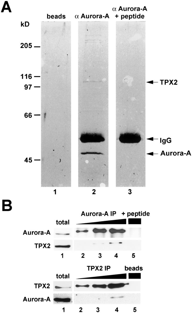
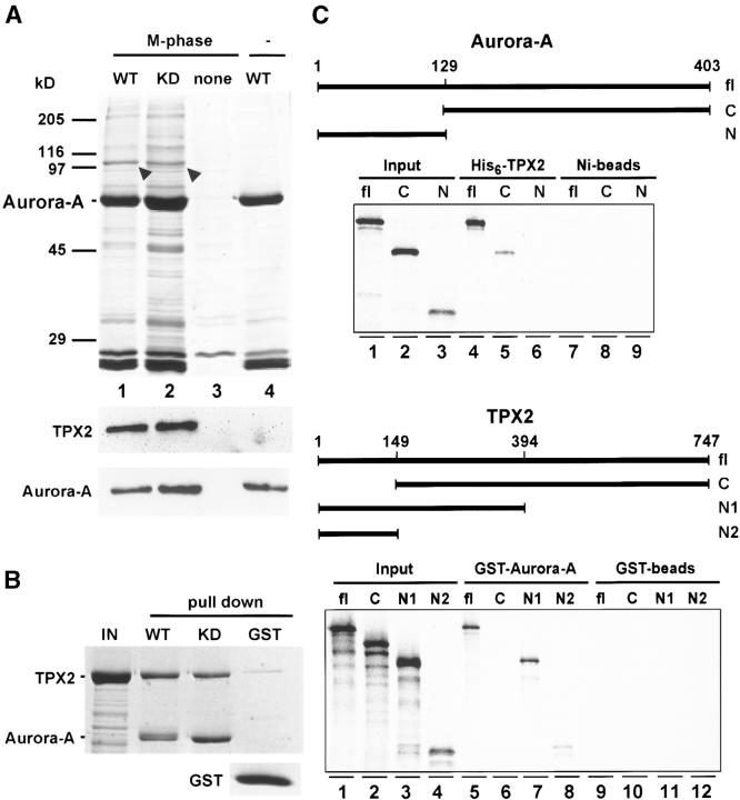
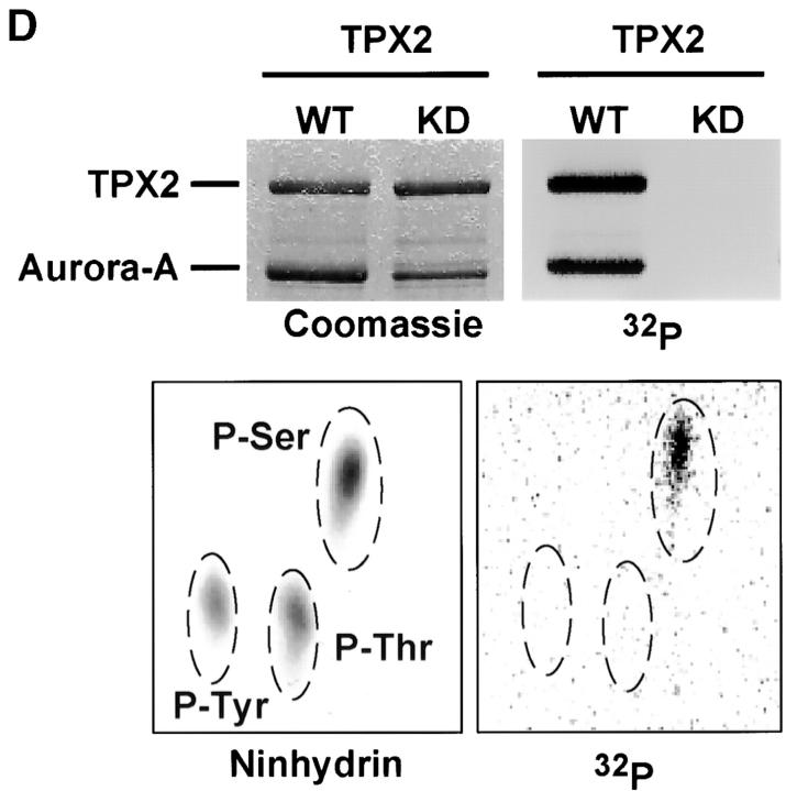
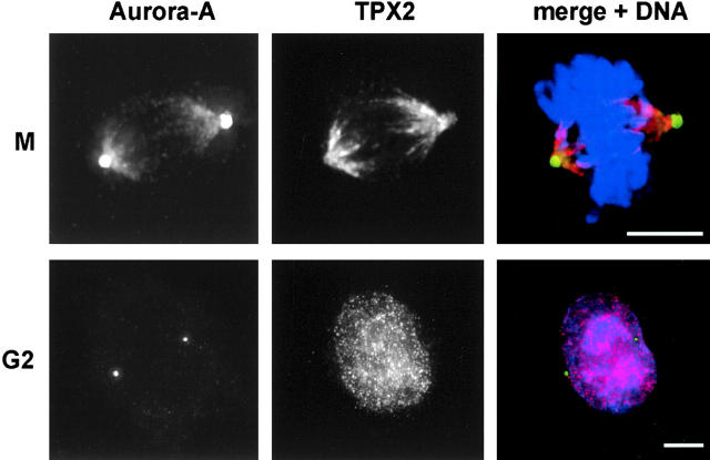
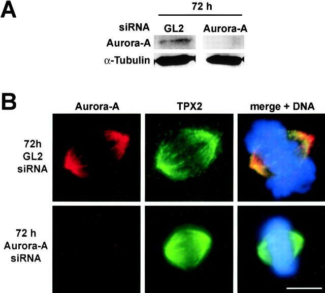
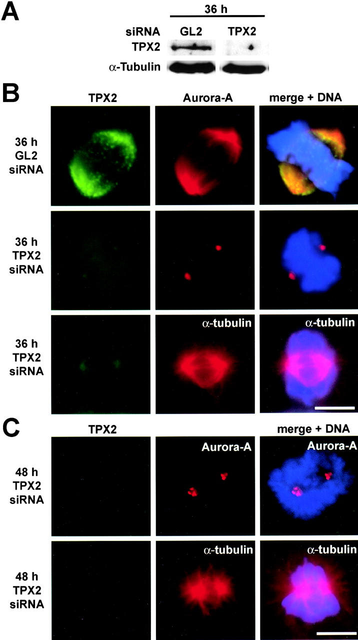
Aurora-A is a serine-threonine kinase implicated in the assembly and maintenance of the mitotic spindle. Here we show that human Aurora-A binds to TPX2, a prominent component of the spindle apparatus. TPX2 was identified by mass spectrometry as a major protein coimmunoprecipitating specifically with Aurora-A from mitotic HeLa cell extracts. Conversely, Aurora-A could be detected in TPX2 immunoprecipitates. This indicates that subpopulations of these two proteins undergo complex formation in vivo. Binding studies demonstrated that the NH2 terminus of TPX2 can directly interact with the COOH-terminal catalytic domain of Aurora-A. Although kinase activity was not required for this interaction, TPX2 was readily phosphorylated by Aurora-A. Upon siRNA-mediated elimination of TPX2 from cells, the association of Aurora-A with the spindle microtubules was abolished, although its association with spindle poles was unaffected. Conversely, depletion of Aurora-A by siRNA had no detectable influence on the localization of TPX2. We propose that human TPX2 is required for targeting Aurora-A kinase to the spindle apparatus. In turn, Aurora-A might regulate the function of TPX2 during spindle assembly.


Figures

Figure 1.
Figure 1.
Identification of TPX2 as an Aurora-A–interacting protein. (A) Coomassie blue–stained gel of Aurora-A immunoprecipitate from mitotic HeLa cell extract (lane 2). For control, immunoprecipitations were also performed with either protein A–coated beads alone (lane 1) or Aurora-A antibody blocked with an excess of antigenic peptide (lane 3). Arrows point to the precipitated 45-kD Aurora-A protein, IgG heavy chain, and the 100-kD protein identified as TPX2 by tryptic peptide fingerprinting. The identity of the high molecular weight proteins coprecipitating with Aurora-A is currently under investigation. (B, top) Increasing amounts of Aurora-A immunoprecipitates (lanes 2–4) were separated by SDS-PAGE and analyzed by Western blotting using antibodies against Aurora-A and TPX2. The control immunoprecipitation was performed with the peptide blocked Aurora-A antibody (lane 5). (B, bottom) TPX2 coimmunoprecipitates were analyzed by Western blotting using antibodies against Aurora-A and TPX2. Beads alone were used for the control precipitation (lane 5). Total cell lysate was analyzed in parallel (lane 1).
Figure 2.
Figure 2.
TPX2 physically interacts with Aurora-A and is phosphorylated on serine. (A, top) Immobilized recombinant Aurora-A WT and KD proteins were incubated for 1 h at 4°C in mitotic HeLa cell extract, washed in LS buffer, and analyzed by SDS-PAGE followed by Coomassie blue staining (lanes 1 and 2). Controls show the results of incubating extract with beads alone (lane 3) and of loading WT Aurora-A protein without extract incubation (lane 4). Arrowheads indicate the prominent 100-kD protein that was identified as TPX2 by Western blotting. (A, bottom) Western blots were performed on the same samples using antibodies against TPX2 and Aurora-A. (B) In vitro binding of recombinant TPX2 (IN) to immobilized GST-tagged Aurora-A WT and KD proteins, or GST for control. (C) Mapping the interaction domains on Aurora-A and TPX2. Fragments of Aurora-A and TPX2, as indicated schematically, were produced by IVT, and binding to His6-TPX2 (top) or GST-Aurora-A (bottom) was assayed. Protein complexes were recovered on Ni-Sepharose beads and glutathione beads, respectively; Ni-Sepharose beads or GST-coated glutathione beads were used for control. Autoradiographs show corresponding amounts of input samples and proteins recovered in protein complexes. (D) In vitro kinase assay was performed with recombinant GST-tagged Aurora-A and recombinant human TPX2 in the presence of [γ-32P]ATP. Both the Coomassie blue–stained gel (left) and the autoradiograph (right) are shown. (D, bottom) Phosphoamino acid analysis of 32P-labeled TPX2 (right); ninhydrin staining (left) shows the migration of phosphoamino acid standards.
Figure 2.
Figure 2.
TPX2 physically interacts with Aurora-A and is phosphorylated on serine. (A, top) Immobilized recombinant Aurora-A WT and KD proteins were incubated for 1 h at 4°C in mitotic HeLa cell extract, washed in LS buffer, and analyzed by SDS-PAGE followed by Coomassie blue staining (lanes 1 and 2). Controls show the results of incubating extract with beads alone (lane 3) and of loading WT Aurora-A protein without extract incubation (lane 4). Arrowheads indicate the prominent 100-kD protein that was identified as TPX2 by Western blotting. (A, bottom) Western blots were performed on the same samples using antibodies against TPX2 and Aurora-A. (B) In vitro binding of recombinant TPX2 (IN) to immobilized GST-tagged Aurora-A WT and KD proteins, or GST for control. (C) Mapping the interaction domains on Aurora-A and TPX2. Fragments of Aurora-A and TPX2, as indicated schematically, were produced by IVT, and binding to His6-TPX2 (top) or GST-Aurora-A (bottom) was assayed. Protein complexes were recovered on Ni-Sepharose beads and glutathione beads, respectively; Ni-Sepharose beads or GST-coated glutathione beads were used for control. Autoradiographs show corresponding amounts of input samples and proteins recovered in protein complexes. (D) In vitro kinase assay was performed with recombinant GST-tagged Aurora-A and recombinant human TPX2 in the presence of [γ-32P]ATP. Both the Coomassie blue–stained gel (left) and the autoradiograph (right) are shown. (D, bottom) Phosphoamino acid analysis of 32P-labeled TPX2 (right); ninhydrin staining (left) shows the migration of phosphoamino acid standards.
Figure 3.
Figure 3.
TPX2 and Aurora-A colocalize on the spindle. Indirect immunofluorescence micrographs of HeLa S3 cells stained for Aurora-A (left) and TPX2 (middle). A merge of the two images together with DAPI staining of DNA is shown in the right panel. The top row shows a representative metaphase cell; the bottom row shows a representative G2 phase cell. Bars, 10 μm.
Figure 4.
Figure 4.
TPX2 localization is independent of Aurora-A. (A) Western blot analysis of total extracts from HeLa cells treated for 72 h with a siRNA duplex targeting Aurora-A or a control duplex (GL2). Filters were probed for Aurora-A and α-tubulin as a loading control. (B) HeLa S3 cells were treated for 72 h with an Aurora-A–specific siRNA duplex (top row) or a control duplex (GL2; bottom row) and then fixed and stained with antibodies against Aurora-A (left) and TPX2 (middle). A merge of the two images together with DAPI staining of DNA is shown on the right. Bar, 10 μm.
Figure 5.
Figure 5.
TPX2 localizes Aurora-A to the spindle. (A) Western blot analysis of total extracts form HeLa cells treated for 36 h with a TPX2-specific siRNA duplex or a control duplex (GL2). Filters were probed for Aurora-A and α-tubulin as a loading control. (B and C) HeLa S3 cells were treated for 36 (A) or 48 h (B) with TPX2-specific siRNA duplex or a control duplex (GL2) and then fixed and stained with the antibodies indicated. Right panels show merged images, including DNA staining by DAPI. Bars, 10 μm.

References

    1. Bischoff, J., and G.D. Plowman. 1999. The Aurora/Ipl1p kinase family: regulators of chromosome segregation and cytokinesis. Trends Cell Biol. 9:454–459. - PubMed
    1. Bischoff, J.R., L. Anderson, Y. Zhu, K. Mossie, L. Ng, B. Souza, B. Schryver, P. Flanagan, F. Clairvoyant, C. Ginther, et al. 1998. A homologue of Drosophila aurora kinase is oncogenic and amplified in human colorectal cancers. EMBO J. 17:3052–3065. - PMC - PubMed
    1. Boyle, W.J., G.P. van der Geer, and T. Hunter. 1991. Phosphopeptide mapping and phosphoamino acid analysis by two-dimensional separation on thin-layer cellulose plates. Methods Enzymol. 201:110–149. - PubMed
    1. Elbashir, S., J. Harborth, W. Lendeckel, A. Yalcin, K. Weber, and T. Tuschl. 2001. Duplexes of 21-nucleotide RNAs mediate RNA interference in cultured mammalian cells. Nature. 411:494–498. - PubMed
    1. Giet, R., and C. Prigent. 1999. Aurora/Ipl1p-related kinases, a new oncogenic family of mitotic serine-threonine kinases. J. Cell Sci. 112:3591–3601. - PubMed

MeSH terms