. 2002 Aug 15;22(16):6980-90.
doi: 10.1523/JNEUROSCI.22-16-06980.2002.

Kalirin Dbl-homology guanine nucleotide exchange factor 1 domain initiates new axon outgrowths via RhoG-mediated mechanisms

Affiliations

Kalirin Dbl-homology guanine nucleotide exchange factor 1 domain initiates new axon outgrowths via RhoG-mediated mechanisms

Victor May et al. J Neurosci. .

Abstract

The large multidomain Kalirin and Trio proteins containing dual Rho GTPase guanine nucleotide exchange factor (GEF) domains have been implicated in the regulation of neuronal fiber extension and pathfinding during development. In mammals, Kalirin is expressed predominantly in the nervous system, whereas Trio, broadly expressed throughout the body, is expressed at a lower level in the nervous system. To evaluate the role of Kalirin in fiber initiation and outgrowth, we microinjected cultured sympathetic neurons with vectors encoding Kalirin or with Kalirin antisense oligonucleotides, and we assessed neuronal fiber growth in a serum-free, satellite cell-free environment. Kalirin antisense oligonucleotides blocked the continued extension of preexisting axons. Kalirin overexpression induced the prolific sprouting of new axonal fibers that grew at the normal rate; the activity of Kalirin was entirely dependent on the activity of the first GEF domain. KalGEF1-induced sprouting of new fibers from lamellipodial structures was accompanied by extensive actin cytoskeleton reorganization. The kalGEF1 phenotype was mimicked by constitutively active RhoG and was blocked by RhoG inhibitors. Constitutively active Rac1, RhoA, and Cdc42 were unable to initiate new axons, whereas dominant-negative Rac1, RhoA, and Cdc42 failed to block axon sprouting. Thus Kalirin, acting via RhoG in a novel manner, plays a central role in establishing the morphological phenotypic diversity that is essential to the connectivity of the developing nervous system.


Figures

Fig. 1.
Fig. 1.
Kalirin and Trio. The structures of two isoforms of rat Kalirin (rKalirin-12; accession number AF232669;rKalirin-9; accession number AF232668), human Trio (hTrio; accession number AAC34245), and dTrio (accession number AB035419) are drawn to scale. Kalirin and Trio possess two distinct Dbl-homology (DH)/pleckstrin-homology (PH) domains typical of GEFs for Rho GTPases along with a putative serine/threonine kinase domain. In addition, both hTrio and rKalirin possess a Sec14p domain, multiple spectrin-like repeats, SH3 domains, and Ig/fibronectin III (FN3) domains. Among the Kalirin isoforms that arise from alternative splicing, Kalirin-12 protein is structurally similar to hTrio. Kalirin-9, like dTrio, is devoid of the kinase domain; Kalirin-7 lacks the second GEF domain, and GEF1 is followed instead by a C-terminal postsynaptic density-95/discs large/zona occludens-1-binding motif mediating Kalirin-7 enrichment in postsynaptic density fractions (Penzes et al., 2000, 2001).
Fig. 2.
Fig. 2.
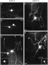
Kalirin initiates SCG fiber outgrowth.A, Control cultured SCG sympathetic neuron 72 hr after microinjection with 200 ng/μl EGFP-plasmid alone; two fibers project from the soma, with one branching a short distance from the soma.B, C, Sequential micrographs of neurons coinjected with EGFP and Kalirin-9 plasmid (200 ng/μl each). New fiber outgrowth is apparent when the 24 and 48 hr postinjection micrographs are compared (B, C). A burst of neuronal fiber initiation and extension is observed in Kalirin-9-expressing neurons between 24 and 48 hr. Cy3 immunofluorescence staining for the myc-epitope was performed at the end of the experiment to demonstrate Kalirin-9 expression in neurons with fiber outgrowth (inset, cell1). D, E, Sequential micrographs of a single neuron coinjected with EGFP and Kalirin-12 plasmids (200 ng/μl each). Like for Kalirin-9, Kalirin-12 expression induced the outgrowth of new fibers and branches. After 24 hr new fibers could be identified by bright EGFP fluorescent growth cones at distal fiber tips (asterisk). Comparison to the same Kalirin-12-expressing neuron 48 hr after injection reveals new fibers (arrowhead) and a high density of growth cones emerging from extensive fiber branching. Kalirin-12 expression in the injected neuron with outgrowth phenotype was demonstrated by staining formyc-epitope; staining was confined to the soma (inset, cell 2).
Fig. 3.
Fig. 3.
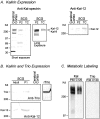
SCG contains more Kalirin than Trio.A, Detergent extracts (30 μg of total protein) of adult rat cerebral cortex, P2 SCG, or 10 d SCG cultures (TC) were fractionated on 4–15% gradient gels, transferred to polyvinylidene difluoride (PVDF) membranes, and visualized by Western blot analysis by using kal-spectrin antibody and enhanced chemiluminescent reagents; a longer exposure is shown also. The bands <200 kDa were not visualized by Kalirin-7-specific antibodies (Penzes et al., 2001). Molecular weight markers are indicated on the left. The same samples were visualized with an antiserum selective for the COOH terminus of Kalirin-12.B, Detergent extracts (30 μg of total protein) of adult or P1 SCG and adult rat cerebral cortex were fractionated on 5% gels and visualized by Western blot analysis with either the Kalirin-12 or Trio antibody; an aliquot of recombinant Trio was analyzed separately to demonstrate recovery and verify its molecular mass. The concentrations of the Kalirin-12 and Trio antibodies were chosen to yield equivalent signals when equimolar amounts of recombinant kalKinase and TrioKinase were analyzed. C, SCG neurons were incubated in defined serum-free medium containing [35S]Met for 30 min and harvested (pulse, 30 min;P30) or chased for an additional 120 min (chase, 120 min; C120) in serum-free medium. Cultures were extracted and immunoprecipitated with antibody to kal-spectrin (Kal) or Trio (Trio). After isolation with protein A-Sepharose beads, the samples were fractionated on 5% gels for fluorography (6d).
Fig. 4.
Fig. 4.
KalGEF1 domain expression is key to the Kalirin-induced fiber outgrowth phenotype. A, Microinjection of sympathetic neuron with 200 ng/μl kalGEF1 expression vector induced robust fiber outgrowth and branching (compare with Fig. 2A). Growth cones at distal fiber terminals appeared as broad lamellipodial sheets (arrowheads). B, C, A neuron injected with kalGEF1 and EGFP (B) and processed immunocytochemically to visualize the myc-epitope of the kalGEF1construct (C) demonstrated kalGEF1 expression in newly formed fibers and terminals. D, E, KalGEF1-induced fiber initiation resulted from extensive actin cytoskeleton reorganization. Micrographs of two different kalGEF1/EGFP-injected neurons (green) were merged with micrographs of the same neurons visualized with TRITC-phalloidin (red). The noninjected sympathetic neurons (D,asterisk) displayed relatively uniform staining for filamentous actin. Expression of kalGEF1 caused a redistribution of filamentous actin to emerging fiber outgrowths (D;arrowheads mark red lamellipodial filigree emerging from green microinjected neuron) and to large aggregates in the perinuclear region of the cell soma (yellow represents actin aggregates fromgreen and red fluorescence overlay). At later stages of fiber development the kalGEF1-injected neurons typically displayed prominent staining for filamentous actin (red) at growth cones (E, arrowheads); aggregates of filamentous actin were still apparent in neuronal soma (asterisk).
Fig. 5.
Fig. 5.
Kalirin GEF2 and kinase domains do not induce fiber outgrowth. Microinjection of kalGEF2, kalKinase, or kal9ΔGEF1 did not induce changes in sympathetic neuronal phenotype.A, A neuron injected with kalGEF2 and EGFP was processed immunocytochemically to visualize the myc-epitope of the kalGEF2 construct; the EGFP image (green) was merged with the Cy3 image (red) for the epitope (yellow, from green andred Cy3 overlay). No changes in phenotype were noted in kalGEF2-expressing neurons when compared with control despite high levels of myc-expression. B, Two kalKinase/EGFP-injected neurons (green) with high levels of myc-epitope expression (yellow) failed to demonstrate an outgrowth phenotype. C, Similarly, expression of a Kalirin-9 GEF1 deletion construct in sympathetic neurons did not elicit the fiber initiation phenotype despite high myc-expression levels (yellow). The patchy staining for kalKinase in the processes was a reproducible observation. Scale bars, 25 μm.
Fig. 6.
Fig. 6.
KalGEF1-induced fiber extensions are axons.A–C, Two sympathetic neurons microinjected with kalGEF1 and EGFP were photographed 24, 48, and 72 hr after injection. Fibers initiated after kalGEF1 expression extended rapidly over time. Note the continued emergence of some new fibers even 48 and 72 hr after injection. D, KalGEF1-induced fibers contained neuropeptide vesicles. Neurons were coinjected with kalGEF1 and NPY-EGFP constructs. Secretory granules containing the NPY-EGFP fusion protein were visualized on the basis of the localization of EGFP; proteins were routed from cellular sites of biosynthesis to growth cones of newly formed fibers (arrowheads; punctategreen fluorescent endings). E, Sholl analysis of Kalirin-initiated fibers and kalGEF1-initiated fibers 2–3 d after microinjection. Control EGFP-injected neurons (CTL;n = 24) demonstrated a slight increase in fiber crossings at distant intervals from the branching of principal fibers. Fiber outgrowths from kalGEF1-injected neurons (n = 14) produced a ninefold increase in fiber crossings (50 μm distance) that diminished to control levels as the Sholl radii exceeded the lengths of the newly formed fibers.Post hoc Student–Newman–Keuls analyses revealed significant differences from CTL at 25–175 μm distances (p < 0.001). Kalirin-injected neurons produced a sixfold increase in intersections (n = 9). Fiber lengths were longer than kalGEF1-injected neurons but variable; differences in the number of fiber crossings compared with control were significant for all radial points (p < 0.001). Data are mean ± SEM, from counting fiber intersections for many neurons. Scale bars, 50 μm.
Fig. 7.
Fig. 7.
Kalirin-induced neuronal fiber outgrowth is mediated by RhoG. A–D, Constitutively active Rho GTPases (Rac1-Q61L, RhoA-Q62L, Cdc42-Q61L, and Rho G12V; all at 200 ng/μl) were microinjected, and the neurons were examined 48 hr later. Only the microinjection of the active RhoG G12V induced the fiber outgrowth phenotype resembling that observed for Kalirin or kalGEF1.E–G, Coinjection of neurons with kalGEF1 (50 ng/μl) and the RhoG competitive inhibitor RhoG F37A (F; 200 ng/μl) or RhoGIP122, a RhoG-GTP binding protein (G; 200 ng/μl), either diminished or blocked, respectively, the fiber outgrowth phenotype observed for kalGEF1 alone (E); the cells were photographed 48 hr after injection. H–J, RhoG F37A expression also blocked Kalirin-12-induced fiber outgrowth. RhoG F37A fluorescence (H; EGFP, green) andmyc-epitope staining for Kalirin-12 (I; Cy3, red) are merged in J; new fiber extensions were never observed despite high levels ofmyc-expression. Scale bar, 50 μm.
Fig. 8.
Fig. 8.
SCG contains RhoG, which binds to KalGEF1.A, Binding of kalGEF1 to nucleotide-depleted Rho proteins. KalGEF1 expressed transiently in pEAK Rapid HEK-293 cells was extracted for use in binding studies. Purified GST-Rho fusion proteins were bound to glutathione agarose beads to which aliquots of kalGEF1-containing extracts were applied. The bound fraction, representing 40× as much material as the input sample, was visualized via its NH2-terminal myc-epitope. The bound GST fusion proteins were visualized by Coomassie staining. Shown are representative data from four independent experiments. The data fromA were quantified, and the ratio of bound kalGEF1 to GST-Rho protein is plotted (bottom panel).B, RhoG activation assay. GST-RhoGIP (10 μg) immobilized to glutathione-Sepharose beads (25 μl) was incubated with extracts from cells expressing EGFP-RhoG G12V (CA RhoG) and EGFP, EGFP-RhoG and EGFP (Control), and EGFP-RhoG and KalGEF1 (KalGEF1). After being washed, the sample was analyzed by Western blot analysis, using a RhoG antiserum.C, Rho family members in SCG. Detergent extracts (30 μg of total protein) of adult rat cerebral cortex, P2 SCG, and adult rat liver were fractionated on 4–15% gels; the proteins were transferred to PVDF membranes and visualized by Western blot analysis by using antisera to the indicated Rho family members. Some Rho subfamilies are similar, and the specificity of the RhoA antibody was not evaluated in these tissues.
Fig. 9.
Fig. 9.
Endogenous Kalirin modulates sympathetic axonal fiber development. Sympathetic axonal fiber extension was arrested in neurons microinjected with Kalirin antisense oligonucleotides; fibers and terminals appeared static over time (A, B). Of 63 neurons that followed after Kalirin antisense oligonucleotide injection, 21 neurons showed axon growth arrest or slight axonal retraction (as in A, B). In contrast, none of the 12 neurons injected with scrambled control oligonucleotides showed growth arrest, instead growing steadily like the neurons in Figures 2A and 7A–C. Kalirin antisense oligonucleotides had no effect on neuronal survival and did not impede constitutively active RhoG-induced neuronal outgrowth (C).
Fig. 10.
Fig. 10.
Endogenous Kalirin maintains dendritic outgrowth and development. A small population of sympathetic neurons developed dendrites identified by MAP2 staining via a Cy3-conjugated secondary antibody (A, inset;asterisk). Characteristic neuronal dendrites ceased further development and demonstrated retraction after injection with Kalirin antisense oligonucleotides (compare A, B withC, D). Data are representative of the nine dendritic profiles that were examined, all of which retracted with a Kalirin antisense oligonucleotide injection. Scale bar (major tick): 15 μm.

Similar articles

Cited by

References

    1. Acebes A, Ferrús A. Cellular and molecular features of axon collaterals and dendrites. Trends Neurosci. 2000;23:557–565. - PubMed
    1. Alam MR, Caldwell BD, Johnson RC, Darlington DN, Mains RE, Eipper BA. Novel proteins that interact with the COOH-terminal cytosolic routing determinants of an integral membrane peptide-processing enzyme. J Biol Chem. 1996;271:28636–28640. - PubMed
    1. Alam MR, Johnson RC, Darlington DN, Hand TA, Mains RE, Eipper BA. Kalirin, a cytosolic protein with spectrin-like and GDP/GTP exchange factor-like domains that interacts with peptidylglycine α-amidating monooxygenase, an integral membrane peptide-processing enzyme. J Biol Chem. 1997;272:12667–12675. - PubMed
    1. Awasaki T, Saito M, Sone M, Suzuki E, Sakai R, Ito K, Hama C. The Drosophila Trio plays an essential role in patterning of axons by regulating their directional extension. Neuron. 2000;26:119–131. - PubMed
    1. Bateman J, Shu H, Van Vactor D. The guanine nucleotide exchange factor Trio mediates axonal development in the Drosophila embryo. Neuron. 2000;26:93–106. - PubMed

Publication types

MeSH terms