. 2002 Aug 15;22(16):7065-79.
doi: 10.1523/JNEUROSCI.22-16-07065.2002.

Regulation of structural plasticity by different channel types in rod and cone photoreceptors

Affiliations

Regulation of structural plasticity by different channel types in rod and cone photoreceptors

Nan Zhang et al. J Neurosci. .

Abstract

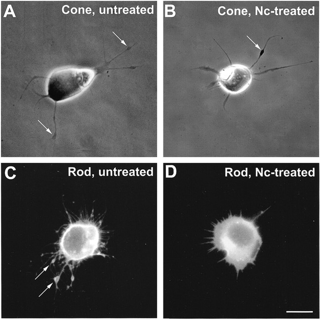
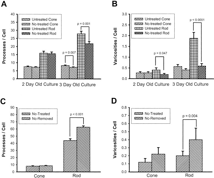
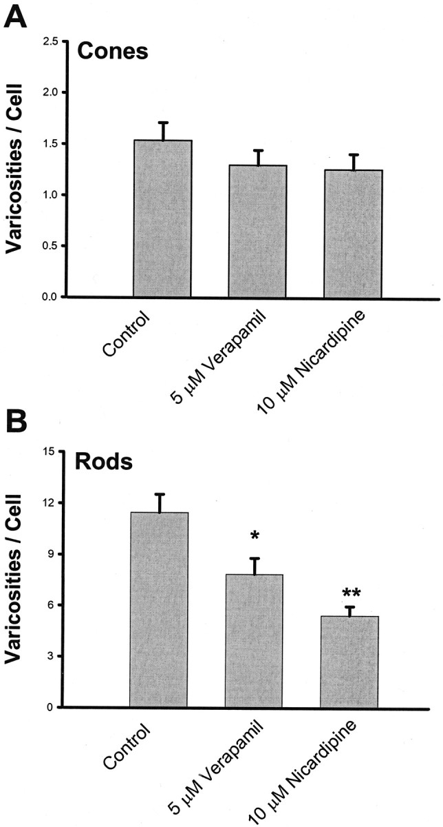
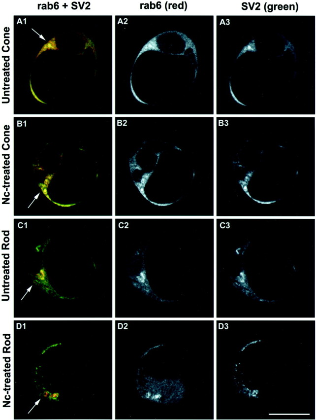
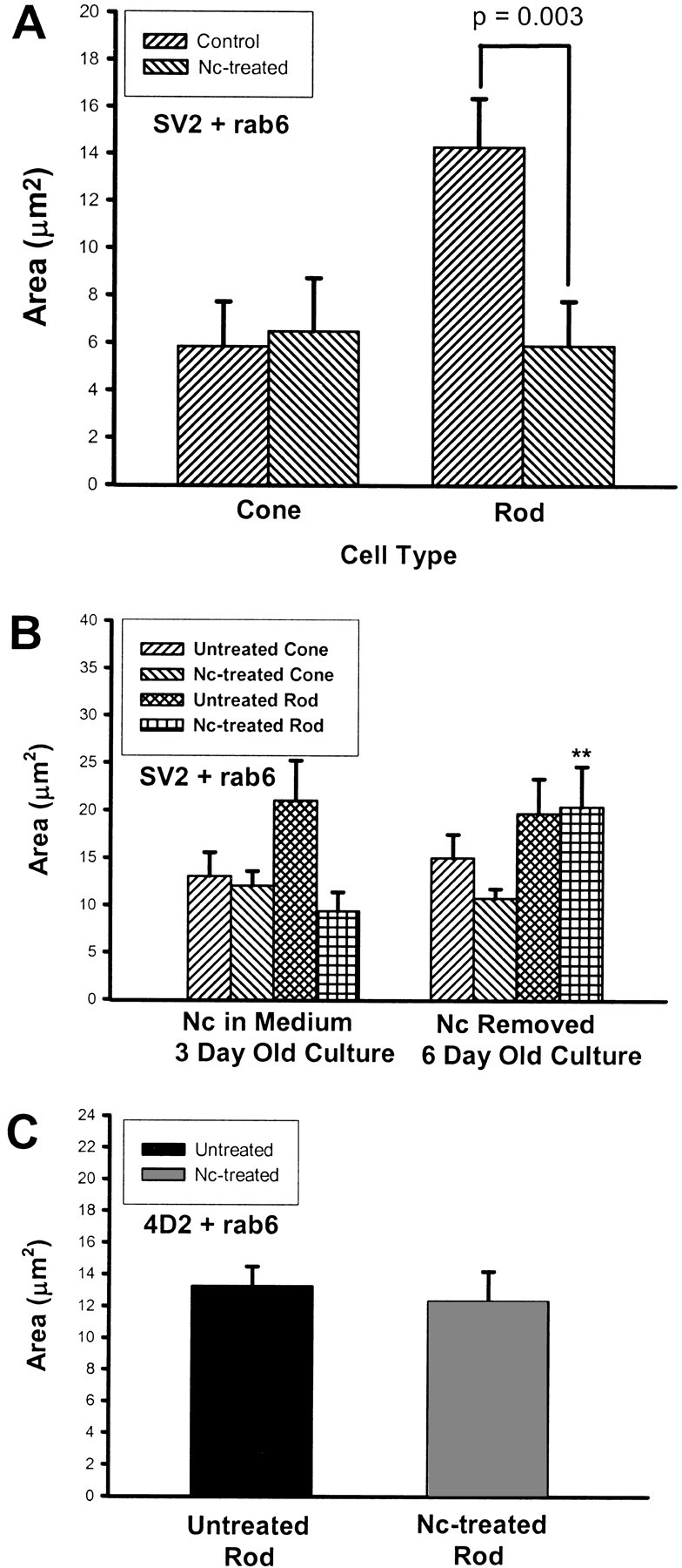
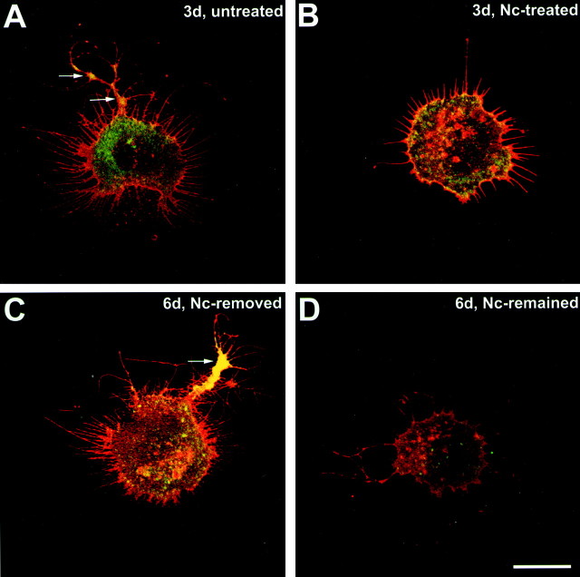
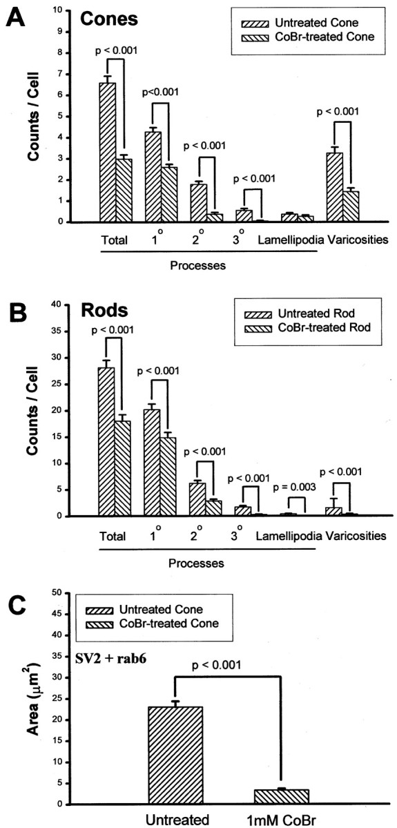
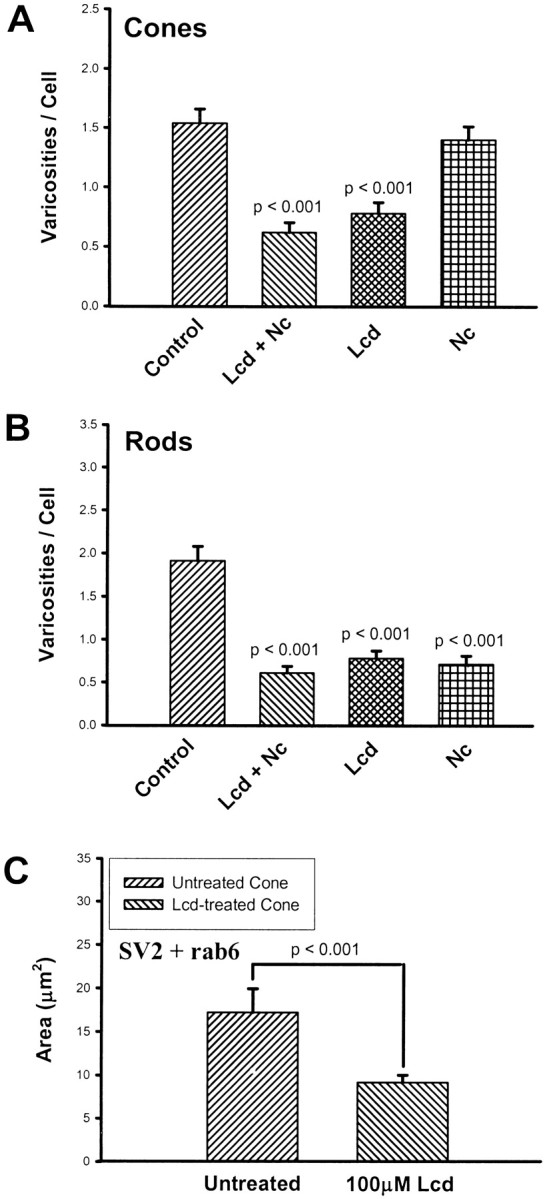
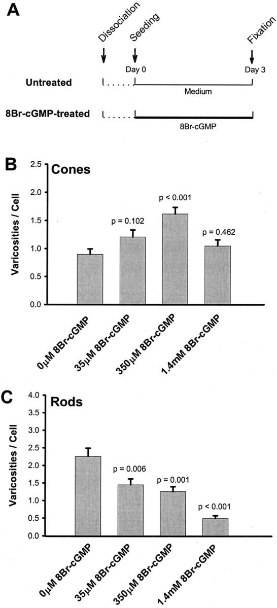
In response to retinal disease and injury, the axon terminals of rod photoreceptors demonstrate dramatic structural plasticity, including axonal retraction, neurite extension, and the development of presynaptic varicosities. Cone cell terminals, however, are relatively inactive. Similar events are observed in primary cultures of salamander photoreceptors. To investigate the mechanisms underlying these disparate presynaptic responses, antagonists to voltage-gated L-type and cGMP-gated channels, known to be present on rod and cone cell terminals, respectively, were used to block calcium influx during critical periods of plasticity in vitro. In rod cells, L-type channel antagonists nicardipine and verapamil inhibited not only the outgrowth of processes and the formation of varicosities, but also the synthesis of vesicle proteins, SV2 and synaptophysin. In contrast, the synthesis of opsin in rod cells was unaffected. In cone cells, L-type channel antagonists caused only modest changes. However, cobalt bromide, which blocks all calcium channels, and l-cis-diltiazem, a potent antagonist of cGMP-gated channels, significantly inhibited varicosity formation and synthesis of SV2 in cone cells. Moreover, the cGMP-gated channel agonist 8-bromo-cGMP caused a significant increase in varicosity formation by cone but not rod cells. Thus voltage-gated L-type channels in rod cells and cGMP-gated channels in cone cells are the primary calcium channels required for structural plasticity and the accompanying upregulation of synaptic vesicle synthesis. The differing responses of rod and cone terminals to injury and disease may be determined by these differences in the regulation of Ca2+ influx.


Figures

Fig. 1.
Fig. 1.
Experimental design for administration of channel blockers. Photoreceptors were cultured with channel blockers for 48 hr after seeding or after 1 d in culture. Some photoreceptors were cultured for an additional 3 d after channel blockers were removed from the culture medium.
Fig. 2.
Fig. 2.
Morphological changes of photoreceptors treated with nicardipine. A, Phase-contrast microscopy of an untreated cone cell. Neuritic processes with branching structures were present. Varicosities (arrows) had formed along the processes. B, Nc-treated cone cell. Processes showed fewer branches, but varicosities (arrow) had still formed along the processes. C, Fluorescence microscopy of an untreated rod cell labeled with rod opsin antibody. The rod cell showed prominent process outgrowth with obvious branching. Multiple varicosities (arrows) had formed along the thicker processes, the neurites. D, Nc-treated rod cell labeled with rod opsin antibody. The rod cell showed fewer processes and no varicosities. Cells were from 3-d-old cultures; Nc treatment started after 1 d in culture. Scale bar, 10 μm.
Fig. 3.
Fig. 3.
Effect of nicardipine (Nc) treatment and removal on the growth of processes and varicosities by photoreceptors as determined by quantitative analysis. A, Growth of neuritic processes. Nc treatment caused a significant decrease in the total number of processes per cell as compared with controls in both cone and rod cells from 3-d-old cultures. B, Growth of varicosities. Nc-treated rod cells from both 2- and 3-d-old cultures showed a significant decrease in the number of varicosities as compared with controls. The decrease in cone cells did not reach statistical significance. C, Growth of neuritic processes after removal of Nc. Nc was removed from the medium, and photoreceptors were cultured for an additional 3 d; there was a prominent growth of processes in rod cells, but no change in the total number of processes from cone cells. D, Growth of varicosities after removal of Nc. After Nc was removed from the medium and photoreceptors were cultured for another 3 d, there was also a prominent increase in the number of varicosities in rod cells. In cone cells, no significant growth of processes or varicosities was seen after removal of Nc. A total of 880 cells in 14 cultures from three animals were analyzed in A and B, and a total of 250 cells in 10 cultures from one animal were analyzed inC and D.
Fig. 4.
Fig. 4.
Effect of verapamil treatment on the growth of varicosities by photoreceptors in 3-d-old cultures. A, Growth of varicosities by cone cells. Neither 5 μm Vrp nor 10 μm Nc caused significant inhibition of varicosity formation in cone cells. B, Growth of varicosities by rod cells. Both 5 μm Vrp and 10 μm Nc caused significant inhibition of varicosity formation in rod cells. A total of 300 cells in eight cultures from one animal were analyzed. *p < 0.05; **p < 0.01.
Fig. 5.
Fig. 5.
Diagram of expected Golgi labeling in the presence and absence of protein synthesis. Left panel, Double staining of the Golgi apparatus by SV2 (green) and rab6 (red). When SV2, an integral membrane protein of all vesicles, is being synthesized, it appears in the Golgi as well as other locations in the cell. Rab6 is present only in thetrans-Golgi network and post-Golgi vesicles. Double staining (yellow) indicates the presence of SV2 and rab6 in the TGN. Yellow stain will only appear when SV2 is being actively synthesized and is passing through the TGN. Right panel, When no SV2 is synthesized, there is little double staining. SV2 may be present in the cell soma but is in low levels in the Golgi apparatus. Rab6 staining occurs in thetrans-Golgi, and a red body appears in the cell soma. Thus, the area of double staining gives an indication of the amount of SV2 synthesis.
Fig. 6.
Fig. 6.
Photoreceptors from 3-d-old cultures double stained with rab6 and SV2 antibodies. Optical sections (1 μm) were obtained at a level showing the Golgi apparatus. Images are displayed in redgreenblue(A1, B1, C1, andD1), red (A2,B2, C2, and D2), andgreen channels (A3, B3,C3, and D3), respectively.A1A3, Untreated cone. The Golgi apparatus and post-Golgi vesicles were labeled by rab6 and SV2 antibodies and appeared yellow (arrow), indicating the synthesis of SV2. B1B3, Nc-treated cone. Nc treatment did not cause an obvious change of rab6 and SV2 staining in the Golgi apparatus, suggesting the continued presence of SV2 in the TGN (arrow).C1C3, Untreated rod. Both rab6 and SV2 antibodies stained the trans-Golgi apparatus, which appears yellow (arrow). SV2 staining (green) is also present in other locations in the cell soma such as the cis-Golgi.D1D3, Nc-treated rod. Nc treatment reduced SV2 staining in the Golgi area. The arrowindicates the Golgi apparatus that showed rab6 label (red), but SV2 staining (green) occurred primarily in areas outside the Golgi apparatus. This suggests that SV2 synthesis had been reduced but that SV2, perhaps in old synaptic vesicles, continues to be present in the cell soma. Scale bar, 10 μm.
Fig. 7.
Fig. 7.
The effects of application and removal of nicardipine (Nc) on SV2 and opsin synthesis in photoreceptors as measured with colocalization analysis. Area is derived from the number of pixels labeled by rab6 and either SV2 or opsin antibodies. A, SV2 and rab6 labeling in cone and rod cells from 3-d-old cultures that had been treated with Nc after 1 d in culture. In rod cells, the Nc treatment significantly reduced the SV2 labeling in the Golgi, resulting in a smaller average area of colocalized staining. There was no change in cone cells. A total of 120 cells in six cultures from three animals were analyzed.B, SV2 and rab6 labeling in cone and rod cells from 6-d-old cultures; Nc had been removed at day 3 (Fig. 1). The SV2 staining increased significantly in Nc-treated rod cells after Nc removal, increasing the areas of colocalization (**p < 0.01), but showed no significant change in Nc-treated cone cells. A total of 80 cells in four cultures from one animal were analyzed. C, Rod opsin (4D2) and rab6 labeling in rod cells from 3-d-old cultures. No change in the area of double labeling was observed in Nc-treated rod cells compared with untreated cells, suggesting that Nc had no effect on opsin synthesis. A total of 40 cells in four cultures from two animals were analyzed.
Fig. 8.
Fig. 8.
Effect of nicardipine treatment on synaptophysin labeling in photoreceptors. Rod cells were stained with rod opsin (red) and synaptophysin (green) antibodies. Confocal microscopic sections (1 μm) from a level containing processes and varicosities are shown. A, An untreated rod cell. The cell had prominent process outgrowth and several varicosities (arrows). Strong synaptophysin staining was present in both the soma and varicosities in the 3-d-old culture. B, Nc-treated rod cell. There was clear inhibition of process outgrowth and varicosity formation and weaker synaptophysin staining in the soma in the 3-d-old culture.C, A rod cell after Nc removal. The cell had resumed process growth and varicosity formation. Although the synaptophysin staining in the soma was still weak, strong synaptophysin staining was present in varicosities (arrow), suggesting a transport of synaptophysin to peripheral structures from the soma after Nc was removed in the 6-d-old culture. D, A rod cell treated with Nc for 5 d after 1 d in Nc-free culture. The prolonged treatment of rod cells with Nc caused a drastic decrease of synaptophysin staining in addition to the inhibition of growth in the 6-d-old culture. Scale bar, 10 μm.
Fig. 9.
Fig. 9.
Effect of nicardipine (Nc) treatment on synaptophysin staining as measured with densitometric analysis. The analysis was performed on 1 μm optical sections taken from a level showing processes and a level showing the Golgi apparatus.A, Experimental design for Nc application and Nc removal. Nc (10 μm) was added to and removed from the culture medium as indicated. B, Densitometry of cone cells. Cone cells did not show a significant decrease of synaptophysin staining intensity in Nc-treated, Nc-removed, and Nc-remained groups as compared with the untreated group at both levels. C, Densitometry of rod cells. Although rod cells from the Nc-treated group showed only a trend toward decreased synaptophysin staining intensity as compared with the untreated group, prolonged Nc treatment (Nc-Remained group) caused a significant decrease in synaptophysin staining intensity at both process and Golgi levels. Removal of Nc from the medium reduced the extent of this decrease (Nc-Removed group). A total of 160 cells in eight cultures from one animal were analyzed.
Fig. 10.
Fig. 10.
Effects of a 2 d application of cobalt bromide (CoBr) on process outgrowth, varicosity formation, and SV2 synthesis in 3-d-old cultures. A, Process outgrowth and varicosity formation in cone cells. Processes and anti-synaptophysin-labeled varicosities were counted. The CoBr application significantly reduced the growth of processes and varicosities in cone cells. A total of 200 cone cells in four cultures from one animal were analyzed. B, Process outgrowth and varicosity formation in rod cells. Processes and anti-synaptophysin-labeled varicosities were counted. The CoBr application also significantly reduced the growth of processes and varicosities in rod cells. A total of 200 rod cells in four cultures from one animal were analyzed. Rod and cone cells were identified by the presence and absence, respectively, of rod opsin antibody labeling.C, SV2 and rab6 labeling of cone cells. Cone cells treated with 1 mm CoBr showed an 85.3% reduction of the area of colocalized staining. A total of 40 cone cells in four cultures from two animals were analyzed.
Fig. 11.
Fig. 11.
Effects of a 2 d application of the cGMP-gated channel antagonistl-cis-diltiazem (Lcd) on varicosity formation and SV2 synthesis in 3-d-old cultures.A, Varicosity formation by cone cells. Varicosities were analyzed with phase-contrast microscopy. Lcd caused a 49.4% decrease in varicosity production; Lcd plus nicardipine (Lcd + Nc) also caused a significant decrease, but Nc alone caused no significant changes in varicosity formation by cone cells. A total of 400 cone cells in 16 cultures from two animals were analyzed.B, Varicosity formation by rod cells. Anti-opsin-stained varicosities were counted. Both Nc and Lcd caused significant decreases in varicosity formation by rod cells. A total of 400 rod cells in 16 cultures from two animals were analyzed. C, SV2 and rab6 immunocytochemical labeling of cone cells. Cone cells treated with Lcd showed a 46.8% reduction in the area of colocalized staining, suggesting a decrease in SV2 synthesis. A total of 40 cone cells in four cultures from one animal were analyzed.
Fig. 12.
Fig. 12.
Effect of cGMP-gated channel agonist 8Br-cGMP (an analog of cGMP) on the varicosity formation of photoreceptors.A, Experimental design for the administration of 8Br-cGMP. 8Br-cGMP was added to the culture medium immediately after cell plating. B, Varicosity formation by cone cells. Varicosities were analyzed with phase-contrast microscopy. 8Br-cGMP, at 350 mm, caused a significant increase, whereas the other two concentrations caused no significant changes in varicosity formation. C, Varicosity formation by rod cells. Anti-opsin-stained varicosities were counted. 8Br-cGMP caused a dose-dependent inhibition of varicosity formation by rod cells. A total of 800 cells in 16 cultures from two animals were analyzed.

Similar articles

Cited by

References

    1. Akopian A, Johnson J, Gabriel R, Brecha N, Witkovsky P. Somatostatin modulates voltage-gated K+ and Ca2+ currents in rod and cone photoreceptors of the salamander retina. J Neurosci. 2000;20:929–936. - PMC - PubMed
    1. Antony C, Cibert C, Géraud G, Santa Maria A, Maro B, Mayau V, Goud B. The small GTP-binding protein rab6 is distributed from medial Golgi to the trans-Golgi network as determined by a confocal microscopic approach. J Cell Sci. 1992;103:785–796. - PubMed
    1. Bech-Hansen NT, Naylor MJ, Maybaum TA, Pearce WG, Koop B, Fishman GA, Mets M, Musarella MA, Boycott KM. Loss-of-function mutations in a calcium-channel α1-subunit gene in Xp11.23 cause incomplete X-linked congenital stationary night blindness. Nat Genet. 1998;19:264–267. - PubMed
    1. Benfey M, Búnger UR, Vidal-Sanz M, Bray GM, Aguayo AJ. Axonal regeneration from GABAergic neurons in the adult rat thalamus. J Neurocytol. 1985;14:279–296. - PubMed
    1. Borst JGG, Sakmann B. Calcium influx and transmitter release in a fast CNS synapse. Nature. 1996;383:431–434. - PubMed

Publication types

MeSH terms

LinkOut - more resources