. 2002 Aug 15;22(16):7097-110.
doi: 10.1523/JNEUROSCI.22-16-07097.2002.

Long-lasting sprouting and gene expression changes induced by the monoclonal antibody IN-1 in the adult spinal cord

Affiliations

Long-lasting sprouting and gene expression changes induced by the monoclonal antibody IN-1 in the adult spinal cord

Florence M Bareyre et al. J Neurosci. .

Abstract

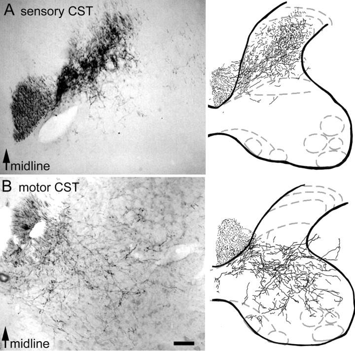
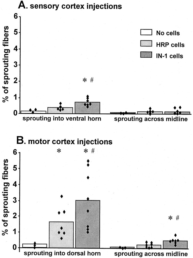
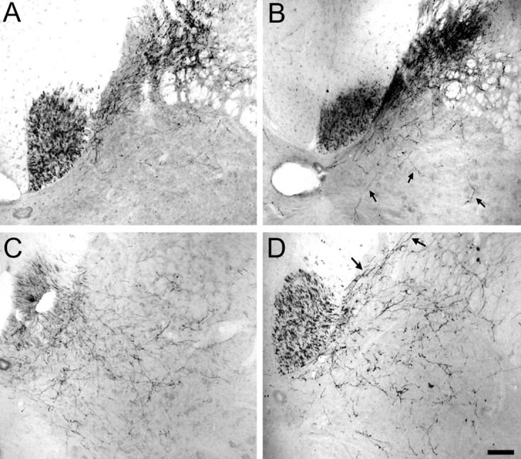
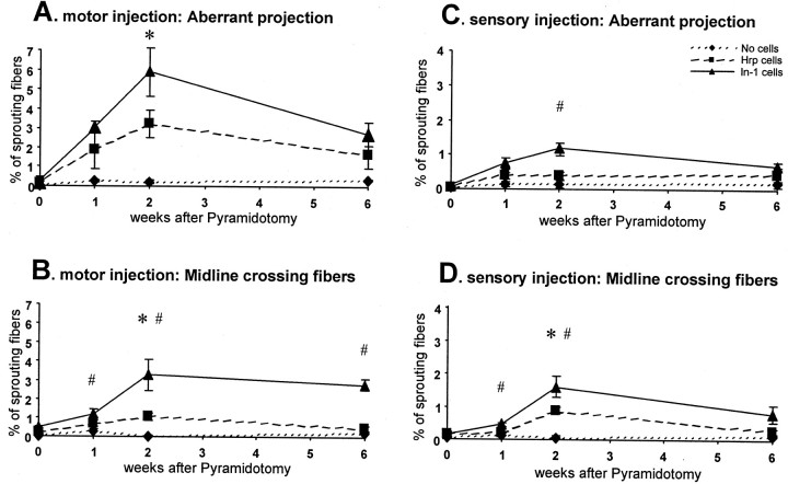
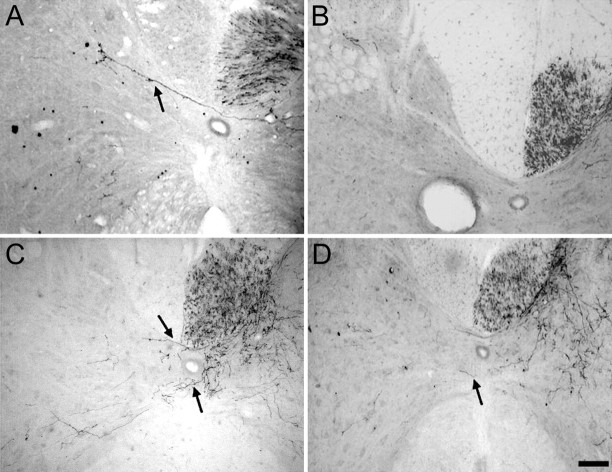
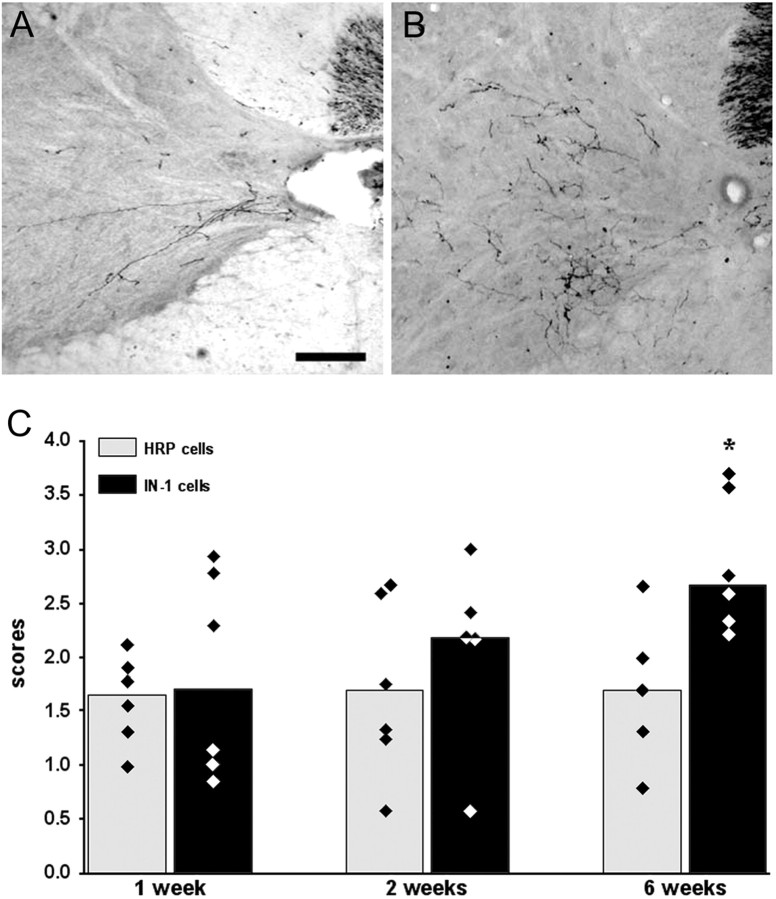
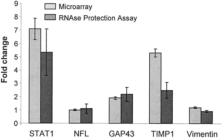
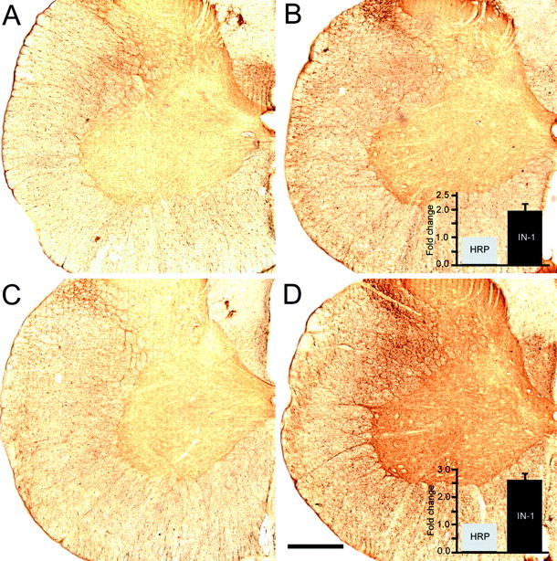
Lesion-induced plasticity of the rat corticospinal tract (CST) decreases postnatally, simultaneously with myelin appearance. In adult rats, compensatory sprouting can be induced by the monoclonal antibody (mAb) IN-1 raised against the growth inhibitory protein Nogo-A. In this study, we examined separately the fate of sensory and motor corticospinal fibers after mAb IN-1 application. Intact adult rats treated with the IN-1 antibody exhibited an increase of aberrant CST projections, i.e., sensory fibers projecting into the ventral horn and motor fibers projecting dorsally. Unilateral lesion of the CST [pyramidotomy (PTX)] in the presence of mAb IN-1 triggered a progressive reorganization of the sprouting of the remaining CST across the midline, with sensory fibers projecting gradually into the denervated dorsal horn and motor fibers projecting into the denervated ventral horn. In unilaterally denervated spinal cords, aberrant sprouts were only transient and disappeared by 6 weeks, whereas midline crossing fibers ending in the appropriate target region were stabilized and persisted over the entire study period. Within the spinal cord, IN-1 antibody treatment was associated with upregulation of growth factors (BDNF, VEGF), growth-related proteins (actin, myosin, GAP-43), and transcription factors (STATs), whereas pyramidotomy induced an enhanced expression of guidance molecules (semaphorins and slits) as well as neurotrophic factors (BDNF, IGFs, BMPs). These gene expression changes may contribute to attraction, guidance, and stabilization of sprouting CST fibers.


Figures

Fig. 1.
Fig. 1.
Photomicrographs and camera lucida reconstructions of corticospinal projections to the cervical spinal cord. A, Projections of sensory CST fibers to the dorsal horn as shown by injections of BDA into the sensory cortex.B, Projections of motor CST fibers to the intermediate layers and the ventral horn as shown by injections of BDA into the motor cortex. Scale bar, 250 μm.
Fig. 2.
Fig. 2.
Percentage of CST fibers sprouting across lamina V or across the midline in the cervical spinal cord in nonlesioned animals. A, Sensory CST projections. B, Motor CST projections. Monoclonal antibody IN-1 induced both sensory and motor fibers to sprout across lamina V when compared with anti-HRP mAb treatment. In addition, IN-1 induced motor fibers to cross the midline of the spinal cord. *IN-1 or HRP antibody-treated rats significantly different from control non-treated rats (p < 0.05). #IN-1 antibody-treated animals significantly different from anti-HRP antibody-treated rats (p < 0.05). Black diamondsindicate individual values.
Fig. 3.
Fig. 3.
Photomicrographs of fibers sprouting across lamina V in nonlesioned animals. A, No invasion of the ventral horn by sensory fibers after anti-HRP treatment. B, Invasion of the ventral horn by sensory fibers (arrows) after IN-1 treatment. C, No invasion of the dorsal horn by motor fibers after anti-HRP treatment. D, Pronounced invasion of the dorsal horn by motor fibers (arrows) after IN-1 treatment. Scale bar, 250 μm.
Fig. 4.
Fig. 4.
Long-term reorganization of motor (A, B) or sensory (C,D) CST fibers in pyramidotomized animals. IN-1 treatment induced more motor fibers to sprout dorsally (A) and more sensory fibers to invade the ventral horn on the intact side 2 weeks after pyramidal tract lesion than anti-HRP antibody treatment (C). IN-1 also induced motor and sensory fibers to cross the midline 1 and 2 weeks after pyramidal tract lesion, in contrast to anti-HRP treatment (B, D). Sensory sprouts retracted, but motor fibers grew across the midline to the denervated cord and were stable at 6 weeks after injury (B). ∗Anti-HRP-treated rats significantly different from control nontreated rats (p < 0.05). #IN-1 antibody-treated rats significantly different from anti-HRP-treated rats (p < 0.05).
Fig. 5.
Fig. 5.
Photomicrographs of fibers sprouting across the spinal cord midline 2 weeks after pyramidotomy. A, Topographic sprouting of a sensory CST fiber (arrow) into the contralateral, denervated dorsal horn after IN-1 mAb treatment. B, Absence of sprouting of sensory fibers after anti-HRP treatment. C, Topographic sprouting of motor CST fibers into the contralateral ventral horn (arrows) after IN-1 mAb treatment. D, Virtual absence of sprouting (very rare fibers) of motor fibers in anti-HRP treatment. Scale bar, 250 μm.
Fig. 6.
Fig. 6.
Photomicrographs of the arbor complexity of motor fibers grown across the midline into the denervated ventral horn at 2 weeks (A) and 6 weeks (B) after pyramidotomy and monoclonal IN-1 antibody treatment.C, Complexity of axonal arbors of motor CST fibers 1, 2, and 6 weeks after pyramidotomy and antibody treatment. Six weeks after the injury, IN-1-treated animals had higher scores than anti-HRP-treated animals. Scores are described in Materials and Methods. ∗IN-1-treated rats significantly different from anti-HRP-treated rats (p < 0.05).Black and white diamonds represent individual values. Scale bar, 200 μm.
Fig. 7.
Fig. 7.
Results from RNase protection assays done on cervical spinal cord tissue and compared with results from the analysis with the Affymetrix gene chip. Five different genes were similarly expressed using both assays in rats treated with mAb IN-1 for 7 d compared with rats treated with an anti-HRP antibody.
Fig. 8.
Fig. 8.
Photomicrographs of GAP-43 immunoreactivity in the denervated cervical spinal cord. IN-1 antibody treatment (B) increased GAP-43 immunoreactivity (twofold by densitometry) in unlesioned animals when compared with unlesioned animals treated with anti-HRP antibody (A). GAP-43 immunoreactivity was also increased (2.5-fold) 2 weeks after pyramidotomy and IN-1 antibody treatment (D) compared with lesioned anti-HRP antibody-treated rats (C). Scale bar, 850 μm.

Similar articles

Cited by

References

    1. Armand J. The origin, course and terminations of corticospinal fibers in various mammals. Prog Brain Res. 1982;57:329–360. - PubMed
    1. Bernstein DR, Stelzner DJ. Plasticity of the corticospinal tract following midthoracic spinal injury in the postnatal rat. J Comp Neurol. 1983;221:382–400. - PubMed
    1. Bregman BS, Kunkel-Bagden E, McAtee M, O'Neill A. Extension of the critical period for developmental plasticity of the corticospinal pathway. J Comp Neurol. 1989;282:355–370. - PubMed
    1. Brosamle C, Schwab ME. Cells of origin, course, and termination patterns of the ventral, uncrossed component of the mature rat corticospinal tract. J Comp Neurol. 1997;386:293–303. - PubMed
    1. Brosamle C, Huber AB, Fiedler M, Skerra A, Schwab ME. Regeneration of lesioned corticospinal tract fibers in the adult rat induced by a recombinant, humanized IN-1 antibody fragment. J Neurosci. 2000;20:8061–8068. - PMC - PubMed

Publication types

MeSH terms

LinkOut - more resources