. 2002 Aug 15;22(16):7132-46.
doi: 10.1523/JNEUROSCI.22-16-07132.2002.

Specification of cerebellar progenitors after heterotopic-heterochronic transplantation to the embryonic CNS in vivo and in vitro

Affiliations

Specification of cerebellar progenitors after heterotopic-heterochronic transplantation to the embryonic CNS in vivo and in vitro

Barbara Carletti et al. J Neurosci. .

Abstract

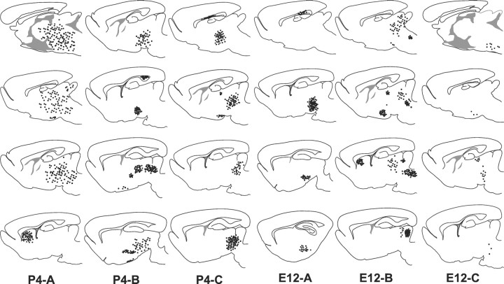
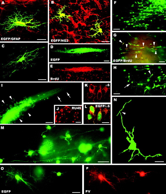
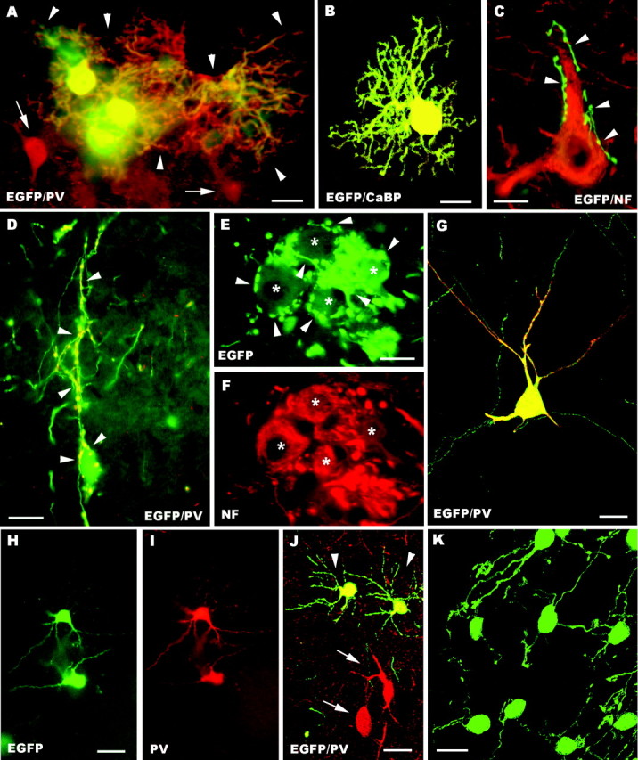
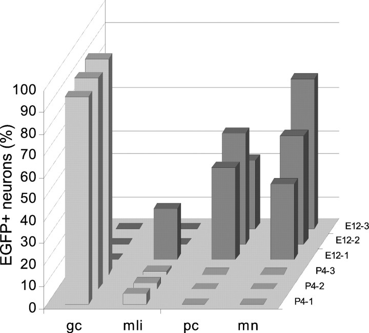
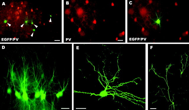
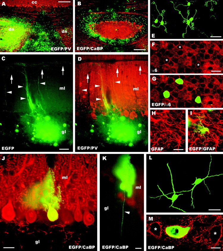
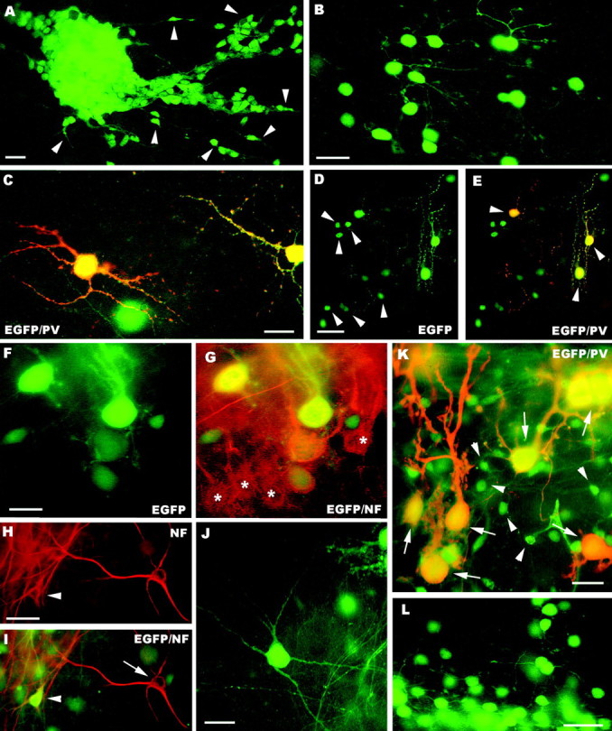
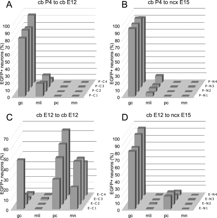
The different cerebellar phenotypes are generated according to a precise time schedule during embryonic and postnatal development. To assess whether the differentiative potential of cerebellar progenitors is progressively restricted in space and time we examined the fate of embryonic day 12 (E12) or postnatal day 4 (P4) cerebellar cells after heterotopic-heterochronic transplantation into the embryonic rat brain in utero or into organotypic CNS explants in vitro. Donor cells, isolated from transgenic mice overexpressing the enhanced-green fluorescent protein under the control of the beta-actin-promoter, engrafted throughout the host brainstem and diencephalon, whereas they rarely incorporated into specific telencephalic structures. In any recipient site, the vast majority of transplanted cells could be recognized as cerebellar phenotypes, and we did not obtain clear evidence that ectopically located cells adopted host-specific identities. Nevertheless, the two donor populations displayed different developmental potentialities. P4 progenitors exclusively generated granule cells and molecular layer interneurons, indicating that they are committed to late-generated cerebellar identities and not responsive to heterotopic-heterochronic environmental cues. In contrast, E12 precursors had the potential to produce all major cerebellar neurons, but the repertoire of adult phenotypes generated by these cells was different in distinct host regions, suggesting that they require instructive environmental information to acquire mature identities. Thus, cerebellar precursors are able to integrate into different foreign brain regions, where they develop mature phenotypes that survive long after transplantation, but they are committed to cerebellar fates at E12. Embryonic progenitors are initially capable, although likely not competent, to generate all cerebellar identities, but their potential is gradually restricted toward late-generated phenotypes.


Figures

Fig. 1.
Fig. 1.
Distribution of postnatal (P4-A–C) and embryonic (E12-A–C) cerebellar cells transplanted to the embryonic rat brain in utero. For each case, four serial sections, modified from Paxinos and Watson (1982), are shown (top to bottom = medial to lateral). Each dotrepresents cellular aggregates or groups of single scattered cells.
Fig. 2.
Fig. 2.
A–P, Fate of postnatal cerebellar cells engrafted in ectopic regions of the embryonic brainin utero. The confocal pictures A–C show glial phenotypes generated by postnatal cerebellar precursors, including GFAP-positive astrocytes (A), NG2-positive immature oligodendrocytes (B, taken from an animal killed at P1), and mature oligodendrocytes properly integrated in host white matter tracts (C). A large aggregate of small, densely packed transplanted cells is shown inD (taken from an animal killed at P1); BrdU labeling of the same microscopic field (E) reveals the intense proliferative activity of these cells (numerous host cells outside the cluster are also labeled). Migrating cells, with typical leading and trailing processes, radiate from such clusters (F) and likely continue to divide, as shown by BrdU incorporation (G, arrowheads point to some double-labeled cells).  At the end of their migratory phase (H), the cells show several short processes radiating from the perikaryon (e.g.,arrowhead) and eventually acquire the typical morphology of granule cells (e.g., arrow). Mature granule cells can be found scattered or clustered in aggregates (arrowheads in I) with bundles of parallel fibers (arrows). The identification of these cells is further supported by the expression of RU49 mRNA (J) as well as α6 subunit of the GABAA receptor (K, L). The typical morphology of granule neurons, with small round cell bodies, a few short clawed dendrites and thin varicose axons, is displayed inM. In addition, the confocal image Nshows an immature granule cell bearing the characteristic T-shaped bifurcation (arrowhead) of the parallel fiber.O and P display parvalbumin-immunopositive molecular layer interneurons.GFAP, Glial fibrillary acidic protein; α6, α6 subunit of the GABAA receptor; BrdU, bromodeoxyuridine; PV, parvalbumin; EGFP, enhanced green fluorescent protein. Scale bars: A, C, K, L, N, 10 μm; B, 15 μm; M, O, P, 30 μm; F–H, J, 50 μm; D, E, I, 100 μm.
Fig. 3.
Fig. 3.
A–K, Fate of embryonic cerebellar cells engrafted in ectopic regions of the embryonic brain in utero. Parvalbumin-immunolabeled Purkinje cells with large dendritic trees (arrowheads) are illustrated inA; arrows point to adjacent parvalbumin-positive host neurons. Another calbindin-immunolabeled transplanted Purkinje cell, bearing less extended dendrites, is shown in the confocal image B. In C a presumptive EGFP-positive Purkinje axon (arrowheads), recognized by the particularly intense fluorescence, terminates on a neurofilament immunostained host neuron. Frequently, Purkinje axons (arrowheads in D point to the parvalbumin-immunolabeled terminal branches) enwrap the somatodendritic  surface of EGFP-positive multipolar neurons. E and Fshow a small cluster of these neurons (asterisks), which are labeled by anti-neurofilament antibodies (F) and covered by strongly fluorescent Purkinje axons (arrowheads in E). The morphological features of EGFP-multipolar neurons, bearing slender dendrites with long ramifications, are illustrated in the confocal pictureG; note that this neuron is also double labeled for parvalbumin. H and I show small parvalbumin-immunopositive neurons, classified as molecular layer interneurons. Arrowheads in J point to another two of such neurons settled in the host striatum; note the different size and morphology shown by transplanted and recipient neurons (arrows). A cluster of transplanted granule cells in the host dorsal cochlear nucleus is displayed by the confocal picture K. NF, Neurofilament SMI32; PV, parvalbumin; CaBP, calbindin; EGFP, enhanced green fluorescent protein. Scale bars: K, 10 μm; C, E, F, 15 μm; B, D, 20 μm;A, 25 μm; G–J, 30 μm.
Fig. 4.
Fig. 4.
Quantitative analysis of the phenotypic repertoire generated by embryonic (dark gray) and postnatal (light gray) cerebellar cells transplanted to the embryonic rat brain in utero. For each experimental set, three representative cases are illustrated (indicated on thez-axis). The different neuron phenotypes are represented as the percentage on the total number of transplanted nerve cells observed in the relevant brain. Granule cells generated by embryonic progenitors in the lower brainstem are not included in these counts that were performed on sections encompassing mesencephalon and forebrain, where no granule neurons were found. gc, Granule cells; mli, molecular layer interneurons;pc, Purkinje cells; mn, multipolar neurons.
Fig. 5.
Fig. 5.
A–F, Fate of embryonic neocortical precursors grafted to the embryonic CNS in utero. Micrograph A shows EGFP-positive embryonic neocortical cells (some are pointed by arrowheads) engrafted in the host neocortex. Some of such neurons (B, C) show the morphology of stellate cells and can be immunolabeled by anti-parvalbumin antibodies. Many of these grafted cells show site-specific morphological features, such as presumptive pyramidal neurons in hippocampal CA1 (D), medium-sized spiny neurons in the striatum (E), and olfactory bulb interneurons (F). PV, Parvalbumin, EGFP, enhanced green fluorescent protein. Scale bars: B–E, 20 μm; A,F, 50 μm.
Fig. 6.
Fig. 6.
A–M, Fate of postnatal (A–I) and embryonic (J–M) cerebellar cells homotopically engrafted in the embryonic cerebellumin utero. Micrograph A, taken from a rat transplanted with postnatal donor cells, shows a survey of the host cerebellum, stained with anti-parvalbumin antibodies (dn, deep cerebellar nuclei; cc,cerebellar cortex): note the large aggregates of EGFP-positive cells in the region of the deep nuclei. Numerous EGFP-positive cells are also located in the granular layer (B), where they develop into mature granule cells. Such neurons display typical ascending axons (arrowheads in C, D) and parallel fibers (arrows) running along the longitudinal axis of the folium. In addition, they bear characteristic short clawed dendrites (E) and express the α6 subunit of the GABAA receptor (F–G) as their host counterparts (asterisks in F indicate the position of the EGFP-positive neurons). Finally, postnatal cerebellar progenitors also develop into some GFAP-positive astrocytes (H, I). Embryonic progenitors produce granule cells (data not shown) and Purkinje cells (J, K) correctly positioned in the Purkinje cell layer, with monoplanar dendritic trees and thin axons  (arrowhead in K) running across the granular layer. In addition, they develop deep nuclei neurons, properly positioned in the central gray matter (L, M). Such transplanted cells acquire the typical multipolar shape (L) and are innervated by Purkinje axons: the confocal picture M shows an EGFP-positive neuron surrounded by calbindin immunopositive Purkinje axon boutons (asterisks point to two adjacent host neurons that are similarly innervated). CaBP, Calbindin;PV, parvalbumin; α6, α6 subunit of the GABAA receptor; EGFP, enhanced green fluorescent protein; ml, molecular layer;gl granular layer. Scale bars: E–G, M, 10 μm; K, 20 μm; J, 25 μm;H, I, 30 μm; L, 40 μm; C, D, 50 μm; B, 150 μm; A, 250 μm.
Fig. 7.
Fig. 7.
A–H, Fate of postnatal (A–E) and embryonic (F–L) cerebellar cells transplanted to cerebellar (A–C, F–J) or neocortical explants (D, E, K, L). In the cerebellar explants, postnatal cerebellar progenitors form dense aggregates (A) with numerous migrating elements (some are pointed byarrowheads). Mature granule cells, bearing typical morphological features, are shown in B, whereas parvalbumin-immunopositive molecular layer interneurons are displayed in C. D and E illustrate the neuronal phenotypes generated by postnatal cerebellar cells grafted  to neocortical explants, granule cells (arrowheads in D) and molecular layer interneurons (arrowheadsin E). The latter are also labeled by anti-parvalbumin antibodies (E). The micrographs Fand G show EGFP-positive Purkinje cells derived from embryonic precursors intermingled with their host counterparts (asterisks in G) in the cerebellar cortex of the explant. Examples of EGFP-positive multipolar neurons in cerebellar explants are illustrated in H–J. Some of these neurons (arrowhead in H andI) are localized in the deep nuclei of the explant, and can be stained by anti-neurofilament antibodies (arrow in I points to an immunolabeled host neuron). In neocortical explants embryonic cerebellar progenitors also generate parvalbumin-immunopositive Purkinje cells (arrows in K) with variably shaped dendritic trees and granule cells (K, L, some are pointed by arrowheads in K).PV, Parvalbumin; NF, neurofilament;EGFP, enhanced green fluorescent protein. Scale bars:B, C, 20 μm; A, F, G, K, 25 μm;L, 30 μm; D, E, H–J, 50 μm.
Fig. 8.
Fig. 8.
A–D, Quantitative analysis of the phenotypic repertoire generated by postnatal (A, B) and embryonic (C, D) cerebellar cells transplanted to cerebellar (A, C) or neocortical (B, D) explants. Each histogram shows the distribution of neuron phenotypes generated by grafted cells in four representative explants from each experimental set (indicated on the z-axis of each histogram). The different cell types are represented as the percentage on the total number of transplanted neurons observed in the relevant explant.gc, Granule cells; mli, molecular layer interneurons; pc, Purkinje cells; mn, multipolar neurons; cb, brainstem-cerebellar explant;ncx, neocortical explant.

Similar articles

Cited by

References

    1. Alder J, Cho NK, Hatten ME. Embryonic precursor cells from the rhombic lip are specified to a cerebellar granule neuron identity. Neuron. 1996;17:389–399. - PubMed
    1. Altman J, Bayer SA. Development of the cerebellar system in relation to its evolution, structure and functions. CRC; New York: 1997.
    1. Altman J, Das G. Autoradiographic and histological studies of postnatal histogenesis. II. A longitudinal investigation of kinetics, migration and transformation of cells incorporating thymidine in infant rat with special reference to postnatal neurogenesis in some brain regions. J Comp Neurol. 1966;126:337–390. - PubMed
    1. Alvarado-Mallart RM. Fate and potentialities of the avian mesencephalic/metencephalic neuroepithelium. J Neurobiol. 1993;24:1341–1355. - PubMed
    1. Anderson DJ. Stem cells and pattern formation in the nervous system: the possible versus the actual. Neuron. 2001;30:19–35. - PubMed

Publication types

MeSH terms

LinkOut - more resources