

Aged rats are impaired on an attentional set-shifting task sensitive to medial frontal cortex damage in young rats
- PMID: 12177232
- PMCID: PMC182583
- DOI: 10.1101/lm.48602
Aged rats are impaired on an attentional set-shifting task sensitive to medial frontal cortex damage in young rats
Abstract
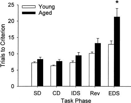
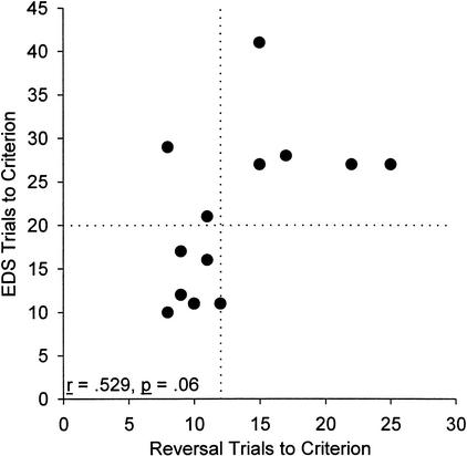
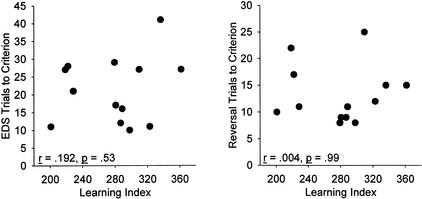
Normal aging is associated with disruption of neural systems that subserve different aspects of cognitive function, particularly in the hippocampus and frontal cortex. Abnormalities in hippocampal function have been well investigated in rodent models of aging, but studies of frontal cortex function in aged rodents are few. We tested young (4-5 mo old) and aged (27-28 mo old) male Long-Evans rats on an attentional set-shifting task modified slightly from previous publication. After training on two problems in which the reward was consistently associated with the same stimulus dimension, and a reversal of one problem, a new problem was presented in which the reward was consistently associated with the previously irrelevant stimulus dimension (extradimensional shift [EDS]). Aged rats as a group were significantly impaired on the EDS, although some individual aged rats performed as well as young rats on this phase. In addition, some aged rats were impaired on the reversal, although a group effect did not reach significance in this phase. Impairment in neither reversal nor EDS was associated with impairments in spatial learning in the Morris water maze. Young rats with neurotoxic lesions of medial frontal cortex are also selectively impaired on the EDS. These results indicate that normal aging in rats is associated with impaired medial frontal cortex function. Furthermore, age-related declines in frontal cortex function are independent of those in hippocampal function. These results provide a possible basis for correlating age-related changes in neurobiological markers in frontal cortex with cognitive decline.
Figures
Comment in
-
It's time to pay attention to attention in aging.Learn Mem. 2002 Jul-Aug;9(4):151-2. doi: 10.1101/lm.52902. Learn Mem. 2002. PMID: 12177227 No abstract available.
References
-
- Albert MS, Stafford JL. CT scan and neuropsychological relationships in aging and dementia. In: Goldstein G, Taske RE, editors. Advances in clinical neuropsychology. Vol. 3. New York: Plenum; 1986. pp. 31–53.
-
- Bachevalier J, Mishkin M. Visual recognition impairment follows ventromedial but not dorsolateral prefrontal lesions in monkeys. Behav Brain Res. 1986;20:249–261. - PubMed
-
- Bartus RT, Fleming D, Johnson HR. Aging in the rhesus monkey: Debilitating effects on short-term memory. J Gerontol. 1978;33:858–871. - PubMed
-
- Bartus RT, Dean RL, III, Fleming DL. Aging in the rhesus monkey: Effects on visual discrimination learning and reversal learning. J Gerontol. 1979;34:209–219. - PubMed
-
- Baxter MG. Cognitive aging in nonhuman primates. In: Hof PR, Mobbs CV, editors. Functional neurobiology of aging. San Diego, CA: Academic Press; 2001. pp. 407–419.
Publication types
MeSH terms
Substances
Grants and funding
LinkOut - more resources
Full Text Sources
Medical