. 2002 Aug 15;30(16):3497-531.
doi: 10.1093/nar/gkf481.

The non-Watson-Crick base pairs and their associated isostericity matrices

Affiliations

The non-Watson-Crick base pairs and their associated isostericity matrices

Neocles B Leontis et al. Nucleic Acids Res. .

Abstract

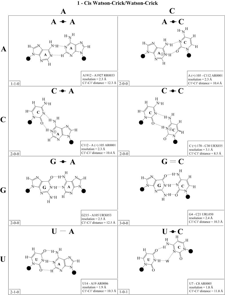
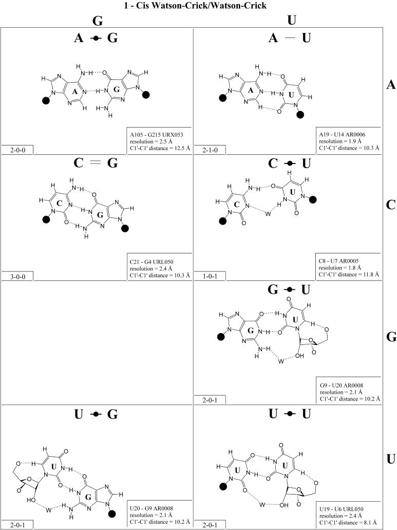
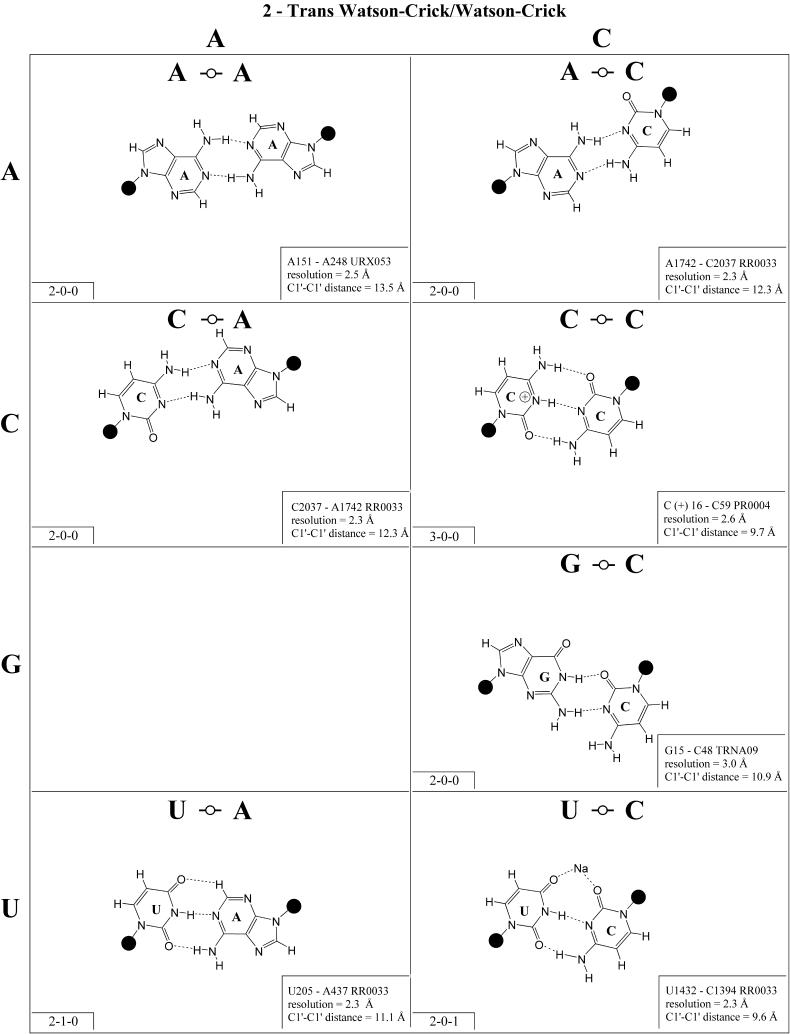
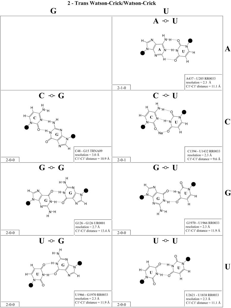
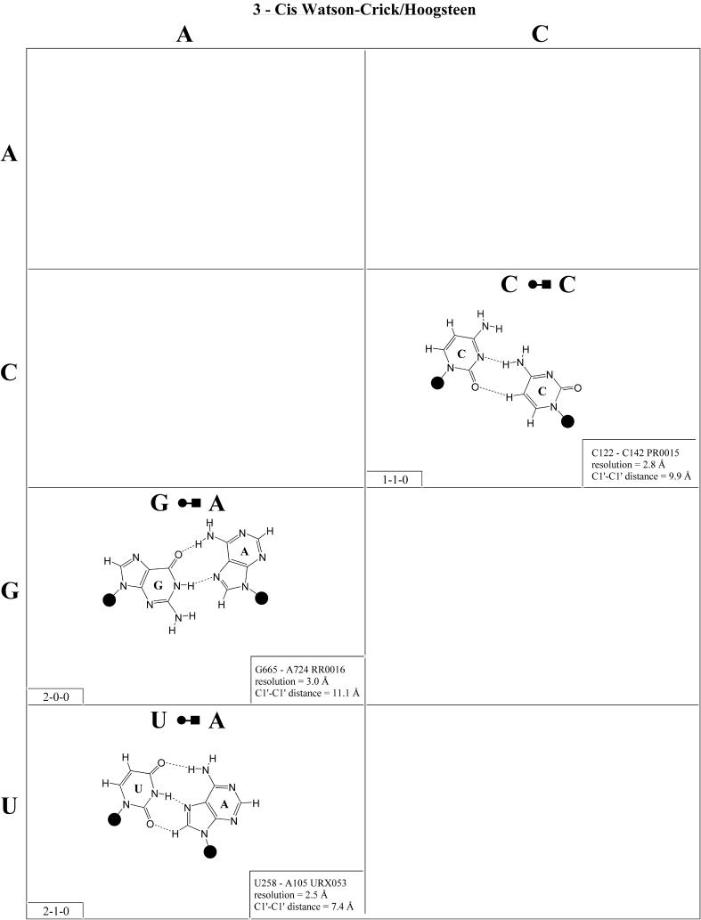
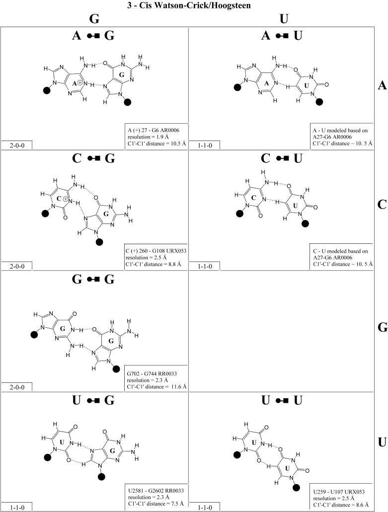
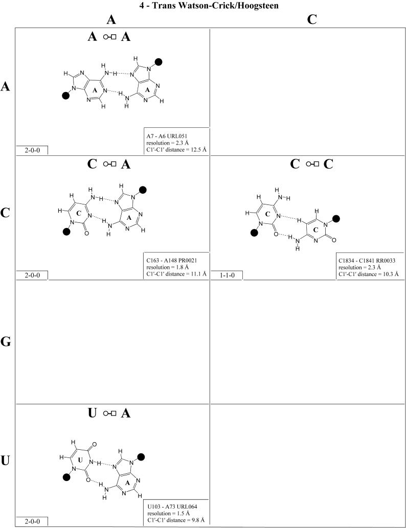
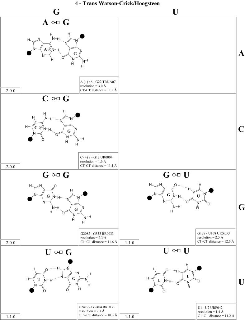
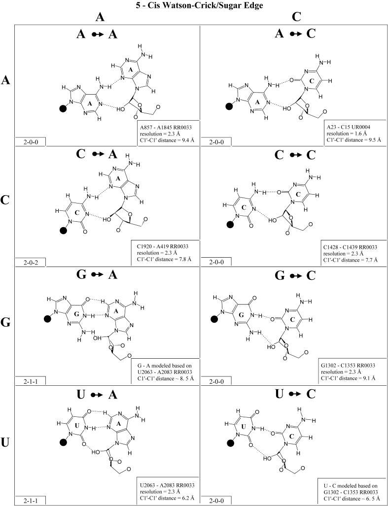
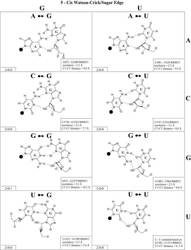
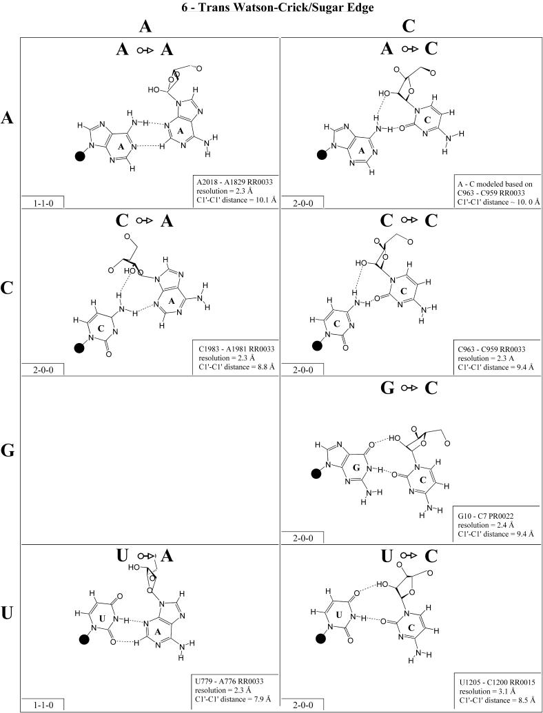
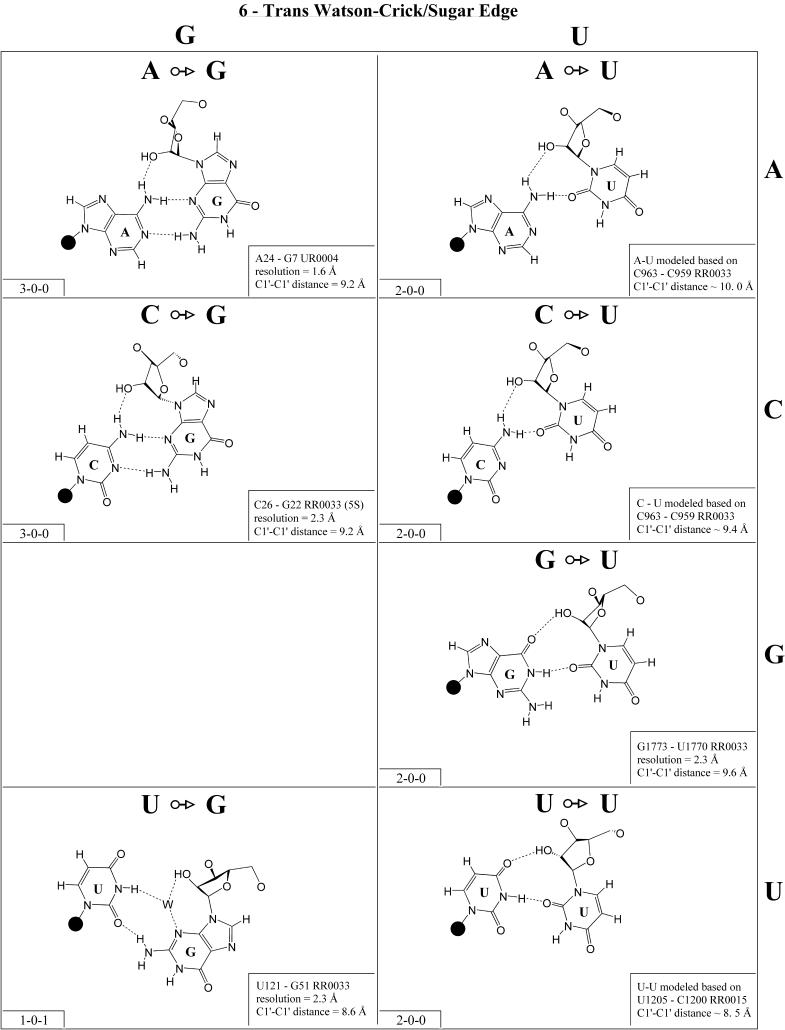
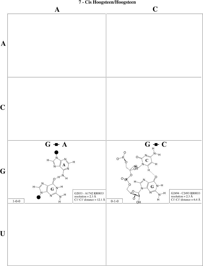
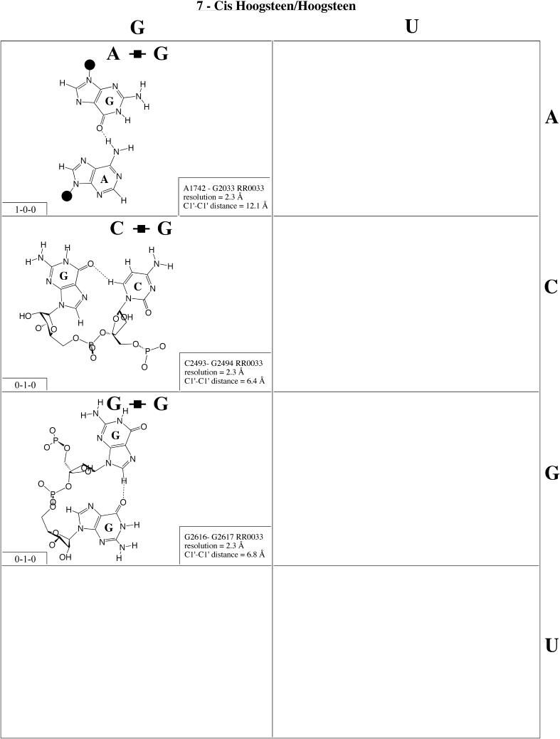
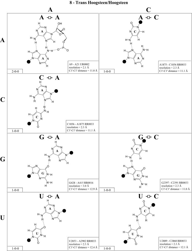
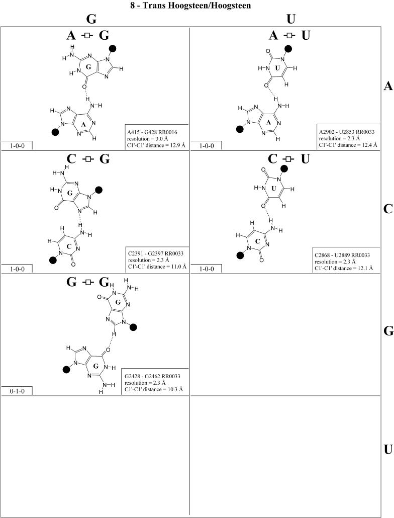
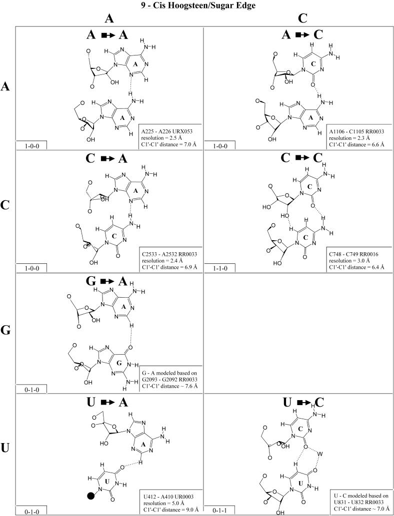
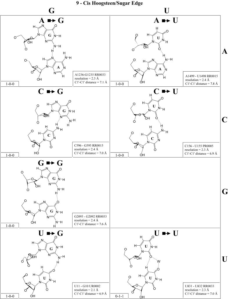
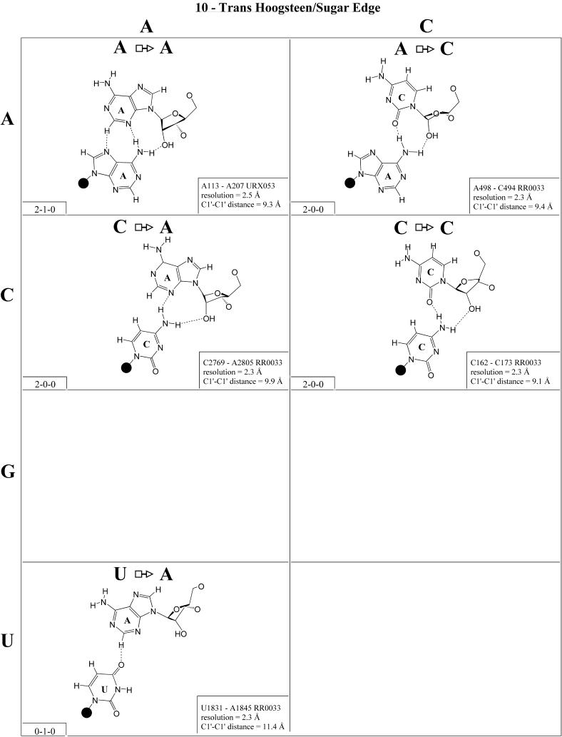
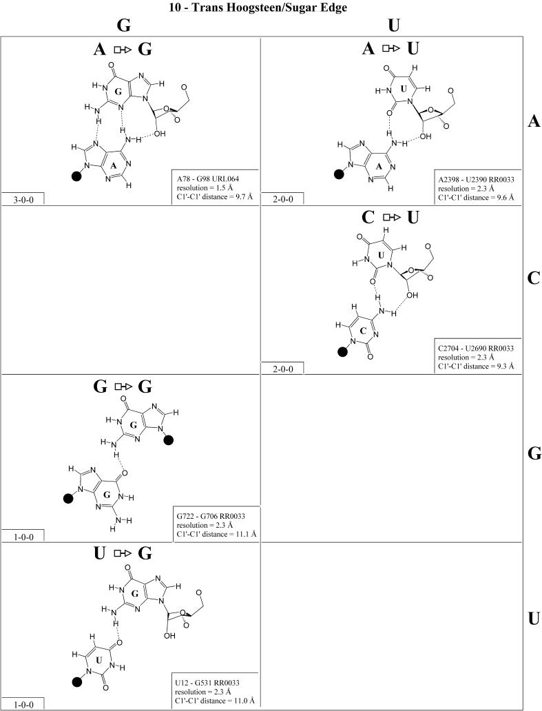
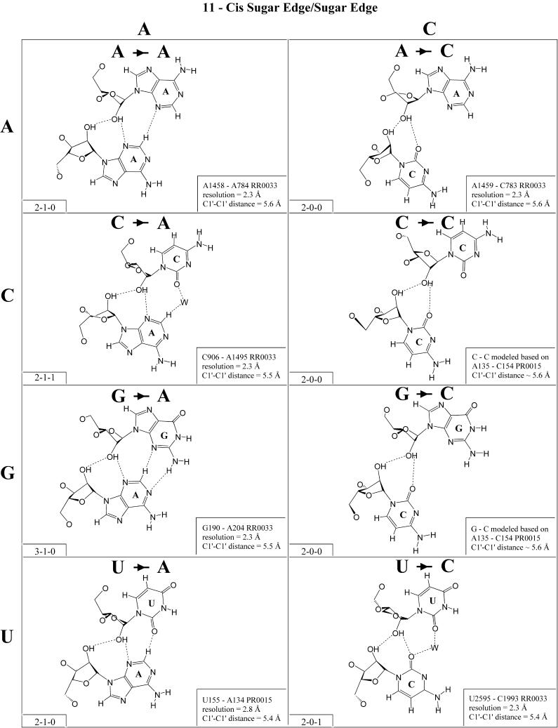
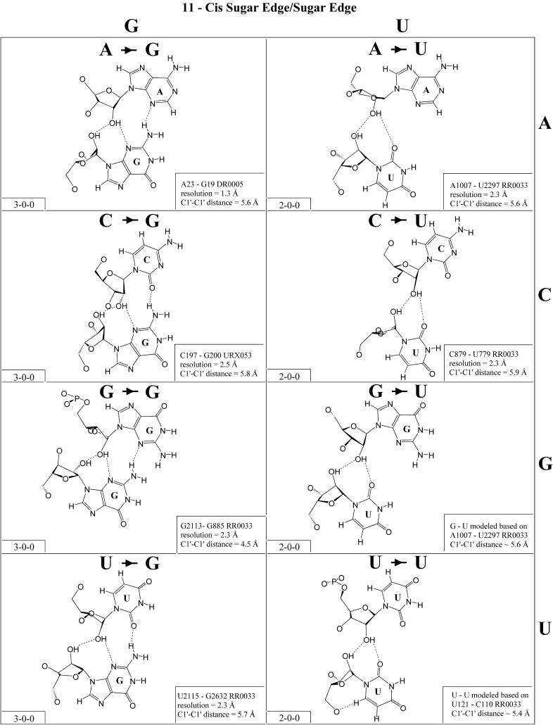
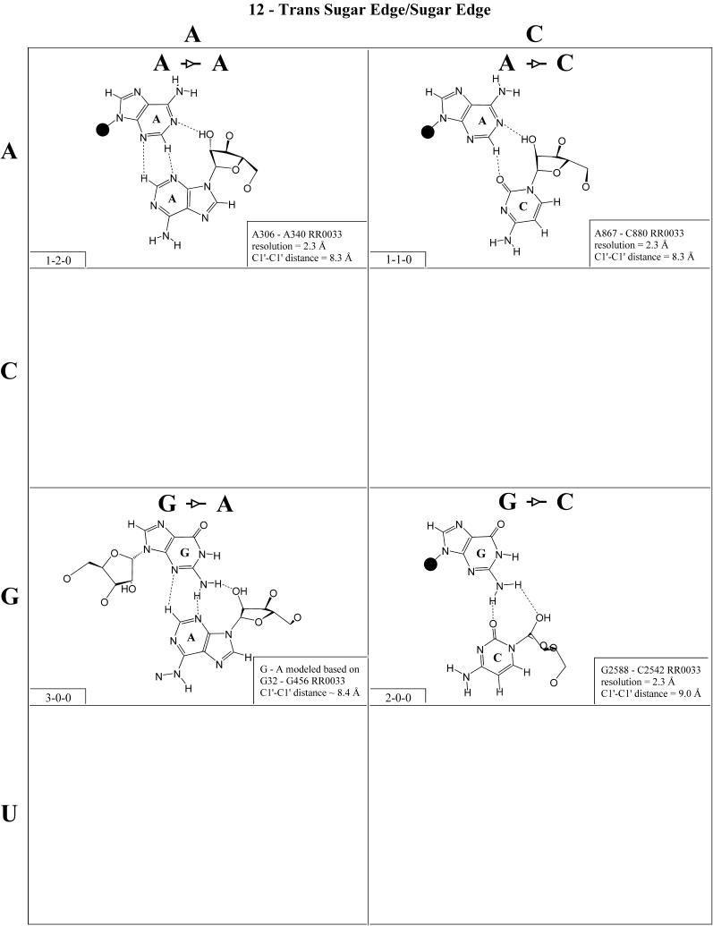
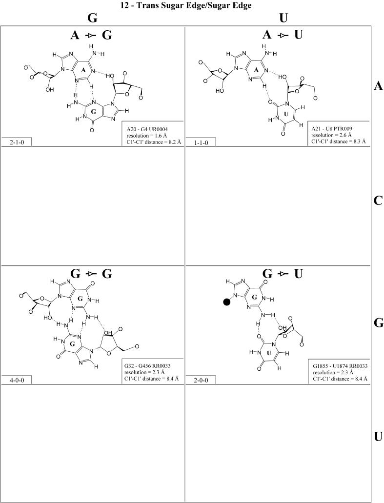
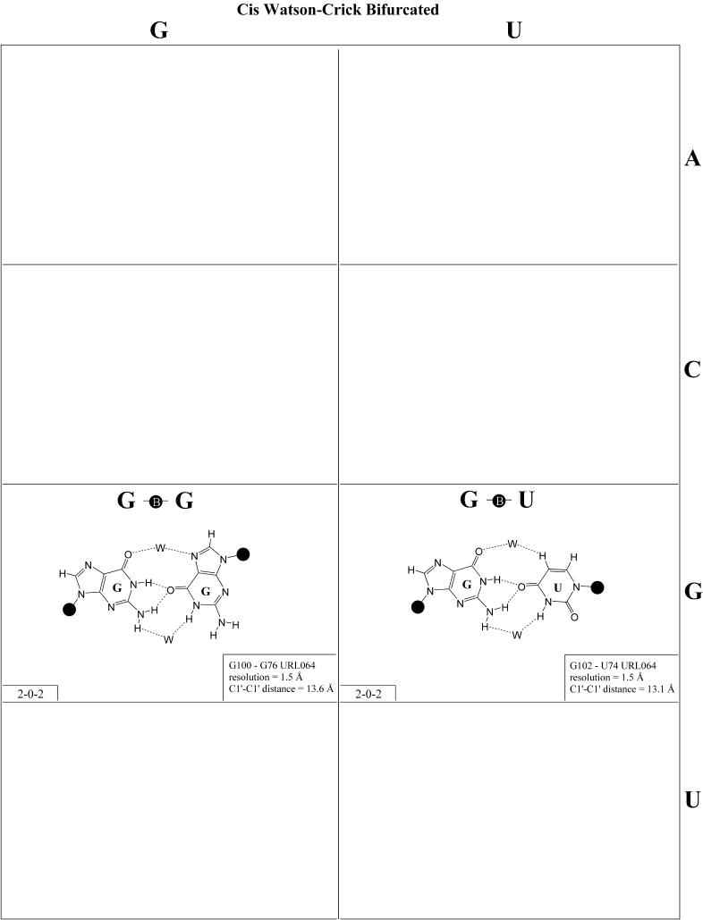
RNA molecules exhibit complex structures in which a large fraction of the bases engage in non-Watson-Crick base pairing, forming motifs that mediate long-range RNA-RNA interactions and create binding sites for proteins and small molecule ligands. The rapidly growing number of three-dimensional RNA structures at atomic resolution requires that databases contain the annotation of such base pairs. An unambiguous and descriptive nomenclature was proposed recently in which RNA base pairs were classified by the base edges participating in the interaction (Watson-Crick, Hoogsteen/CH or sugar edge) and the orientation of the glycosidic bonds relative to the hydrogen bonds (cis or trans). Twelve basic geometric families were identified and all 12 have been observed in crystal structures. For each base pairing family, we present here the 4 x 4 'isostericity matrices' summarizing the geometric relationships between the 16 pairwise combinations of the four standard bases, A, C, G and U. Whenever available, a representative example of each observed base pair from X-ray crystal structures (3.0 A resolution or better) is provided or, otherwise, theoretically plausible models. This format makes apparent the recurrent geometric patterns that are observed and helps identify isosteric pairs that co-vary or interchange in sequences of homologous molecules while maintaining conserved three-dimensional motifs.


Figures

Figure 1
Figure 1
(Left) Identification of edges in the RNA bases. (Right) cis versus trans orientation of glycosidic bonds.
Figure 2
Figure 2
(Opposite and above) 4 × 4 matrix displaying observed base pairs belonging to the cis Watson–Crick/Watson–Crick family. The canonical Watson–Crick pairs comprise the diagonal of the matrix. Symbols used in Figures 2–14 employ circles to designate Watson–Crick edges, squares for Hoogsteen or pyrimidine CH edges, and triangles for sugar edges. Solid symbols indicate cis base pairs and open symbols trans base pairs. In the lower left-hand corner of each panel in Figures 2–14, numbers describing the hydrogen bonding are provided. The first is the number of observed or potential hydrogen bonds between two nitrogen- or oxygen-containing groups, i.e. normal hydrogen bonds. The second is the number of hydrogen bonds involving polarized C-H groups (i.e. AH2, RH8, YH5 or YH6). The third is the number of bridging water molecules.
Figure 2
Figure 2
(Opposite and above) 4 × 4 matrix displaying observed base pairs belonging to the cis Watson–Crick/Watson–Crick family. The canonical Watson–Crick pairs comprise the diagonal of the matrix. Symbols used in Figures 2–14 employ circles to designate Watson–Crick edges, squares for Hoogsteen or pyrimidine CH edges, and triangles for sugar edges. Solid symbols indicate cis base pairs and open symbols trans base pairs. In the lower left-hand corner of each panel in Figures 2–14, numbers describing the hydrogen bonding are provided. The first is the number of observed or potential hydrogen bonds between two nitrogen- or oxygen-containing groups, i.e. normal hydrogen bonds. The second is the number of hydrogen bonds involving polarized C-H groups (i.e. AH2, RH8, YH5 or YH6). The third is the number of bridging water molecules.
Figure 3
Figure 3
(Opposite and above) Observed base pairs of the trans Watson–Crick/Watson–Crick family. The pairing displays a 2-fold rotational symmetry. Thus, the matrix is symmetric.
Figure 3
Figure 3
(Opposite and above) Observed base pairs of the trans Watson–Crick/Watson–Crick family. The pairing displays a 2-fold rotational symmetry. Thus, the matrix is symmetric.
Figure 4
Figure 4
(Opposite and above) Observed and modeled base pairs of the cis Watson–Crick/Hoogsteen family.
Figure 4
Figure 4
(Opposite and above) Observed and modeled base pairs of the cis Watson–Crick/Hoogsteen family.
Figure 5
Figure 5
(Opposite and above) Observed base pairs of the trans Watson–Crick/Hoogsteen family.
Figure 5
Figure 5
(Opposite and above) Observed base pairs of the trans Watson–Crick/Hoogsteen family.
Figure 6
Figure 6
(Opposite and above) Observed and modeled base pairs of the cis Watson–Crick/sugar edge family.
Figure 6
Figure 6
(Opposite and above) Observed and modeled base pairs of the cis Watson–Crick/sugar edge family.
Figure 7
Figure 7
(Opposite and above) Observed and modeled base pairs of the trans Watson–Crick/sugar edge family.
Figure 7
Figure 7
(Opposite and above) Observed and modeled base pairs of the trans Watson–Crick/sugar edge family.
Figure 8
Figure 8
(Opposite and above) Observed base pairs of the cis Hoogsteen/Hoogsteen family.
Figure 8
Figure 8
(Opposite and above) Observed base pairs of the cis Hoogsteen/Hoogsteen family.
Figure 9
Figure 9
(Opposite and above) Observed base pairs of the trans Hoogsteen/Hoogsteen family. The pairing displays a 2-fold rotational symmetry. Thus, the matrix is symmetric.
Figure 9
Figure 9
(Opposite and above) Observed base pairs of the trans Hoogsteen/Hoogsteen family. The pairing displays a 2-fold rotational symmetry. Thus, the matrix is symmetric.
Figure 10
Figure 10
(Opposite and above) Observed and modeled base pairs of the cis Hoogsteen/sugar edge family.
Figure 10
Figure 10
(Opposite and above) Observed and modeled base pairs of the cis Hoogsteen/sugar edge family.
Figure 11
Figure 11
(Opposite and above) Observed base pairs of the trans Hoogsteen/sugar edge family.
Figure 11
Figure 11
(Opposite and above) Observed base pairs of the trans Hoogsteen/sugar edge family.
Figure 12
Figure 12
(Opposite and above) Observed and modeled base pairs of the cis sugar edge/sugar edge family.
Figure 12
Figure 12
(Opposite and above) Observed and modeled base pairs of the cis sugar edge/sugar edge family.
Figure 13
Figure 13
(Opposite and above) Observed and modeled base pairs of the trans sugar edge/sugar edge family.
Figure 13
Figure 13
(Opposite and above) Observed and modeled base pairs of the trans sugar edge/sugar edge family.
Figure 14
Figure 14
(Opposite and above) Observed and modeled base pairs of the cis Watson–Crick bifurcated family.
Figure 14
Figure 14
(Opposite and above) Observed and modeled base pairs of the cis Watson–Crick bifurcated family.

Similar articles

Cited by

References

    1. Woese C.R., Winker,S. and Gutell,R.R. (1990) Architecture of ribosomal RNA: constraints on the sequence of “tetra-loops”. Proc. Natl Acad. Sci. USA, 87, 8467–8471. - PMC - PubMed
    1. Leontis N.B. and Westhof,E. (1998) A common motif organizes the structure of multi-helix loops in 16 S and 23 S ribosomal RNAs. J. Mol. Biol., 283, 571–583. - PubMed
    1. Nissen P., Ippolito,J.A., Ban,N., Moore,P.B. and Steitz,T.A. (2001) RNA tertiary interactions in the large ribosomal subunit: the A-minor motif. Proc. Natl Acad. Sci. USA, 98, 4899–4903. - PMC - PubMed
    1. Moore P.B. (1999) Structural motifs in RNA. Annu. Rev. Biochem., 68, 287–300. - PubMed
    1. Hoogsteen K. (1963) The crystal and molecular structure of a hydrogen-bonded complex between 1-methylthymine and 9-methyladenine. Acta Crystallogr., 16, 907–916.

Publication types